Cardiac Output: Physiology and Regulation
 Call Kai
 Learn
 Practice Test
 Spaced Repetition
 Match
 Flashcards
 Knowt Play
1/22

Earn XP
Description and Tags
Vocabulary practice flashcards covering cardiac output, autonomic and hormonal regulation of heart rate, and length-tension and contractility mechanisms regulating stroke volume.
23 Terms
Cardiac Output (Q˙)
The volume of blood pumped out by the heart every minute, calculated from Cardiac Output=Heart Rate×Stroke Volume and measured in ml⋅min−1 or l⋅min−1. (the little dot on the q means per unit of time like per minute), sometimes it gets abbreviated to CO
Stroke Volume (SV)
The volume of blood ejected by the heart per beat, measured in millilitres (ml) or litres (l).
Heart Rate (HR)
The frequency of cardiac contractions expressed in beats per minute (beats⋅min−1), functioning alongside stroke volume to determine cardiac output.
Vagus Nerve
The parasympathetic pathway whose nerve endings innervate the atria at the SA and AV nodes and secrete acetylcholine to slow the heart rate.
Acetylcholine
The neurotransmitter secreted by parasympathetic vagus nerve endings that hyperpolarises SA node cell membranes and slows the depolarising drift, decreasing heart rate.
Norepinephrine
The sympathetic neurotransmitter released by cardiac nerves that binds to β receptors on the SA and AV nodes and myocardium, increasing heart rate and contractile force.
Circulating Epinephrine
An adrenal hormone carried in the bloodstream that acts on myocardial β receptors to elevate heart rate and contractility, but lags behind direct neural stimulation.
Pacemaker Potential
The spontaneous depolarising drift of SA node cell membranes from resting potential toward the threshold of −40mV, preceding the generation of an action potential.
Muscarinic (M) Receptors
Receptors located on atrial tissue that bind parasympathetic acetylcholine to decrease pacemaker activity and lower heart rate.
Beta (β) Adrenergic Receptors
Receptors located on the atria and ventricles that bind sympathetic norepinephrine and circulating epinephrine to augment heart rate, conduction velocity, and contractility.
Starling's Law of the Heart
The principle stating that stretching cardiac muscle fibres via increased end-diastolic filling causes them to contract more forcefully, directly increasing stroke volume (also called the Frank-Starling mechanism). - the more the heart stretches the stronger the contraction
End-Diastolic Volume (EDV)
The volume of blood filling the ventricles prior to contraction, which increases with venous return and determines the resting stretch of cardiac muscle fibres.
Venous Return
The volume of blood flowing back from the circulation into the heart, which directly determines ventricular end-diastolic filling and muscle stretch.
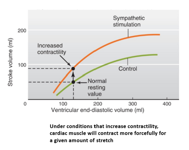
Contractility
The intrinsic force of cardiac muscle contraction for a given amount of stretch, augmented by sympathetic nerve activity (norepinephrine) and circulating epinephrine.
Afterload
The arterial pressure against which the ventricles must pump to eject blood, serving as one of the two primary opposing factors regulating stroke volume.
Atrioventricular (AV) Node Delay
The physiological pause in conduction between atrial and ventricular contraction, which is shortened under sympathetic stimulation to accommodate elevated heart rates.
why does the cardiac output increase diagram all steps?

stroke volume:
what 2 things regulate the stroke volume?
what 2 processes regulate the force of contraction?
the force by which muscle cells contract + the arterial pressure against which they have to eject the blood
the length-tension properties of cardiac muscle cells + the effects of hormonal influence on the contractility of the cardiac muscle
why does parasympathetic stimulation decrease the HR?
why does sympathetic stimulation increase the HR?
causes hyperpolarisation so voltage go further away from action potential (more negative) and so the depolorising drift happens slower and so it takes longer to reach the action potential
causes depolorisation so voltage goes closer to the action potential (more positive) and so the depolorising drift becomes faster and so it takes less time to reach the action potential
why is there a maximum to cardiac output?
as its a combo of heart rate and SV, HR is linear with cardiac output but SV is different and decreases after a while (due to shorter filling time as hr increases) and so cardiac output can’t keep on increasing

typical values for cardiac output

what 2 things does cardiac output depend on?
heart rate and stroke volume
talk about how diastolic and systolic phase changes during both light to moderate exercise and heavy to severe exercise?
during light to moderate exercise systolic and diastolic pressure increases
during heavy to severe exercise systolic pressure increases and diastolic pressure decreases