Lymphoproliferative Disorders
 Call Kai
 Learn
 Practice Test
 Spaced Repetition
 Match
 Flashcards
 Knowt Play
1/26

There's no tags or description
Looks like no tags are added yet.
27 Terms
Lymphoproliferative Disorders
B and T lymphocyte malignant proliferation
Chronic, slow progression
Mainly affects elderly
related to compromised immune systems
Diagnosed w/ flowcytometry and chromosomal analysis
Chronic Lymphocytic Leukemia (CLL)
B lymph proliferation
May be due to chromosomal abnormalities
Small lymphs accumulate in spleen, lymph nodes, BM→ spills out into peripheral circulation
CLL Lab/ peripheral findings
Key: mature lymphs but are nonfunctional
High WBC counts
90% lymphs
Infection-prone due to immunocompromised
10% develop into AIHA (autoimmune hemolytic anemia)
Spherocytes, nRBCs
Smudge cells
fragile remnants lacking identifiable membrane or nuclear structure
WBC: >100,000/ cu mm
M:E ratio 10-20:1
Peripheral blood is almost exclusively small lymphocytes with a few lymphoblasts
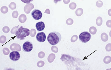
CLL symptoms
Fatigue
pallor
weight loss
CLL disease progression/ genes
BCL2: Anti-apoptosis gene in 80% of cases
Dysfunctional B cells live longer
RAI staging system
Common hypogammaglobinemia
CLL may lead to
splenomegaly
lymphadenopathy
anemia
thromboneutropenia
altered immune funtion
AIHA
CLL treatments
Irradiation
drugs
aklylating agents
Monoclonal Abs
Allogenic stem cell transplant
Hair Cell Leukemia (HCL) + marker
Rare B cell malignancy (2%)
“Hairy” mononuclear cells
Fragile
hair-like projections off cytoplasm
Spongy, looks like chromatin
Diagnose with cytochemical stain: tartrate resistant acid phosphatase (TRAP)
CD103: hairy cell marker
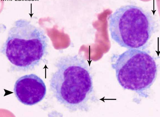
TRAP (tartrate acid phosphatase)
Diagnoses HCL
Hair cells STRONGLY positive, other cells may be somewhat positive
Hairy cells remain stained when tartrate acid is added but other cells become negative
HCL symptoms
Pancytopenia
splenomegaly
bleeding
infection
anemia
Dry tap: BM too fibrous and BM cannot be aspirated
HCL treatment
Therapeutic splenectomy
Improves cytopenias, hypersplenism, physical symptoms
Interferon alpha
2-chlorodeoxyadenosine (2-CdA)→ had remissions
Sezary Syndrome
T-cell lymphoma associated w/ mycosis fungoides
Cutaneous manifestation
Infiltration of abnormal lymphs
May affect spleen, BM, lymph nodes
Deletions on chromosomes 10 and 17, or duplications on 8 and 17
Sezary peripheral blood
Large cells (8-20 um)
Nucleus: ovoid, clefted, or folded
Chromatin: smooth, homogenous
may look like monocytes

Prolymphocytic Leukemia
Variant of CLL
Majority of circulating prolymphocytes
has more cytoplasm than mature lymphocytes
Symptoms:
Splenomegaly
Liver involvement
Elevated WBC count

Hodgkin’s Lymphoma
Cervical lymph nodes are firm to the touch and does not disappear
Hypermetabolism:
Low grade fever
weight loss
Lymph node biopsy determines diagnosis
Hodgkin’s Lymphoma peripheral smear
Presence of Reed-Sternberg cell
Large, multinucleated
looks like “owl’s eye”
May involve liver, spleen, BM

Non- Hodgkin’s Lymphomas
3x more common than Hodgkin’s
May present as painless cervical lymph node involvement
Treatment: radiation, chemotherapy
Disease may spready to:
GI system
Respiratory system
skin
liver
spleen
Non- Hodgkin’s Lymphoma peripheral smear
Lymphoma cells may be present
Diagnosis based on histological types of lymphocytic cells

Normal Plasma Cell
Evolves from B lymphocyte
Medium sized
off center nucleus
well-defined golgi
cytoplasm: blue/cornflower color
Chromatin: organized in a pinwheel structure
Makes IGs
4 polypeptide chains
2 heavy chains
Gamma (γ), alpha (α), mu (µ), epsilon (ε), and
delta (δ)
2 light chains
Kappa (κ) and lambda (λ)
IEP (immunoelectrophoresis) separates IGs via ABs directed against each fraction and ran against electric field
Multiple Myeloma (MM)
Abnormal proliferation of plasma cells
Accumulates in BM and other locations
Mono clonal
Purposeless production of one specific Ab
Excess of IgG
Usually in older ages
Translocations on
t11:14
t4:14
MM BM findings + other factors
Plasma cell accumulation in BM
Contributing factors:
Atomic radiation exposure
Organic solvents exposure
Textile toxins exposure
basically other exposure to chemicals, pesticides, herbicides
May also be caused by chromosome abnormalities

MM pathophysiology
Acceleration of plasma cells in BM
Caused by cytokine IL-6
Colorless inclusions: Russell bodies, or other crystal inclusions'
in IgA myeloma: Flame cells (pictured) may be seen
Deep pink cytoplasm
Activation of bone resorption factors or osteoclasts
Increased osteoclast activity→ bone loss
Increased serum calcium→ kidney failure/ kidney stones
Production of abnormal monoclonal protein

MM diagnosis
Increased plasma cells in BM and may be in peripheral blood
Rouleaux formation
Nonspecific binding of RBCs
Reduced zeta potential from abnormal proteins
Falsely decreased RBC count
False increased MCV and MCHC
Increased ESR
Bence-Jones protein in urine
Precipitates out when heated, different from other proteins

MM symptoms
Anemia→ fatigue
Excess calcium→ excess thirst/ urination, nausea
Plasma cell acceleration→ pain bone, back, ribs
Calcium from bones into circulation→ bone fractures
Vertebrae compression→ weakness and numbness in legs
MM screening and treatment
CVC
BM procedures
urinalysis
protein panel
Serum protein electrophoresis
Serum beta microglobulin
chemotherapy/ radiation
Some may show punched out lesions on skull on initial radiography
Plasma cell leukemia
Complication of MM
Increased plasma cells in circulating blood
Waldenstrom’s Macroglobulinemia
Overproduction of IgMs from abnormal lymphocytes
Interferes with coag factors by coating platelets→ less functional
Peripheral blood: rouleaux, plasmacytoid lymphocytes
Cryoglobulins found in some patients
May head to Raynaud’s phenomenon
Causes bleeding
Waldenstrom’s Macroglobulinemia Treatment
Chemotherapy
Stem cell transplantation
Plasmapheresis