ECG/EKG & ACS
 Call Kai
 Learn
 Practice Test
 Spaced Repetition
 Match
 Flashcards
 Knowt Play
1/33

There's no tags or description
Looks like no tags are added yet.
34 Terms
normal conduction pathway
SA node (pacemaker)
“sinus rhythm” = electrical impulse begins in the SA/sinus node (P wave should be present)
atrial depolarization → atrial contraction
AV node (brief delay before transmitting impulse to ventricles)
allows time for atrial contraction/filling before ventricular contraction
Bundle of His/AV bundle
Right + left bundle branches
Purkinje fibers
ventricular depolarization spreads form apex upward
ventricles contract and eject blood
bundle branch block
delayed/blocked conduction through a right or left bundle branch
may produce “bunny ears” on ECG
junctional rhythm
pacemaker activity originates around the AV junction rather than the SA node
P wave may be absent or inverted
ECG
measures summed electrical activity of the heart recorded from the body surface
Lead II: rhythm monitoring (especially atrial rhythms → A-fib, PACs)

3-lead vs 5-lead vs 12-lead
3-lead: 3 electrodes, basic continuous rhythm/rate, cannot adequately diagnose MI
5-lead: 5 electrodes, continuous telemetry, limited ST-segment evaluation
Limb Leads I, II, III, aVR, aVL, aVF, one V-lead for the chest
12-lead: 10 electrodes → 12 views, diagnostic cardiac work up, not practical for continuous monitoring
ischemia/ACS/MI
Limb Leads I, II, III, aVR, aVL, aVF, and Chest Leads V1–V6

ECG paper basics
one small box: 0.04 sec
one large box: 0.20 sec
standard strip: 6 seconds (30 large boxes)
vertical axis: measures voltage (0.1 mV → small box; 0.5 mV large box)
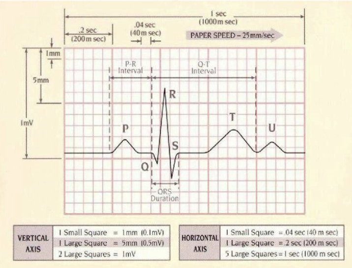
PQRST
P wave: atrial depolarization
QRS complex: ventricular depolarization
T wave: ventricular repolarization
PR interval: atrial impulse → through AV node
ST segment: indicates myocardial ischemia/injury
QT interval: ventricular depolarization → repolarization
PQRST normal intervals
P wave: 0.08-0.10 sec
PR interval: 0.12-0.20 sec
QRS: 0.06-0.10 sec
QTc: ≤0.44 sec
rhythm analysis steps
Calculate HR
count # of QRS/R waves in 6 sec or count boxes between R waves x 0.04 then 60 divided by that #
Determine regular vs irregular
measure R-to-R intervals
Look for P waves
Measure PR interval
Measure QRS
Assess ST segment
Examine T wave
peaked with hyperkalemia
Measure QT interval
RATE → RHYTHM → P → PR → QRS → ST → T → QT

normal sinus rhythm
60-100 bpm
NSR
1 P for every QRS (narrow)

dysrhythmias nursing priorities
FIRST LOOK AT PATIENT
then check lead placement
disconnected lead
artifact
actual arrest
cardiac dysrhythmias etiology & risk factors
etiology
delayed after depolarization
heart block
abnormal pacemaker (ectopic foci)
reentry circuit activity
risk factors
excess caffeine, stress, tobacco use
Digitalis (digoxin), Quinidine overdose
high BP & CAD
heart muscle damage after MI, valve disease
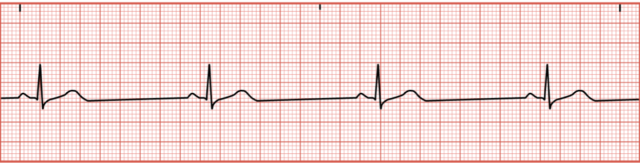
sinus bradycardia
<60 bpm, NSR (1 P for every QRS)
causes: athlete, vasovagal response, PSNS stimulation, MI/heart dz, hypothermia, ↑ ICP, low hormones
vasovagal triggers: straining during defecation, vomiting, intubation, mechanical ventilation
sxs: (act on these) CHEST PRESSURE/pain, dyspnea, fatigue, low BP, dizziness, seizure, fainting (syncope)
tx: fall precautions, reverse the cause, medications, stimulate SNS, suppress PSNS
IV ATROPINE every 3-5 min, do NOT exceed 3 mg
consider cardiac pacemaker to support perfusion


sinus tachycardia
>100 bpm, regular rhythm, 1 P:1 QRS (may be hidden with T wave d/t speed)
causes: normal physiological response (fever, exercise, anxiety, dehydration, pain), fluid volume loss/excess, MI, shock, hyperthyroidism, stress, hypoxia, anemia, hemorrhage, hypotension, heart dz, L-sided heart failure, cardiac tamponade, meds (atropine, catecholamines, theophylline, CNS stimulants, caffeine, nicotine)
sxs: chest pressure/pain, dyspnea, palpitations, lightheadedness
tx: (only if sxs)
stable: evaluate rhythm, consider O2, establish IV, ID/tx reversible causes (adenosine → SVT/BB)
unstable: 12-lead ECG, administer meds, synchronized cardioversion, consider cardiac ablation

cardioversion vs. defibrillation
cardioversion: synchronized, shock synchronized with R WAVE, usually pulse
unstable tachyarrhythmias, A-fib/flutter
defibrillation: unsynchronized, shock delivered immediately, no pulse
VF and pulseless VT
atrial dysrhythmias
SA node dominates (HR > 150 bpm)
causes: cardiac dz, electrolyte imbalance, emotions, exercise, COPD, meds, nicotine
sxs: skipped beats, pulse deficit
tx: adress underlying cause (K loss), meds, cardioversion, lifestyle mods

atrial fibrillation
no distinct P waves (irregular R-R spacing), no PR, atrial rate = 300-600 bpm, ventricular rate (untx) = 100-180 bpm
decrease CO d/t loss of atrial kick (20% ventricular filling)
premature ventricular contraction (PVC) early, full pause after, no pulse (irregular rhythm)
causes: HEART DISEASE (HF, constrictive pericarditis, ischemic/rheumatic heart disease, mitral stenosis, atrial irritation, complication of CABG/valve replacement), COPD, PE, HTN, DM, thyroid dz, sepsis
risk factors: uncontrolled BG, hyperthyroidism, kidney dz, OSA, smoking, alcohol, sedentary lifestyle, over 60
sxs: HEART PALPITATIONS, fatigue, dyspnea, lightheadedness, chest pain
tx: synchronized cardioversion, meds (metoprolol, diltiazem/CCB, digoxin), anticoagulant (reduce CVA and MI risk)


atrial flutter
SAWTOOTH, atrial rate 240-360 bpm, no PR, ventricular rate depends on degree AV block
P:QRS (2:1, 4:1, 6:1, variable)
causes: MI, comp. CABG/valve replacement, antiarrhythmic meds (BB, CCB, amiodorone, digoxin)
risk factors (same as a-fib): uncontrolled BG, hyperthyroidism, kidney dz, OSA, smoking, alcohol, sedentary lifestyle, over 60
sxs: heart palpitations, fatigue, dyspnea, lightheadedness, chest pain
tx: synchronized cardioversion, meds (BB, CCB + sodium channel blocker or amiodorone → not for older)
heart blocks
AV junction either slow or block signal from reaching ventricles
1st degree
2nd degree (Mobitz I/Wenckebach) & 2nd degree (Mobitz II)
3rd degree (Complete heart block) = most severe
tx: monitor, consider meds pt taking, intervene IMMEDIATELY


first-degree heart block
60-100 bpm, regular rhythm, PR >0.20 sec
AV conduction delayed → every impulse still reaches ventricles
causes: MI, electrolye (hypokalemia, hypomagnesemia), infections, hypothyroidism, autoimmune (RA, lupus), male, age > 60
antiarrhythmic meds (sodium channel blockers, BB, CCB, potassium channel blockers, cardiac glycosides, magnesium)
athletes d/t higher resting vagal tone → lower HR
sxs: none
tx: none

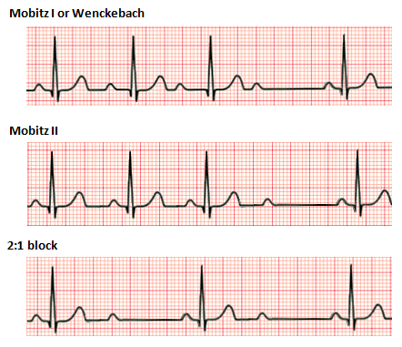
second-degree heart block (Mobitz I/Wenckebach)
PR interval progressively longer → QRS drops
atrial regular, ventricular irregular
causes: severe CAD, anterior wall MI, acute myocarditis, digoxin toxicity
sxs: none
tx: monitor/observe, atropine/isoproterenol


second-degree heart block (Mobitz II)
PR interval constant, QRS suddenly dropped
may progress to third-degree block
causes: extensive anterior wall MI, degenerative changes in His-Purkinje system, massive calcification of mitral/aortic valve anulus
sxs: none (rare: dizziness, lightheadedness, palpitations, syncope)
tx: atropine/isoproterenol, pacemaker therapy


third-degree heart block (complete heart block)
complete failure of comm. between atria and ventricles (severely impair CO/perfusion)
atrial 60-100 bpm, ventricular 15-60 bpm, no PR
causes: inferior or anterior wall MI, congenital abnormality, rheumatic fever
sxs: fainting, dizziness, extreme fatigue, chest pain, dyspnea
tx: IMMEDIATE PACEMAKER

ventricular dysrhythmias
MOST SERIOUS d/t CO severely reduced
QRS wide and bizarre, foci within ventricles cause them to contract
one or two beats ok → series can be fatal
causes: cardiac dz, CHF, MI, atrial enlargement, caffeine, electrolyte imbalance (low K)
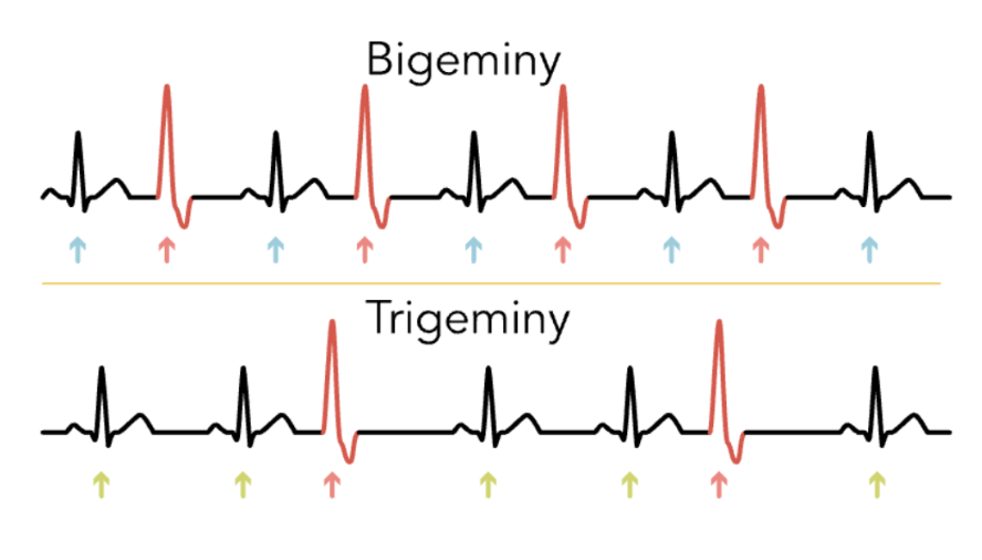
premature ventricular contraction (PVC)
an early ventricular beat before the next expected normal sinus beat
premature, wide/bizarre QRS, followed by compensatory pause, unifocal (look similar) or multifocal (shapes differ d/t K imbalance)
couplet (2 PVCs in a row), triplet (3 PVCs in a row)
causes: cardiac muscle injury, chronic lung dz, electrolyte imbalance, caffeine, alcohol
sxs: palpitations (fluttering or skipped beats), chest pain, dyspnea, lightheaded, syncope


ventricular tachycardia
150-250 bpm, regular rhythm (varies), no P waves, QRS > 0.12 (distorted)
causes: severe heart disease, MI, digoxin toxicity, prolonged QT, electrolyte imbalance, acidosis
sxs: acute MI (angina/chest pain), no pulse, altered LOC, pupil dilation → can lead to cardiac arrest
tx: pulse (cardioversion) or pulseless (CPR/defib/amiodarone), pacemaker/ICD, meds (lidocaine, pronestyl)
< 30 sec: nonsustained V-tach
> 30 sec: sustained V-tach


ventricular fibrillation
LETHAL RHYTHM
most common cause of death w/ CAD
no P wave, PR interval R-R interval, QRS
multiple ectopic in Purkinje network
no HR/pulse/BP/respirations
tx: CPR, meds (epi, amiodarone → not for older); shock (only if rhythm to shock them out of)


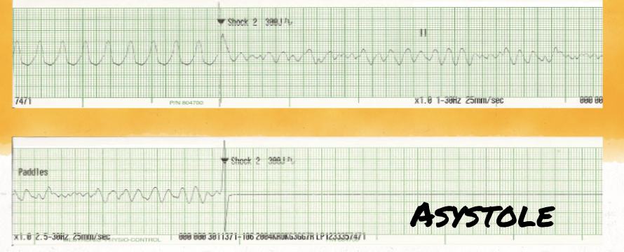
PEA and asystole
PEA: electrical rhythm appears on monitor → NO palpable pulse/CO
death of cells/mitochondria
reversible causes:
5 H’s: hypovolemia, hypoxia, hydrogen ion (acidosis), hypo/hyperkalemia, hypothermia
5 T’s: tension pneumothorax, tamponade, toxins, thrombosis (pulmonary/coronary)
asystole: no effective cardiac electrical activity or CO
acute coronary syndrome
group of disorders caused by reduced/blocked coronary blood flow
unstable angina, NSTEMI, STEMI
patho: atherosclerotic plaque → plaque rupture → platelets + clot formation → coronary artery becomes narrowed → ↓ myocardial oxygen → ischemia → injury → infarction
ischemia, injury, infarction
ischemia: decreased oxygen/blood supply to myocardium (ST depression)
injury: more severe myocardial compromise (ST elevation)
infarction: irreversible myocardial tissue death (pathologic Q wave)
MI patho
primary: coronary artery occlusion
causes: coronary atherosclerosis, vasospasm, platelet aggregation, emboli, A-fib, LV mural thrombus, infective endocarditis vegetations
low O2 → anaerobic metabolism → angina
STEMI, NSTEMI, unstable angina
Condition | Main ECG/marker clue |
|---|---|
STEMI | ST elevation |
NSTEMI | ST depression or T-wave inversion + elevated cardiac markers |
Unstable angina | ACS symptoms without elevated markers in slide algorithm |
cardiac markers
most sensitive: cardiac troponin (cTnI, cTnT)
CK-MB: increases 4-6 hr after MI (less specific)
myoglobin: rapid increases 1-3 hr (not cardiac-specific)
BNP/NT-proBNP: heart failure
hs-CRP: inflammation/LT CV risk
ACS emergency care (IMPORTANT)
main goal: ACUTE REPERFUSION
baseline cardiac rhythm + serial ECG, vital signs, IV access/labs, oxygen if <90%, chewable aspirin, nitrates, morphine/opiaye PRN, reperfusion therapy, IV heparin
other therapies after: BB, ACE-I, statins, clopidogrel/Plavix, aspirin, SGLT-2/GLP-1 for other comoboridities