Eye Function

0.0(0)
Studied by 0 people
call kaiCall Kai
learnLearn
examPractice Test
spaced repetitionSpaced Repetition
heart puzzleMatch
flashcardsFlashcards
GameKnowt Play
Card Sorting

1/41

encourage image

There's no tags or description

Looks like no tags are added yet.

42 Terms

1
New cards

directions of eye

  • temporal (what would be lateral)

  • nasal (what would be medial)

  • use of anterior and posterior (like human anatomy, instead of cranial/caudal)


2
New cards

regions of the eye

  1. orbit

  2. eyeball

  3. ocular adnexa (extraocular mm, eyelids, lacrimal apparatus)


3
New cards

cornea

cupula in frontal part of eye

<p>cupula in frontal part of eye</p>
4
New cards

sclera

the tough, white outer layer of the eyeball that covers most of its surface and protects the inner eye

<p><span style="color: rgb(141, 14, 173);">the tough, white outer layer of the eyeball that covers most of its surface and protects the inner eye</span></p>
5
New cards

limbus

circular transition zone in the eye that marks the border between the transparent cornea and the opaque sclera

6
New cards

choroid

blood-vessel-rich layer at the back of the eye

  • vascular tunic


7
New cards

ciliary body

produces aqueous humor and helps focus

<p>produces aqueous humor and helps focus</p>
8
New cards

iris

colored part, regulates how much light enters eye

<p>colored part, regulates how much light enters eye</p>
9
New cards

pupil

the round, black opening in the center of the iris that lets light enter the eye so it can reach the lens and retina.

<p>the round, black opening in the center of the iris that lets light enter the eye so it can reach the lens and retina.</p>
10
New cards

fibrous tunic

  • cornea

  • sclera

  • limbus


<ul><li><p><span style="color: blue;">cornea</span></p></li><li><p><span style="color: blue;">sclera</span></p></li><li><p><span style="color: blue;">limbus</span></p></li></ul><p></p>
11
New cards

vascular tunic

choroid

iris and pupil

ciliary body

<p><span style="color: red;">choroid</span></p><p><span style="color: red;">iris and pupil</span></p><p><span style="color: red;">ciliary body</span></p>
12
New cards

nervous tunic

  • retina (photosensitive and non-photosensitive)

  • optic nerve


<ul><li><p><span style="color: rgb(225, 205, 149);">retina (photosensitive and non-photosensitive)</span></p></li><li><p><span style="color: rgb(225, 205, 149);">optic nerve</span></p></li></ul><p></p>
13
New cards

lens

knowt flashcard image
14
New cards

anterior chamber

anterior chamber

between cornea and iris

aqueous humor

<p>anterior chamber</p><p>between cornea and iris</p><p>aqueous humor</p>
15
New cards

posterior chamber

posterior chamber

aqueous humor

between iris and front of lens

<p>posterior chamber</p><p>aqueous humor </p><p>between iris and front of lens</p>
16
New cards

vitreous chamber

  • posterior to lens

  • vitreous body/humor

  • the largest compartment in the eye, located behind the lens and in front of the retina


<ul><li><p>posterior to lens</p></li><li><p>vitreous body/humor</p></li><li><p>the largest compartment in the eye, located behind the lens and in front of the retina</p></li></ul><p></p>
17
New cards

epithelium of the cornea

  • stratified squamous nonkeratinized

  • regenerative


<ul><li><p>stratified squamous nonkeratinized</p></li><li><p>regenerative</p></li></ul><p></p>
18
New cards

basement membrane of epithelium

  • barrier to infection, does not regenerate if damaged

(bowmans membrane)

<ul><li><p>barrier to infection, does not regenerate if damaged</p></li></ul><p>(bowmans membrane)</p>
19
New cards

corneal stroma

•Collagen fibrils (type V) & ground substance with sulfated proteoglycans.

•consists of alternating lamellae of collagen fibers and fibroblast.

•This orthogonal array of highly regular fibrils is responsible for the transparency

<p><span><strong>•Collagen fibrils (type V)</strong> &amp; ground substance with sulfated proteoglycans.</span></p><p><span>•consists of alternating lamellae of collagen fibers and fibroblast.</span></p><p><span>•This orthogonal array of highly regular fibrils is responsible for the transparency</span></p>
20
New cards

descemets membrane

regenerates

<p>regenerates</p>
21
New cards

corneal endothelium

  • metabolic exchange occurs, repair is limited

  • low cuboidal epithelium


<ul><li><p>metabolic exchange occurs, repair is limited</p></li><li><p>low cuboidal epithelium</p></li></ul><p></p>
22
New cards

corneoscleral limbus

located at the iridocorneal angle:

•Transition zone between the cornea and sclera

•Canal of Schlemm drains aqueous humor out of the eye and into venous system

•Corneal stem cells live here

angular aqueous plexus
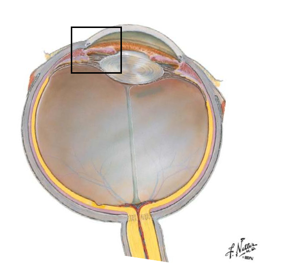

<p><span>located at the iridocorneal angle:</span></p><p><span>•Transition zone between the cornea and sclera</span></p><p><span>•Canal of Schlemm drains aqueous humor out of the eye and into venous system</span></p><p><span>•Corneal stem cells live here</span></p><p><span>angular aqueous plexus </span></p><p></p>
23
New cards

sclera

•An opaque layer that consists predominantly of dense irregular connective tissue

•Relatively avascular

•Receives insertions of the extrinsic ocular muscle

<p><span>•An opaque layer that consists predominantly of dense irregular connective tissue</span></p><p><span>•Relatively avascular</span></p><p><span>•Receives insertions of the <strong>extrinsic ocular muscle</strong></span></p>
24
New cards

ciliary body

where does the iris arise from?

25
New cards

stroma of iris

dense irregular CT with melanocytes and fibroblasts

has dilator and sphincter mm which change the size of the pupil in response to light

26
New cards

miosis

contraction

27
New cards

mydriasis

dilation

28
New cards

dilator pupillae m

innervated by sympathetic nerves, dilates pupil in response to dim light.

29
New cards

sphincter pupillae m

innervated by parasympathetic nerves, constricts pupil in response to bright light.

30
New cards

ciliary body

lens accomodation and production of the aqueous humor in the posterior chamber

31
New cards

muscle contraction

relaxation of zonule fibers, lens becomes round and accommodates for shorter distances

<p><span>relaxation of zonule fibers, lens becomes round and accommodates for shorter distances</span></p>
32
New cards

muscle relaxation

zonule fibers tense, lens flattens, accommodates for longer distances

<p><span>zonule fibers tense, lens flattens, accommodates for longer distances</span></p>
33
New cards

fundus

back of eye

structures are viewed during routine examination (fundoscopic exam)

•Optic disc

•Fovea centralis and macula lutea (primates)

•Blood vessels of retina and choroid

•Tapetum lucidum

•Retina

<p>back of eye</p><p>structures are viewed during routine examination (fundoscopic exam)</p><p><span>•Optic disc</span></p><p><span>•Fovea centralis and macula lutea (primates)</span></p><p><span>•Blood vessels of retina and choroid</span></p><p><span>•Tapetum lucidum</span></p><p><span>•Retina</span></p>
34
New cards
<p>tapetum lucidum</p>

tapetum lucidum

  • vascular layer


<ul><li><p>vascular layer </p></li></ul><p></p>
35
New cards

optic nerve

sensory innervation of the eye - vision

36
New cards

layers of retina

  1. nerve fiber layer

  2. ganglion cells

  3. inner plexiform layer

  4. inner nuclear layer

  5. outer plexiform layer

  6. outer nuclear layer

  7. layer of cones and rods

  8. retinal pigement epithelium

  9. lamina vitrea (Bruchs membrane)

  10. choroid


37
New cards

retinal pigment epithelium

• absorbs light; prevents glare.

•Site of the blood retina barrier - isolates retinal cells from blood-borne substances - elaborate tight junctions between adjacent RPE cells.

38
New cards

photoreceptors

convert light signals into nerve impulses.

39
New cards

rods

•contain rhodopsin, function best in darkness (Rod may synapse with several bipolar cells).

<p><span>•contain <strong>rhodopsin, </strong>function best in darkness (Rod may synapse with several bipolar cells).</span></p>
40
New cards

cones

contain iodopsin, function best in light, but produce greater visual acuity (Each cone synapses with a single bipolar neuron).
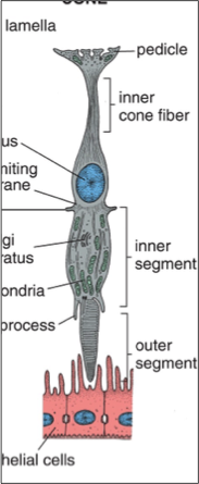
<p><span>contain <strong>iodopsin, </strong>function best in light, but produce greater visual acuity (Each cone synapses with a single bipolar neuron).</span></p>
41
New cards

optic disk

Ganglion cell axons converge at the optic disc to form the optic nerve (CN II) (blind spot)

<p><span>Ganglion cell axons converge at the <strong>optic disc </strong>to form the <strong>optic nerve (CN II) </strong>(blind spot)</span></p>
42
New cards

fovea centralis

•Small depression the retina where photoreceptors are maximally exposed to light

•Area of greatest visual acuity bc most light hits photoreceptors

  • most mammals have a cone and ganglion dense streak
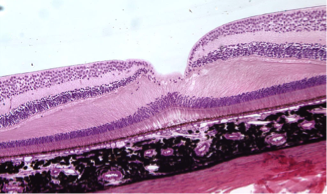

<p><span>•Small depression the retina where photoreceptors are maximally exposed to light</span></p><p><span>•Area of greatest visual <strong>acuity </strong>bc most light hits photoreceptors </span></p><ul><li><p>most mammals have a cone and ganglion dense streak </p></li></ul><p></p>



