1.2 The heart (electrical activity) --> Part 2
 Call Kai
 Learn
Learn Practice Test
 Spaced Repetition
 Match
Match Flashcards
 Knowt Play
1/35

Earn XP
Name | Mastery | Learn | Test | Matching | Spaced | Call with Kai | Chat |
|---|
No analytics yet
Send a link to your students to track their progress
36 Terms
Electrical activity of the heart
A part of the cardiovascular system that controls when the heart beats and pumps blood.
Another name for the electrical activity of the heart
The conduction system
Key components of the conduction system
Sino-atrial (SA) node
Atrioventricular (AV) node
The bundle of His
Purkinje fibres
Another spelling of ‘Purkinje’
Purkyne
Sinoatrial node
Often referred to as the heart’s peacemaker, located in the upper wall of the right atrium.
Functions of the sinoatrial node
Regulates the heart rate in line with the body’s demand by sending out electrical impulses
The impulses which travel across the muscle cells in the atria, cause them to contract
Atrioventricular node
A cluster of cells located at the bottom of the right atrium, near the ventricles.
Functions of the atrioventricular node
Receives impulses from the SA node and delays it
The delay ensures the atria have completely emptied their blood into the ventricles, before the ventricles contract
Bundle of His
A specialised pathway of fibres extending from the AV node down into the interventricular septum
Functions of bundle of his
It is the only electrical bridge between the upper and lower heart chambers
The impulses travel down the bundle of his from the AV node
The bundle of his splits into right and left branches to carry the impulses down to the ventricles
Purkinje fibres
A complex, branching network of fibres that fan upward and outward from the bundle branches, into the muscular walls of both ventricles
Functions of the Purkinje fibres
They rapidly transmit the impulses throughout the entire ventricular muscle mass
Allows the heart’s conduction system to create synchronised contractions of its ventricles
Therefore, they are essential for maintaining a consistent heart rhythm
Atrial depolarisation
The effect the SA node has on the atria, causing them to contract by providing an electrical stimulus across them
Ventricular depolarisation
The effect the AV node has on the ventricles, causing them to contract by providing an electrical stimulus
Atrial and ventricular repolarisation
Occurs during a brief time period following depolarisation and is the return of the heart’s electrical impulse to a baseline value
ECG
Electrocardiogram
Electrocardiogram
A test that that checks how well the heart is working by measuring the electric signals that travel through the heart.
How electrocardiogram works
During a test, small sticky patches called electrodes are placed on the arm, chest and legs
These electrodes pick up the electrical signals and send them to a machine that makes a pattern of the heart’s activity
The pattern helps the doctor see if the heart is healthy or not
What ECG measures
Heart rate, rhythm and timing of electrical impulses
What ECG shows electrical impulses as
Waves
The waves ECG shows
P, Q, R, S and T waves
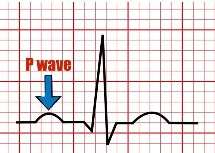
P wave
Represents the electrical impulse that triggers atrial contraction
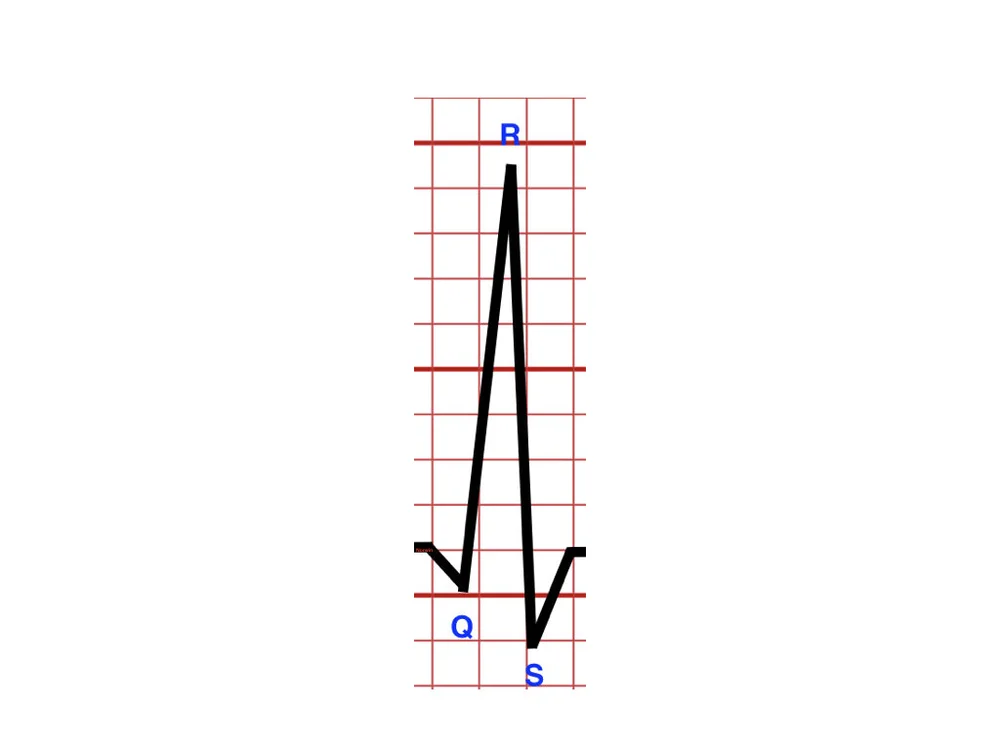
QRS complex
Represents the electrical activity of the ventricles contracting
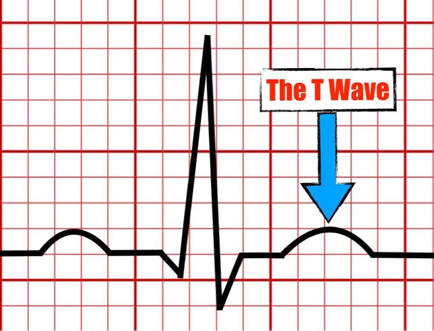
T wave
Represents the heart’s full electrical reset, preparing it for the next cardiac cycle

ST segment
Marks the end of ventricular contraction and the beginning of the heart’s recovery phase

Q-T interval
It measures the time it takes for the ventricles to contract and then start to recover

T-P interval
It measures the time when the ventricles are relaxed and filling up with blood. It is the period between the recovery phase (T wave) and the next electrical signal starting (P wave).
If the wave is irregular, absent or abnormal, it may suggest issues with the atrial contraction.
P wave abnormalities
If the wave is long or distorted, it can suggest issues with ventricular conduction or a blockage within the bundle of His.
QRS complex abnormalities
If raised or reduced, it can indicate ischemia or heart attack.
ST segment abnormalities
If the wave is inverted or unusually tall, it can signal electrolyte imbalances or ischemia.
T wave abnormalities
How heart conditions like; arrhythmias and fibrillation, are diagnosed using ECG
Patterns are studied in different conditions and compared to the standard ECG.
How the heart condition fibrillation is stopped
Passing a strong electric current through the chest wall.
Then the heart stops for up to 5 seconds after which it begins to beat in a controlled way.
How the heart rate can be modified
Heart rate can be modified by the nervous system and hormones.
How the nervous system modifies heart rate
Sympathetic Nervous System: Fight or flight increases heart rate
Parasympathetic Nervous System: Rest and digest decreases heart rate
How hormones modifies heart rate
Adrenaline and thyroxine both increases heart rate