Clin Med Exam 4 Arrythmia, Clin Med Exam 4 Atrial and Misc
 Call Kai
 Learn
 Practice Test
 Spaced Repetition
 Match
 Flashcards
 Knowt Play
1/134

There's no tags or description
Looks like no tags are added yet.
135 Terms
Essential for diagnosis
Fast, wide QRS complex on ECG
Associated with ischemic heart disease, particularly in older patients
In the absence of reversible cause, implantable cardioverter defibrillator (ICD) is recommended if meaningful life expectancy is > 1 year.
What is a PVC
Premature ventricular complex
Premature beat originating from the ventricle (not a problem but a warning)
Wide, bizarre QRS
Compensatory pause
Common, usually does not require treatment
Most ventricular arrhythmias occur after a PVC
Ventricular Tachycardia is simply
≧ 3 consecutive ventricular premature
Classifications for VT
Sustained
Monomorphic
Polymorphic
What is sustained VT
Lasts ≥ 30 seconds
What is monomorphic VT
Electrical impulse originates in one of the ventricles → same QRS morphology from beat to beat
What is polymorphic VT?
Electrical impulses originate in multiple areas leading to variable QRS morphologies from beat to beat (e.x., Torsade de pointes)
VT vs Vfib
VT is regular irregular and Vfib is irregular irregular
What is nonsustained VT
< 30 seconds, self-terminating
What is sustained VT:
> 30 seconds or requiring intervention; HR > 100 bpm
What is the prognosis for VT
Without heart disease: nonsustained VT is usually benign
With structural heart disease: nonsustained VT linked to higher risk of symptomatic VT and sudden death, especially > 48 hrs post-MI
Epidemiology for VT
Incidence increases with age
AMAB > AFAB due to higher incidence in CAD
15% of patients with CAD have VT
5-10% of patients presenting with acute myocardial infarction have VT within the first few days of presentation
Rare in children, but can occur with inherited channelopathies or structural heart disease
15 per 100,000 individuals have idiopathic VT
Etiologies & Associations for VT
Common in ischemic heart disease, acute MI, dilated cardiomyopathy, valvular heart disease, and heart failure
Prolonged QT interval
Electrolyte abnormalities: hypokalemia, hypomagnesemia, hypocalcemia
Digoxin toxicity
Proarrhythmic effects of some antiarrhythmics
Torsades de Pointes
"Twisting of the points"
Form of polymorphic ventricular tachycardia that appears to be twisting around a baseline
Prolonged repolarization and early after depolarization plus triggering activity
Accelerated Idioventricular Rhythm
Wide-complex, 60-120 bpm, gradual onset
Seen post-infarction or after thrombolytic reperfusion
Usually benign; treat only if hemodynamic compromise or serious arrhythmias
Risk Factors for VT
Ischemic heart disease, ST elevation MI
Structural heart disease, cardiomyopathy
Sustained ventricular tachycardia
Hypokalemia
Hypotension
History of smoking
Male gender
Clinical manifestations for VT
Can be asymptomatic (particularly if non-sustained)
Palpitations, chest pain, dyspnea
Dizziness, fatigue, diaphoresis, pallor
Syncope and Presyncope
If unstable, due to lack of perfusion → hypotension, altered mental status, hemodynamic instability/collapse and cardiac arrest
Diagnosis for VT
ECG
Labs:
Metabolic panel - check electrolytes for hypokalemia and hypomagnesemia
Troponin
BNP
Digoxin Level
Drug screen
SVT vs Vtach (anatomy and clinical significance)
SVT:
- rhythms that originate from the atrial
- less dangerous b/c they are somewhat controlled by the refractory nature of the AV node, thus not every single depolarization actually travels into the ventricles from the artria
Vtach:
- rhythms that originate from the ventricles
- more dangerous (often fatal)
Acute management of VT
Determine treatment based on hemodynamic stability and arrhythmia duration.
Unstable with pulse: synchronized cardioversion
Unstable without pulse: start CPR and defibrillation (unsynchronized cardioversion)
Correct reversible causes (i.e., electrolyte abnormalities)
How do you determine tx for VT
based on hemodynamic stability and arrhythmia duration
Structural Heart effected by VT
Usually benign; syncope uncommon
Often due to triggered activity from RV/LV outflow tract or His-Purkinje system
Treat with short-acting IV β-blocker or verapamil
Structural Heart Disease effected by VT
If unstable (hypotension, HF, ischemia): immediate synchronized DC cardioversion (100 - 200 J)
If VT recurs: IV amiodarone (150 mg bolus → 1 mg/min x 6h → 0.5 mg/min x 18 hrs); watch for hypotension
What is tx for stable sustained VT
IV amiodarone, lidocaine, or procainamide
Escalate to cardioversion if ineffective or symptoms worsen
What is the acute management for polymorphic VT
Empiric IV magnesium (1-2 g)
Recurrent cases: increase HR with isoproterenol (up to 20 mcg/min) or atrial pacing (90-120 bpm)
If QT normal: evaluate for ischemia and revascularize if needed
What is acute management for recurrent VT
Consider ventricular overdrive pacing via pacemaker or ICD
What is the long-term management for high risk occurrence
if no reversible cause (e.g., MI, electrolytes, drugs).
What is long term management for structurally normal heat
Typical outflow tract or fascicular VT: try β-blocker or nondihydropyridine CCB
Catheter ablation effective if meds fail
What is the long term management for structural heart disease
LV dysfunction: ICD recommended if life expectancy >1 year.
β-blockers are first-line.
Amiodarone/sotalol reduce VT episodes and ICD shocks but don't improve mortality.
Catheter ablation for refractory or intolerant cases.
Consider ablation as first-line in ischemic cardiomyopathy due to antiarrhythmic side effects.
Eti for Torsades de Pointes
Prolonged QT Interval: Medications such as digoxin, class 1a antiarrhythmics, class III antiarrhythmics, macrolide antibiotics, antipsychotics, antidepressants, antiemetics
Electrolyte Abnormalities: hypokalemia, hypomagnesemia, hypocalcemia
Clinical manifestation for Torsades de Pointes
Palpitations
Dizziness
Fatigue
Dyspnea
Chest pain
Presyncope & Syncope
Cardiac arrest
Tx for torsades de pointes
IV magnesium sulfate
Treat underlying electrolyte abnormalities
Discontinue/adjust QT prolonging drugs
Vfib
Ineffective ventricular contractions → decreased cardiac output → hemodynamic collapse
Chaotic, irregular "fibrillation" waves of varying amplitude WITHOUT useful contractions
No identifiable P waves, QRS complexes, or T waves
Rate 150-500 bpm
Coarse vs. Fine
Vfib RF
Ischemic heart disease, ST elevation MIs
Structural heart disease, cardiomyopathy
Hypokalemia
Hypotension
History of smoking
Male gender
Vfib Clinical Manifestation
Chest pain, shortness of breath
Unresponsive
Pulseless
Syncope and sudden collapse from cardiac arrest
Sudden death
Vfib Tx
defibrillation
WHAT IS A FIB?
Supraventricular arrhythmia that originates above the ventricles.
The heart's muscle fibers contract at different times, resulting in an inconsistent, quivering heartbeat.
Essential Diagnosis of afib
Presents as an irregularly irregular heart rhythm on examination and ECG
RF for afib
Prevention of stroke should be considered in all patients with risk factors for stroke (e.g., HF, HTN, 65+, DM, prior hx of stroke or TIA, and vascular disease
Management of afib?
Heart rate control with beta-blocker or calcium channel blockers generally required.
Rhythm control in symptomatic pts
Patho of afib
High Rate + Bad Squeeze
Dumb AV node get confused by bad SA node
Disorganized waves of atrial depolarization
Regular impulses of sinus node overwhelmed by rapid electrical discharges from various sources (automatic foci, multiple reentry phenomena)
What results from the AV node trying to filter impulses
rapid ventricular response
Pathway for afib flow
Suboptimal ventricular filling → decreased cardiac output → symptoms can develop → possible hemodynamic compromise
Patho mechanisms of afib
stretched myocardium
Ischemia
Inflammation
↑SNS stimulation
Electrolyte Imbalance
Main theories for origins of afib
Multiple Wavelet Hypothesis
Automatic Focus Theory
Multiple Wavelet Hypothesis
AF is believed to be a random propagation of multiple electrical wavelets across the atria
Abnormal atrial tissue resulting in slow conduction, shortened refractory periods, and increased atrial mass favor higher number of self-perpetuating "daughter wavelets."
Automatic Focus Theory
AF persists only in isolated regions of atrial myocardium.
Isolation of this source can eliminate AF - seen with ablation treatment
The pulmonary veins appear to be the most frequent source of these automatic foci
What is the progression of Afib
Paroxysmal
Persistent
Permanent
What is paroxysmal afib?
Intermittent rhythm, may revert back to sinus rhythm spontaneously
Persistent AF
AFib lasting longer than 7 days; requires intervention to convert back to sinus rhythm
permanent afib
Long-standing atrial fibrillation, cardioversion unsuccessful
Epi for Afib
More afib because ppl live longer '
More common in men than women
What is holiday heart
Afib associated with acute alcohol excess and withdrawal
For regular to moderate drinkers abstinence from alcohol reduces recurrence of Afib by about 50%
Common comorbidities
Rheumatic Heart Disease
Valvular Heart Disease
Dilated Cardiomyopathy
Atrial Septal Defect
Hypertension
Chest trauma
Thoracic or cardiac surgery
Congestive Heart Failure
Coronary Artery Disease
Myocardial Infarction
Wolff-Parkinson White Syndrome
Non cardiac rf for afib
Thyrotoxicosis or Hyperthyroidism
Obstructive sleep apnea
Pulmonary disease (pneumonia, pulmonary embolism)
Medications (beta-adrenergic agonists, inotropes, bisphosphonates)
Excessive alcohol (Holiday Heart)
Pheochromocytoma
Illicit substances
Electrolyte imbalances
Clinical Manifestations of a-fib
Often Asymptomatic
Fatigue; exercise intolerance
Dyspnea
Palpitations
Lightheadedness
Weakness
Chest pain
Hemodynamic shock
Diagnostics tools for afib
ECG
Echocardiogram
Misc:
- tox screening
- thyroid testing
- electrolytes (BMP or CMP)
- CBC
- Holter monitoring

What do we expect to see on an ECG for afib
Irregularly irregular pattern on ECG
- RR interval no repetitive pattern
- No distinct P waves
- Can be rapid rate or bradycardia
Chaotic electrical activity results in a chaotic wave form between the QRS complexes
P waves are absent
Replaced with "f" waves
Rapid electrical activity overwhelms the AV node causing impulses to enter the ventricular conduction system at irregular points
Irregular R to R intervals
Echocardiogram considerations for afib
Should be done on all patients with new diagnosis of atrial fibrillation
Tx goals for Afib
Reduce symptoms
Prevent thromboembolism
Prevent cardiomyopathy
1st line pharm tx for afib
If EF sucks:
IV beta-blockers (esmolol, propranolol, and metoprolol)
IV calcium channel blockers (diltiazem and verapamil)
Hemodynamically unstable afib tx
Hospitalization
1st line tx
Urgent electrical cardioversion if indicated
IF symptoms are persistent and started 2 days ago what is indicated
echocardiogram prior to cardioversion to exclude left atrial thrombus
Hemodynamically Stable management for afib
Hospitalized is generally not necessary unless there is an acute event causing it (i.e., MI)
Early Rhythm Control is now emphasized over rate control
Rate control and anticoagulation is appropriate
1st line: beta blocker or calcium channel blocker
Subsequent management for afib requires what outside of lifestyle changes
Anticoagulation
What to look out for in afib in relation to coagulation
Lone AFib: under age 65 y/o need no antithrombotic tx
Transient AFib:
If associated with MI or pneumonia anticoagulation may be necessary
If post-surgery (CABG) or associated with hyperthyroidism long-term anticoagulation is not necessary
CHA2DS2-VASC SCORE
0 = then anticoagulation generally doesn't need to be considered
1 = oral anticoagulation should be considered, taking into account risk, benefit, and patient preferences
Oral anticoagulation is indicated for:
- Men with score of 2
- Women with score of 3
Should you use aspirin as prolonged tx of afib
Not for long term management
What form can anticoagulation be in
Can be in the form of vitamin K antagonist (warfarin) or newer agents including direct-acting oral anticoagulants (DOACs)
Which is preferred: DOAC or Warfarin
DOAC is better at preventing strokes
Contraindications for DOAC usage
Rheumatic mitral stenosis that is severe
Mechanical heart valves
Recent bioprosthetic valve implanted (3-6 months)
Drug interactions
Diet sensitive
What monitoring is required for Warfarin
PT/INR (measures at how clottable the blood is); must be between 2-3
What doe we do for rate control for afib
Considered background treatment in nearly all patients with afib
Generally done with beta blockers or CCB
Rhythm Control for afib
Initial Cardioversion:
Elective cardioversion followed by anticoagulation (at least 3 weeks)
Pharmacologic cardioversion: ibutilide or procainamide
Long term rhythm control:
Amiodarone
Propafenone, flecainide, dronedarone, dofetilide, sotalol
Recurrent Paroxysmal AFib
Pts with rare but symptomatic episodes of AFib
Recurrent paroxysmal afib tx
Pill-in-pocket treatment
On demand pharmacologic cardioversion:
Flecainide (200-300 mg) or Propafenone (450-600 mg)
BB or nondihydropyridine CCB
Refractory AFib
Considered refractory if it causes persistent symptoms or limits activity despite attempts at rate or rhythm control.
Refractory AFib tx
If antiarrhythmic or rate control fail then try:
Catheter ablation of foci around the pulmonary veins
Surgical ablation
Complications of Afib
Left Ventricular Dysfunction
Heart failure
Myocardial ischemia
Thrombus formation and embolization leading to:
Stroke
Pulmonary embolism
Atrial flutter
Rapid, regular tachycardia presenting classically with 2 to 1 block in the AV node and ventricular heart rate of 150 bpm.

What is seen with the EKG for Aflutter
ECG shows "sawtooth" pattern of atrial activity (rate 300 bpm)
usually with transmission of every second, third, or fourth impulse through the AV node to the ventricles
What leads are aflutter seen
II, III, and AVF
What should be an evaluated risk for aflutter
Stroke risk should be considered equivalent to that with atrial fibrillation
What is the DEFINITIVE tx for aflutter
Catheter ablation is highly successful and is considered the definitive treatment for typical atrial flutter
General considerations for Aflutter
Less common than AFib
May occur in patients with structurally normal hearts but is more commonly seen in patients with:
COPD
Valvular or structural heart disease
Atrial Septal Defect (ASD)
Surgically repaired congenital heart disease
What is the patho for Aflutter
Typical or cavotricuspid isthmus (CTI) is the most common type of atrial flutter; rhythm originates in the right atrium at the level of the tricuspid valve annulus
Atypical atrial flutter is independent of the CTI and the origin of the arrhythmia can be in the right atrium or left atrium
What is the Eti for AFlutter
the presence of the reentry mechanism for initiation of the tachycardia and the area of cells that repolarize differently, like scar tissue allowing an electrical loop to form and continue to stimulate the atria muscle
What are clinical manifestations for AFlutter
Palpitations
Fatigue
Dizziness
Symptoms and Signs of heart failure: dyspnea, exertional intolerance, edema
What is the diagnostic workup for A-flutter
Physical Exam, Vitals, and ECG
Echocardiogram
Labs
Holter monitor
What labs are we looking at for the diagnostics for aflutter
evaluate for electrolyte imbalances, abnormal thyroid function, infection, anemia, and hypoxia
What is Holter monitor
should be done if arrhythmia is suspected but not seen on an ECG
Tx for Aflutter
Rate control
Rhythm control
Anticoagulation due to embolization risk
Use CHA2DS2-VASC score
Score of 1 or more may need anticoagulation
Rate control in aflutter
Electrical cardioversion
Pharmacological cardioversion - Ibutilide
Catheter ablation
Rate control pharm tx for aflutter
B-blocker, diltiazem, or verapamil (class I)
ATRIOVENTRICULAR BLOCK patho
blockage or delay in electrical signal stimulating contraction between atria and ventricles
causes for av blocks
●Congenital heart disease
●Heart disease (infiltrative/dilated cardiomyopathies, muscular dystrophy, lyme disease, MI, myocarditis, endocarditis, hyperkalemia, high vagal tone)
●Iatrogenic: beta blockers, CCB, cardiac glycosides, post-cardiac surgery, post-catheter ablation
●Lev's Disease: Idiopathic fibrosis and calcification of heart's electrical conduction system
Complications for AV blocks
Heart failure secondary to bradycardia; sudden cardiac death

First degree AV block ECG
Delayed PR interval
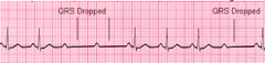
2nd degree (Type 1 Mobitz) ECG
PR interval lengthens with each beat until blocked completely resulting in a dropped beat
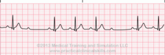
2nd Degree-Type 2 (Mobitz II) ecg
Prolonged PR interval (>200 ms). Block commonly in bundle of his; no progressive lengthening of PR interval but shows dropped beat