Advanced Cell Bio Exam 1
 Call Kai
 Learn
Learn Practice Test
 Spaced Repetition
 Match
Match Flashcards
 Knowt Play
1/86

There's no tags or description
Looks like no tags are added yet.
Name | Mastery | Learn | Test | Matching | Spaced | Call with Kai | Chat |
|---|
No analytics yet
Send a link to your students to track their progress
87 Terms
1. Brightfield microscopy
Uses visible light that passes through a specimen. Images typically have a bright background, and stains can make transparent cells easier to see. It is useful for observing cell shapes, tissues, and larger structures.
2. Fluorescence microscopy
Uses fluorescent molecules that absorb light at one wavelength and emit light at another. It highlights specific proteins or cell structures, often against a dark background. Some fluorescence techniques can image living cells.
3. Transmission electron microscopy (TEM)
Sends electrons through an extremely thin specimen. It produces detailed, grayscale images of internal cell structures, including membranes and organelles. Samples must be specially prepared and cannot be alive during imaging.
4. Scanning electron microscopy (SEM)
Scans a specimen's surface with electrons. It produces detailed images with a three-dimensional appearance, making it useful for studying surface texture and shape.
Magnification:
How much larger an object appears than its actual size.
Resolution:
The ability to distinguish two closely spaced objects as separate structures.
Micrograph
An image produced using a microscope
Fluorophore
A molecule that emits light after absorbing light
Direct staining
Using a fluorescent chemical that binds to a cell component
Immunolabeling
Using antibodies to identify a particular protein or structure
GFP
Green fluorescent protein, which can be attached genetically to a protein to track its location
1. Reception
A signaling molecule (ligand) binds to its receptor.
2. Transduction
The signal is converted into intracellular events through a signaling cascade.
3. Response
The cell changes its activity, such as altering enzyme function or gene expression.
Endocrine signaling
Signals such as hormones travel through the bloodstream to distant target cells. It allows long-distance communication but can take longer to reach a target than local signaling.
Neuronal (synaptic) signaling
Neurons release neurotransmitters at synapses onto nearby target cells. Electrical signals travel along the neuron, while neurotransmitters cross the synaptic cleft, allowing rapid, targeted communication.
Paracrine signaling
A cell releases signaling molecules that affect nearby cells. It is useful for local coordination, such as communication between cells in a tissue.
Autocrine signaling
A cell releases a signal that binds to receptors on that same cell. This can help regulate or reinforce the cell's own activity.
Juxtacrine signaling
Requires direct contact between cells, typically through a membrane-bound ligand and receptor. Notch–Delta signaling is an important example in animal development.
ligand
a molecule that binds to a receptor and initiates or changes its activity
Receptor
detect signals and help the cell respond.
Cell-surface receptors
bind signals outside the cell and transmit the message across the plasma membrane.
Intracellular receptors
bind signals that can cross the membrane, often small, nonpolar or lipid-soluble molecules.
Relay
Passes a signal from one component to another.
Transduction
Converts the signal into a different form inside the cell.
Amplification
Allows one activated component to activate many downstream molecules.
Integration
Combines information from multiple signaling pathways.
Feedback
Later steps influence earlier steps, either increasing or decreasing activity.
Distribution
Sends the signal toward multiple targets to produce different cellular effects.
Second messengers
are small intracellular molecules or ions that help transmit and amplify signals.
A. Ion-channel-coupled receptors
Ligand binding opens or closes an ion channel. Ions move across the membrane, changing the cell's electrical state. These receptors are especially important in neurons.

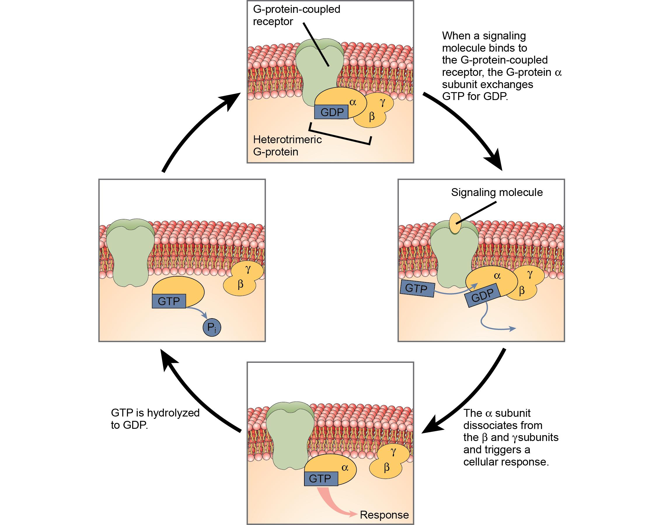
B. G-protein-coupled receptors (GPCRs)
Ligand binding activates a G protein. The Gα subunit exchanges GDP for GTP and separates functionally from Gβγ, allowing both to influence downstream targets. Gα eventually hydrolyzes GTP to GDP, helping turn off the signal.
C. Enzyme-coupled receptors
Ligand binding activates enzymatic activity or associated enzymes. Receptor tyrosine kinases (RTKs), for example, often dimerize and phosphorylate tyrosine residues, creating docking sites for downstream signaling proteins

Phosphorylation
is the addition of a phosphate group to a molecule, commonly a protein. Protein kinases add phosphate groups; protein phosphatases remove them.
Autophosphorylation:
A receptor or protein phosphorylates itself or another subunit of the same protein complex.
Ras:
A small GTP-binding protein that acts as a molecular switch in many growth-related signaling pathways.
MAP kinase cascade:
A series of kinase activations that relay and amplify signals, often affecting cell growth and gene expression.
Scaffold protein:
Holds signaling components together to help organize and coordinate their interactions.
Posttranslational modification (PTM):
A chemical or structural change made to a protein after it has been produced.
Fast responses:
Often change the activity of existing proteins, ion channels, or enzymes. They can occur in milliseconds to seconds and are useful when an immediate response is needed.
Slow responses:
Often involve changes in gene expression and protein production. They may take minutes to hours or longer but can produce longer-lasting changes.
Receptor inactivation:
The receptor remains present but can no longer signal effectively.
Receptor sequestration:
The receptor is removed from the cell surface temporarily, limiting access to its ligand.
Receptor downregulation:
The receptor is internalized and degraded, reducing the number of available receptors.
Signaling protein inactivation:
A component farther down the pathway is switched off.
Negative feedback:
Later steps in the pathway help inhibit earlier steps.
The packaging sequence is:
DNA double helix: The DNA molecule itself.
Nucleosome: DNA wrapped around a histone protein complex.
Chromatin fiber: Nucleosomes organized into a more compact structure.
Chromatin loops and domains: DNA is arranged in three-dimensional loops and organized regions.
Highly condensed chromosomes: DNA becomes especially compact during cell division.
DNA double helix:
The DNA molecule itself.
Nucleosome:
DNA wrapped around a histone protein complex.
Chromatin fiber:
Nucleosomes organized into a more compact structure.
Chromatin loops and domains:
DNA is arranged in three-dimensional loops and organized regions.
Highly condensed chromosomes:
DNA becomes especially compact during cell division.
linker DNA
The DNA between adjacent nucleosomes is called
Histone H1
is the linker histone. It binds near the DNA entry and exit points of a nucleosome and helps stabilize and compact chromatin; it is not one of the eight core histone proteins.
Histones have a _____ charge overall
positive, while DNA's phosphate backbone is negatively charged. These opposite charges help the two interact.
Chromatid
One of the copies of a replicated chromosome.
Sister chromatids
The two copies of a replicated chromosome, held together until they separate.
Nucleosome
DNA wrapped around a histone octamer.
Euchromatin:
Generally less condensed, more accessible to transcription machinery, and more likely to contain actively transcribed genes.
Heterochromatin:
Generally more condensed and less accessible to transcription machinery, so genes in these regions are often less active.
Constitutive heterochromatin:
Usually remains highly condensed, such as many repetitive DNA regions.
Facultative heterochromatin:
Can become more open when particular genes need to be expressed.
Chromatin loops:
Bring different DNA regions into proximity.
Topologically associating domains (TADs):
Regions where DNA sequences interact with one another more frequently than with sequences outside the region.
A/B compartments:
Larger-scale groupings of chromatin with different interaction patterns. A compartments are generally more active and open; B compartments are generally less active and more condensed.
Lamina-associated domains (LADs):
Chromatin regions associated with the nuclear lamina, often linked to reduced gene activity.
Acetylation:
Often associated with more accessible chromatin and increased transcription.
Methylation:
Can be associated with either increased or decreased transcription, depending on which residue is modified and how many methyl groups are added
DNA methylation:
Often associated with transcriptional repression at particular regulatory regions, especially when promoter CpG sites are methylated.
Epigenetics
examines changes in gene activity that occur without changing the underlying DNA sequence. Histone modifications, DNA methylation, and chromatin organization are important mechanisms.
General transcription factors:
Help assemble the machinery needed to initiate transcription.
Activators:
Bind regulatory DNA sequences such as enhancers and increase transcription.
Repressors:
Bind regulatory regions or interact with other proteins to reduce transcription.
Mediator:
A protein complex that helps connect regulatory proteins with the transcription machinery.
Chromatin-remodeling complexes:
Move or remove nucleosomes to change DNA accessibility.
Histone-modifying enzymes:
Modify histones in ways that can promote or restrict access to DNA.
Nuclear lamina
Supports the nuclear envelope and helps organize chromatin.
Nucleolus
Produces ribosomal RNA and assembles ribosomal subunits.
Nuclear pore complex (NPC)
Protein complex that controls transport through a nuclear pore.
Which amino acids can be phosphorylated?
Serine, Threonine, Tyrosine
Negatively charged amino acids
Aspartate (Asp, D)
Glutamate (Glu, E)
1. Primary structure —
sequence
The linear order of amino acids joined by peptide bonds.
Remember: The sequence determines which side chains are present and influences how the protein folds.
2. Secondary structure —
local folding
Alpha-helices and beta-sheets form through hydrogen bonds between atoms in the polypeptide backbone, not directly between side chains.
3. Tertiary structure —
overall 3D shape
The complete three-dimensional folding of one polypeptide, influenced by side-chain interactions such as hydrophobic interactions, hydrogen bonds, ionic interactions, and disulfide bonds.
4. Quaternary structure —
multiple chains
The arrangement of multiple polypeptide chains into one functional protein complex. Hemoglobin, which contains four subunits, is a classic example.
nucleosome sliding
histone octamers are spread out for better access.
nucleosome ejection
removes a histone octamer for better access.