Respiratory Part 2
 Call Kai
 Learn
 Practice Test
 Spaced Repetition
 Match
 Flashcards
 Knowt Play
1/20

There's no tags or description
Looks like no tags are added yet.
21 Terms
Primary bronchi
Arise after division of the trachea and then passes into the lungs

Right primary bronchus
Wider and runs more superior then trifurcates at hilum
More susceptible to aspiration of foreign bodies
Left primary bronchus
Passes inferolateraly to the arch of the aorta and anterior to esophagus
Bifurcates at the hilum
Lobar (secondary) bronchi
Intrapulmonary, two on the left and three on the right to each lobe of the lung
Segmental (tertiary) bronchi
Intrapulmonary, 10 on the right and 8-10 on the left
Pyramidal-shaped bronchopulmonary segments of the lung that are surgically resectable because each has its own pulmonary artery and are drained by veins that run through the adjacent interlobar CT
Primary Bronchi histo
C shaped cartilage and an incomplete layer of smooth muscle deep to mucosa
Lobar and segmental bronchi histo
Surrounded by alveoli
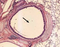
Conducting (primary) bronchiole characteristics
Cartilage, plates, and glands are absent in all bronchioles
disease conditions: muscles contraction, fluid, mucus collection
Conducting (primary) bronchiole histo

Terminal bronchioles characteristics
largely clara cells and simple cuboidal
goblet cells are absent
surrounded by lung tissue

Respiratory bronchioles characteristics
Transition zone is involved in both air conduction and gas exchange
contatins alternating thick and thin walls
Respiratory bronchioles histology
High amont of elastic and reticular fibers in CT
Thin wall: made of alveoli outpouchings
Thick walls: made of cuboidal cells, Clara cells, and smooth muscle

Alveolar ducts
Continuation of respiratory duct
Walls consist of alveoli
Alveolar ducts terminate at alveolar sacs
gas exchange

Alveolar sacs
Arise from alveolar ducts
Common space surrounded by clusters of alveoli
Alveoli are seperated by interalveolar septa that contain blood capillaries

Alveoli
site of gas exchange
surrounded by network of capillaries that bring blood close to gas exchange
300-600- millionalveoli in an adult
surrounded by visceral pleura
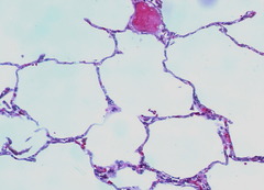
Type 1 pneumocyte
covers 95% of surface
squamous cell joined by tight junctions
facilitate air exchange-rapid diffusion
terminally differentiated
Type 2 pneumocyte
Cuboidal cells at septal junctions
stem cell for type 1 pneumocyte
secretes lipid and protein surfactant that decreases surface tension mixes with alveolar fluid that establishes a liquid air interface and excess is resorbed by type 2 cells
modulates immune response
Alveolar septum thin portion
most gas exchange occurs here
air-blood barrier
Type 1 pneumocyte, surfactant, capillary endothelium
Basal lmina fusion stabilizes alveolar and capillary interface
Air blood barrier
formed by cells and cel products across gas must diffuse between alveolar and capillary compartments
Alveolar septum thick portion
Location of both types of pneumocytes, fibroblasts, WBC
abdundant elastic and reticular fibers
Alveolar macrophages phagocytize particulate matter found in lumen
Alveolar pores
septal wall can be interrupted by small pores that connect adjacent alveoli
not visible histologically
equalize pressure and allow collateral air flow if alveoli is blocked