Muscular System Exam- Macey
 Call Kai
 Learn
 Practice Test
 Spaced Repetition
 Match
 Flashcards
 Knowt Play
1/105

There's no tags or description
Looks like no tags are added yet.
106 Terms
trapezius

pectoralis major

deltoid

latissimus dorsi

Brachialis

biceps brachii
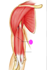
Brachioradialis

triceps brachii

rectus abdominis

external oblique
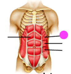
internal oblique

transversus abdominis

gluteus maximus

gluteus medius

Sartorius

adductor muscle
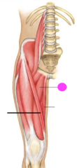
Gracilis
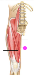
biceps femoris
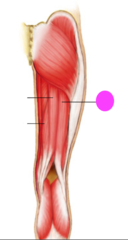
Semimembranosus
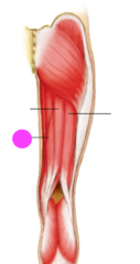
semitendinosus

vastus medius

vastus lateralis

rectus femoris
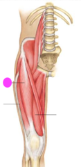
Gastrocnemius

4 characteristics of muscke tissue
excitability, contractibility, extensibility, elasticity
excitability
respond to stimulus
contractibility
shorten when stimulated
extensibility
legnthen or stretch
Elasticity
bounce back to their original shape after being stretched
3 main types of muscle tissue
cardiac, smooth, skeletal
cardiac muscle
found in heart, striated, involuntarily controlled
smooth muscle
found in walls of internal organs, non striated, involuntarily controlled
skeletal muscle
attach to bones of skeleton, striated, voluntary movement
Endomysium
surrounds each skeletal muscle fiber
Epimysium
surrounds the skeletal muscle and binds multiple fascicles together
Perimysium
surrounds each fasicle
name of the neurotransmitter that is released by a motor neuron into the neuromuscular junction?
acetylcholine (Ach)
role of tendon
connects muscle to bone
role of an aponeurosis
connects to the muscle to another muscle
role of troponin
binding sites for actin, topomyosin, and calcium (holds topomyosin in place)
role of tropomyosin
spiraling around the helix
structure that separates one sarcomere from the next
Z-line
in a muscle contraction, what happens during a power stroke
ADP and phosphate group are released and cross bridge pulls thin filament
A bands are dark because they contain which protein filament?
actin and myosin overlap
I-bands
light bands
A-Band
dark bands
I-bands are light because why?
they only contain thin filaments or Actin
components of myofibril
myosin and actin
myosin
thick filaments
Actin
thin filament
what is the functional unit of skeletal muscle tissue
sarcomere
sarcoplasmic reticulum
membranous channels that surrounds each muscle fiber
Role of sarcoplasmic reticulum
stores calcium ions needed for muslce contractions, corresponds to the enodplasmic reticulum of other cells
transverse tubules
membranous channels that extend deep into the fiber
explain what happens at a neuromuscular junction
the synapse where a motor neruon axon and skeletal muscle fiber meet
role of acetylcholinesterase
destroys any remaining Ach in the neurotransmitter junction
1st event of muscle relaxation
enzyme acetylcholinesterase detsroys any remaining Ach in the neruotransmitter junction
2nd event of muscle relaxation
calcium ions are moved back into the sarcoplasmic reticulum
role of creatine phosphate
energy is stored in the phosphate group
agonist
prime mover, muscle primarily respondsible for an action
antagonist
muscle that works against the action
Synergist
muscles that contract and assits the agonist
if oxygen is not present in the cell, after glycolysis what process will a skeletal muscle cell resort to?
lactic acid fermentation
What is the specific role of calcium during a muscle contraction?
binds to actin and causes it to change shape so that it can interact with myosin
1st event of muscle contraction
motor impulse is initiated in the brain
2nd event of muscle contraction
impulse travels down the spinal cord inot a motor nerve ending
3rd event of muscle contraction
nerve ending membrane depolarizes and calcium ions rush in
4th event of muscle contraction
calcium causes the release of Ach into the synaptic cleft of the neuromuscular junction
5th event of muscle contraction
Ach binds to receptors on the motor end plate of a muscle fiber
6th event of muscle contraction
skeletal muscle fiber depolarize; action potential travels along the surface and deep within via transverse tubules
7th event of muscle contraction
when the impulse reaches the sarcoplasmic reticulum, calcium ions are released into the sarcoplasm
8th event of muscle contraction
calcium ions bind to troponin on actin filaments
9th event of muscle contraction
troponin is pulled aside so that binding sites on actin are exposed
10th Event of Muscle Contraction
myosin head binds to actin (forming cross bridges)
11th event of muscle contraction
ADP and phosphate group are released and cross bridge pull thin filaments (power stroke)
12th event of muscle contraction
sracomeres shorten and muscle contracts
13th event of muscle contraction
ATP binds to myosin, breaking the cross bridge
14th event of muscle contraction
ATP is hydrolyzed and the myosin head returns to its original position
sarolemma

sarcoplasmic reticulum

Myofibril

sliding filament step 1
a muscle contraction starts in the brain, where a signal is sent to the motor neuron. The combination of the motor neuron and the skeletal muscle fibers make up a motor unit
sliding filament step 2
the impulse travels into the transverse tubules where it causes calcium to be released from the sarcoplasmic reticulum
sliding filament step 3
calcium binds to the actin and causes it to change shape so that it can interact with myosin
sliding filament step 4
the change in shape allows myosin heads to form cross bridges between the actin and myosin
sliding filament step 5
energy from ATP is used to create a "power stroke" between the 2 filaments. the actin filament slides inward and shortens, or contracts, the whole muscle
mitochondria
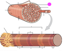
muscle fiber

Nucleus

light I band
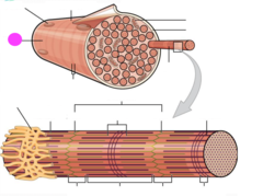
Dark A band

Sarcomere

Thin filament (actin)

H zone

z line

Thick filament (myosin)

I bands

A Bands

M line

fibularis longus
