Haematopoiesis to Haemogram
 Call Kai
 Learn
Learn Practice Test
 Spaced Repetition
 Match
Match Flashcards
 Knowt Play
1/36

There's no tags or description
Looks like no tags are added yet.
Name | Mastery | Learn | Test | Matching | Spaced | Call with Kai | Chat |
|---|
No analytics yet
Send a link to your students to track their progress
37 Terms
haematopoesis
Haematopoesis is the production and development of the different blood cell lineages
• Erythrocytes
• Platelets (Thrombocytes)
Leukocytes
• Granulocytes:
Neutrophils and/or Heterophils
Eosinophils
Basophils
• Monocytes -macrophage family
• Lymphocytes
• Mast cells

Haematopoesis and site of it - foetus
Blood is dynamic- some cell populations turn over more than once within a day Erythrocytes generally have longer half lives (usually over one month- depending on species)
» Initially haematopoesis starts in the yolk sac and the aorta-gonad-mesonephros (AGM) of the embryo
» Production shifts to the liver and spleen by mid-gestation
» Bone marrow and lymphoid organs from mid-gestation onwards. - neonate
juvenile to adult:
Fat begins to replace the haematopoetic tissue as the animal ages
Haematopoesis persists in flat bones and proximal humerus and femur
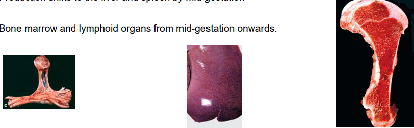
assessment of haematopoesis
Bone marrow sampling
• Performed when we are worried about blood cell production like
• Non-regenerative anaemia
• Thrombocytopenia
• Neutropenia
• Without adequate explanation from other clinical signs
Location may vary according to species
• Dog and cat- Proximal humerus or femur
• Horse- sternum
this is the overview of bone narrow stem cell will go through each pathway

Erythropoesis and Thrombopoesis

Erythroblastic island
red cell production happens in islands
in the spleen
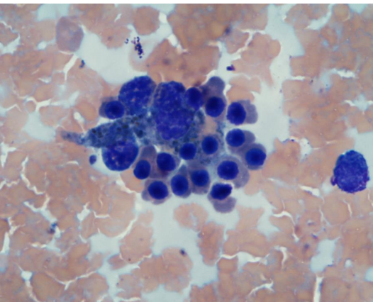
rubriblast
large cell
Earliest recognisable stage of Erythropoesis and Thrombopoesis
» Deep basophilic cytoplasm
» Nucleus round, finely granular chromatin
» One or more nucleoli

Prorubricytes
Nucleoli no longer visible
» Slightly coarser chromatin
» N:C ratio slightly less than rubriblasts
» Changes may often be subtle- so frequently rubriblasts and prorubricytes are considered together as ‘early erythroid’
rubricytes
Basophilic -> Polychromatophilic (purple)-> Metarubricyte
» Last of the stages with a nucleus
• Chromatinbecomes progressively more clumped/mature in appearance
» Cytoplasm develops from basophilic to the red colour consistent with mature erythrocytes

Polychromatophil
Slightly larger than mature erythrocytes
» No nucleus retained
On Giemsa/Romanowksy stain- more purple in appearance than the red mature erythrocyte
• Reflects retention of ribonucleic acid material in the cytoplasm
» May be enumerated precisely on New Methylene Blue stain

Control of Erythropoesis
» Erythropoetin (EPO) is the principal growth factor
• Produced in the kidney (adult)
• Produced in the liver (foetus and small amount in the adult)
• Produced by bone marrow macrophages and erythroid progenitor cells
» Many other hormones will enhance erythropoiesis if EPO is present
» Inhibitors include TGF-b, TNF-a, IFN-g, IL-6 - not expected to remember them just for info
Myelopoesis
the bottom one - black one discussed already

myelopoesis
a- myeloblast
b- promyelocyte

Myelocyte
Stage at where it becomes recognisable as to the lineage which the cell will finally differentiate to
» Large cells
» High N:C ratio
» Thick bean shaped nucleus

Metamyelocyte -> Band -> Mature
» Cells become smaller in size with each division
» Nucleus becomes more elonogated -> band shaped (horseshoe or S)
• – Nucleus thinnest part is less than 1/3 the thickness of the thickest part
» Ultimately the cells mature to lobed polymorphonuclear cells (PMNs) or granulocytes
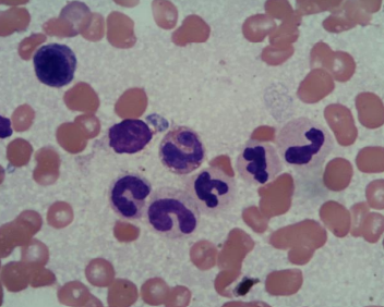
control of Granulopoesis and Monocyte production
Promoters
• IL-3, GM-CSF, G-CSF
• Eosinophils- additionally IL-5
• Basophils also but mainly IL-3
• Mast cells SCF
• Monocytes – IL-3, GM-CSF, M-CSF
• (Monocyte to Macrophage M-CSF)
Inhibition
• Incompletely understood for most cell lines
Thrombopoesis

Thrombopoesis
Megakaryoblast to Promegakaryocytes
Promegakaryocyte to Megakaryocyte

Control of thrombopoesis
Thrombopoetin (TPO) is the main driver
• Most production is in the liver (endothelial cells)
• Also in the kidney and bone marrow stromal cells
• TPO production is usually quite constant
• IL-6 (and other factors) may also stimulate
Inhibition
• PF4, TGF-b, IL-4, TNF-a
Lymphopoesis
» T cells
• IL-7
» NK Cells
• IL-7
• IL-15
» B Cells
• IL-4
• IL-7

Haematopoesis - simplified

Haematology of the patient
» Evaluation of the red cell population(s)
» Evaluation of the white cell populations
» Evaluation of the platelets/thrombocytes
» Identification of abnormalities e.g.
• Infectious agents- intra or extracellular
• Leukaemic cell populations

Core Methods of Analysing the Haemogram
» Spun PCV
• Centrifuge
• Microhaematocrit tubes and plug material
• PCV reader
» Blood Smear (including morphologic description)
• Glass slides
• Microhaematocrit tube
• Stain eg DiffQuik or RapiDiff (automated stainer in a commercial laboratory)
• Microscope (with x100obj- usually oil immersion)
• Cell counter
» Haematology Analyser
» Special stains

PCV Packed Cell Volume
» Centrifugation of a well mixed anticoagulated sample to produce layers
• Red cells
• Platelets and Leukocytes
• Plasma
» Generally considered more reliable- ‘gold standard’ if equipment is well maintained.

Examples of In Clinic Haematology Analysers (IDEXX and Abaxis)

Centrifugal Method- QBC Autoread
Uses the separation of the different cell layers by centrifugal force
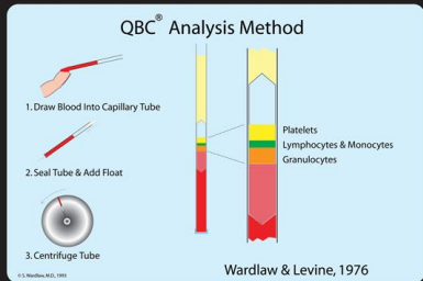

QBC Autoread
Impedance Counters
» Use the change in electrical charge generated by the passage of the cell to allow counting
» Different sizes of cells will generate different change
» May combine lysing agents and impedance counting to improve the differentiation of cell populations

Impedance Counter Histogram
» Graphical representation of the distribution of cell populations
» Can be challenging to reliably distinguish
» Total white cell count is usually a reliable value


Light scatter- flow cytometry
Light Scatter Plot example- Erythrocytes


Light Scatter plot example- White cells
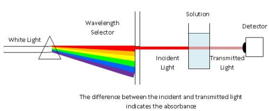
Haemoglobin measurement
great for mammals not so much for non traditional companion animals
Interferences
» Interferences most often affect haemoglobin measurement- red cell parameters
» Lipaemia in the sample may precipitate haemolysis in vitro

Quick checks for reliability
For majority of mammals
• 3xHgb value = Hct
• Hct and PCV are within 3%age points of each other
Do the cell numbers and the smear appearance match

Erythrocyte Parameters
what can we measure?
red cells


White cell parameters
Platelet parameters

‘Normal’ species or breed variations
» The list is not exhaustive
» Microcytosis
• Akita
• Shiba Inu
» Poor platelet/erythrocyte separation in
• Felines- cats often have large platelets and small erythrocytes
• CKCS- a breed hereditary macrothrombocytopenia
• Cairn and Norfolk terriers- a breed hereditary macrothrombocytopenia
Errors with Haematology Analysis
» Pre-Analytical
• Incorrect Labelling
• Inadequate filling of tube
» Analytical
• ‘Short’ sampling
• Sample clots
» Post-Analytical
• Incorrect reporting/assessment of results
Factors that may cause issues with haematology analysis
» Clotted sample
• Improper or inadequate mixing of blood sample and anticoagulant
• Activation of platelets at time of sampling
• If able to be measured then likely to have interference with platelet measurements
• Possible interference with white and red cell measurements also
» Inadequate mixing of sample
» Presence of red cell morphology abnormalities e.g.
• Heinz bodies
• Increase Hgb
• If disassociate then could interfere with platelet measurements
• Red cell fragmentation
• May cause interference with platelet measurements
» Agglutination of erythrocytes
• IMHA (immune mediated haemolytic anaemia