Lab #6 Muscles and Movements
 Call Kai
 Learn
 Practice Test
 Spaced Repetition
 Match
 Flashcards
 Knowt Play
1/90

There's no tags or description
Looks like no tags are added yet.
91 Terms

orbicularis oculi
Orbicularis oculi
Origin: bones and ligments around orbit Insertion: tissue of eyelid Action: closes eye, produces blinking, squinting and draws eyebrows

orbicularis oris
Orbicularis Oris
Origin: maxilla and mandible Insertion: encircles mouth Action: closes lips; purses and protrudes

Temporalis
Temporalis
Origin: Temporal fossa Insertion: mandible Action: closes jaw; elevates and retracts mandible

sternocleidomastoid
Sternocleidomastoid
Origin: (2) manubrium of sternum, and medial portion of clavicle Insertion: Temporal bone and occipital bone Action: flexion of neck; rotate head toward shoulder on opposite side

Trapezius
Trapezius
Origin: occipital bone and spine/vertebrae Insertion: scapula and lateral third of clavicle Action: extends hear; raises, rotates, and retracts (adducts) scapula and stabilizes it

Deltoid
Deltoid
Origin: Lateral 1/3 of clavicle Insertion: humerus Action: whole prime mover of arm abduction

Masseter
Masseter
Origin: zygomatic bone Insertion: angle of mandible Action: Prime mover of jaw closure; elevates mandible

Pectoralis major
Pectoralis Major
Origin: clavicle, sternum, cartilage of ribs (1-6) and external oblique muscle Insertion: humerus Action: Prime mover of arm flexion; adducts medially rotates arm

Pectoralis minor
Pectoralis minor
Origin: anterior surface of ribs near costal cartilage Insertion: scapula Action: draws scapula forward and inferiorly; draws rib cage superiorly

Intercostals
Intercostals
Origin: above and below ribs Insertion: above and below ribs Action: pulls ribs together and apart to elevate and depress ribcage

Rectus Abdominis
Rectus abdominis
Origin: pubic crest and symphysis Insertion: xiphoid process and costal cartilage of ribs Action: flexes the vertebral column, increases abdominal pressure, stabilized pelvis during walking

External oblique
External oblique
Origin: anterior lower 8 ribs Insertion: pubic crest and iliac crest Action: compresses abdominal wall; trunk rotation and lateral flexion

(the deeper layer, not the superficial layer)
Internal oblique
Internal oblique
Origin: iliac crest Insertion: pubic crest and costal cartilage of last 3 ribs Action: same as external oblique

(deepest layer of abdominal region muscles)
Transversus Abdominis
Transversus Abdominis
Origin: iliac crest, lumbar, and cartilage of ribs (last 5-6) Insertion: pubic crest Action: compresses abdominal contents

Latissimus dorsi
Latissimus dorsi
Origin: (lower 6) thoracic vertebrae, lumbar vertebrae, (last 4) ribs, iliac crest Insertion: intertubercular sulcus of humerus Action: prime mover of arm extension; adducts and medially rotates arm
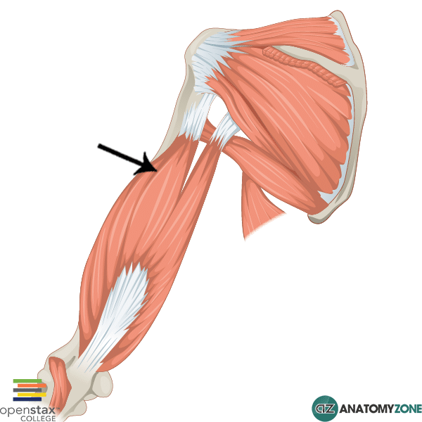
Triceps brachii
Triceps brachii
Origin: glenoid cavity, humerus Insertion: olecranon of ulna Action: powerful forearm extensor

Biceps brachii
Biceps brachii
Origin: glenoid cavity, shoulder joint Insertion: lateral aspect of olecranon of ulna Action: flexion (powerful) and supination of forearm

Brachialis
Brachialis
Origin: distal portion of anterior humerus Insertion: coronoid process of ulna Action: flexor of forearm

Extensor carpi raadialis Longus
Extensor Carpi radialis Longus
Origin:lateral ridge of humerus Insertion: base of metacarpal II Action: extends and abducts hand

(pronated arm view)
Extensor digitorum
Extensor digitorum
Origin: lateral humerus Insertion: 4 tendons into distal phalanges of fingers III-V Action: prime mover of finger extension; extends hand

(supinated arm view)
Flexor digitorum superficialis

(supinated view of arm)
Flexor Carpi radialis
Flexor Carpi radialis
Origin: Medial epocondyle of the humerus Insertion: base of metacarpals of II and III Action: to flex and radially deviate (abduct) the wrist

Gluteus maximus
Gluteus Maximus
Origin: dorsal ilium, sacrum, and coccyx Insertion: gluteal tuberosity of femur and iliotibial tract Action: powerful thigh extensor, laterally rotates and abducts thigh

Gluteus medius
Gluteus Medius
Origin: Upper lateral surface of ilium Insertion: Greater Trochanter of femur Action: abducts and medially rotates thigh; steading pelvis during walking

Sartorius
Sartorius
Origin: anterior superior iliac spine Insertion: medial aspect of proximal tibia Action: flexes, abducts, and laterally rotates thigh; flexes leg (helps effect cross-legged position)

Gracilis
Gracilis
Origin: inferior ramus and body of pubis Insertion: medial surface of tibia just inferior to medial condyle Action: adducts thigh; flexes and medially rotates leg

Quadriceps femoris

Rectus femoris
Rectus femoris
Origin: anterior inferior iliac spine and superior margin of acetabulum Insertion: tibial tuberosity and patella Action: extends leg and flexes thigh

Vastus Lateralis
Vastus lateralis
Origin: greater trochanter Insertion: tibial tuberosity and patella Action: extends leg and stabilizes knee

Vastus Medialis
Vastus medialis
Origin: linea aspera and intertrochanteric line Insertion: tibial tuberosity and patella Action: extends leg; stabilizes patella

Vastus intermedius
Vastus intermedius
Origin: anterior and lateral surface of femur Insertion: tibial tuberosity and patella Action: Extends leg

Biceps femoris
Biceps femoris
Origin: ischial tuberosity and distal femur Insertion: head of fibula and lateral tibia Action: extends thigh; laterally rotates leg, flexes leg

(posterior view)
Semimembranous
Semimembranous
Origin: ischial tuberosity Insertion: medial tibia; lateral femus’s Action: extends thigh; flexes legm medially rotates leg

Gastrocnemius
Gastrocnemius
Origin: two heads from medial and lateral condyles of femur Insertion: calcaneus Action: plantar flexes foot when leg is extended

Flexor digitorum Longus
Flexor digitorum Longus
Origin: posterior tibia Insertion: phalanges II-V Action: flexes toes; plantar flexes and inverts foot

Extensor digitorum Longus
Extensor digitorum Longus
Origin: lateral tibia/fibula Insertion: phalanges II-V Action: prime mover of toe extension, dorsiflexes foot

Tibialis Anterior
Tibialis anterior
Origin: lateral/upper tibia Insertion: metatarsal I Action: prime mover of dorsiflexion; inverts foot, supports longitudinal arch of foot

Dorsiflexion

(downward movement)
Plantar flexion

(downward position)
pronation

(upward position)
supination

abduction

(movement inwards)
adduction
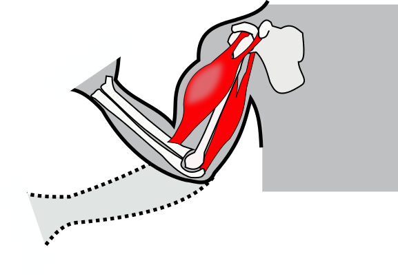
flexion
Origin
the fixed, stationary more stable end of a muscle; typically more proximal (located closer to the torso or center); acts as an anchor during movement
Insertion
the mobile, moveable end of a muscle; typically located distally, is pulled toward the origin to create action

Epimysium
Epimysium
Connective tissue covering the entire muscle

(the section in green)
Fascicle
Fascicle
Bundle of muscle fibers surrounded by perimysium

(green)
Perimysium
Perimysium
Connective tissue surrounding a fascicle

(most inner section at the top right)
Muscle fiber
Muscle fiber
a single muscle cell

(the portion between individual muscle fibers)
endomysium
Endomysium
Connective tissue covering a single muscle fiber