rad w6 (2. 3, 5)
 Call Kai
 Learn
 Practice Test
 Spaced Repetition
 Match
 Flashcards
 Knowt Play
1/13

Earn XP
Description and Tags
indire
14 Terms
SKIPPED BASIC ATOM STUFF
Electron
max 7 shells
closest shell to nuc = highest E
Ion = atom that gained/lost an e- = electrically unbalanced
Ionization
proc of converting atom > ion
need sufficient E to remove an e-
when e- removed from atom = ion pair results
Ion pair
1 pos ion (atom that e- was removed from
1 neg ion (ejected e-)
they cont react w other unstable electrically charged ions until an electrically stable neutral atom is formed
Radiation
emission & trasmission of E thru space/substance in form of wave/particle
Ionizing radiation
radiation that can prod ions by remove/adding e- to an atom
2 groups = particulate radiation + electromag radiation
Particulate radiation (particles)
pertain to mass, travel straight, high speed, KE
electrons (beta particles or cathode rays)
Cathode rays = originate from xray tube
BE ABLE TO LABEL CATH/ANODE DIAGRAM
Electromagnetic radiation
propagation of wavelike E (no mass) thru space/matter
E spead accompanied by oscillating electric + mag fields that are at right angles to each other
man made/occur natural
diff E lvls (electromag spectrum)
ionizing or non-ionizing (high E are ionizing
travel as wave/particle


Wave concept
velocity (wave speed)
wavelength (crest>crest distance)**
freq (# waves in certain amt of time)
freq (# waves in certain amt of time) & wavelength (crest>crest) INVERSELY RELATED
when one go up other go down
long wavelength = low freq
short wavelength = high freq
X-Radiation
high E, ionizing electromagnetic radiation
possess props of both WAVES + PARTICLES
vs
X-ray
photons
no mass
speed of light
no electrical charge
travel in WAVES (short/high freq)
penetrating powers
xrays are absorbed by matter
Tubehead has…
metal housing
insulating oil = (allow not overheat)
tubehead seal = (alum/leaded glass) (filter xray beam)
xray tube = (xray generating sys)
transformer = (step up & step down) (control voltage)
aluminum disks = (behind collimator) (filter xrays)
lead collimator = (quality xray) (restrict beam size)
PID = (aim & shape beam)
1. Step-down transformer
Decreases voltage.
Increases current to heat the cathode filament.
Helps release electrons through thermionic emission.
2. Step-up transformer
Increases voltage (kVp).
Accelerates electrons from the cathode (−) to the anode (+).
Gives electrons enough energy to produce X-rays.
SEE IMAGE FOR MACHINE BREAKDOWN
generally
electricity is E used to make xrays
electrical E = flow of electron thru conductor = electrical current
need flow from cathode > anode

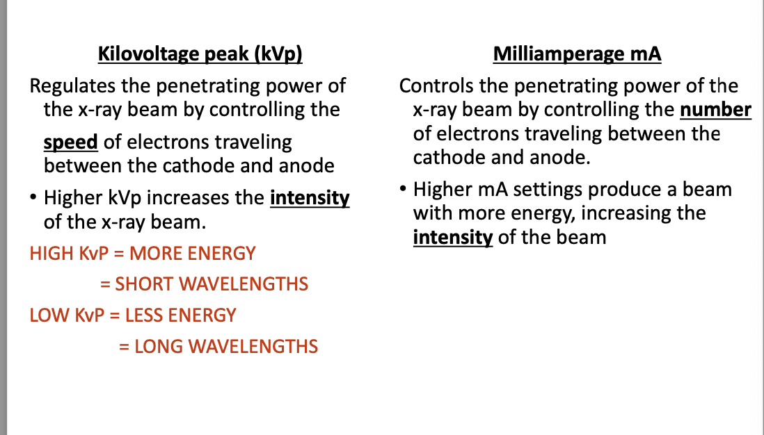
mA (milliamperage)
measurement of # of e- moving thru a conductor
SPEED, DENSITY
total quant of xrays produced
higher mA = darker image
kVp = kilovoltage peak
highest voltage during exposure
control SPEED of e- when travel from cathode > anode
e- speed determine PENETRATING POWER & wavelength
inverse^^
timer
regulate exposure time
like mA control film blackening (DENSITY)
general/braking radiation
sudden stopping of high speed e- after hit tungsten target
when e- hit nuc of tungsten atom = xray photon prod (gen rad)
lower E bc e- slow down when get close to nuc
70% of xrays prod at anode
the lost energy = xray photon (green line in image)
green line in image


types of xrays produced
Characteristic rad
prod when high speed e- dislodges an inner shell (K) electron from tungsten atom (cause IONIZATION)
remaining orbiting e- rearrange to fill vacancy
only small % of xray prod
Primary rad
xray beam prod at target of anode & exit tubehead
primary/useful beam
always interact w matter - mostly what we see in dent
Secondary rad
x-radiation created when prim beam interact w/ matter (ex: head)
less penetrating than prim
Scattered rad
form of SECONDARY deflected from its path (by interact w matter)
result of xray photon deflected in all directions travelling scattered
DETRIMENTAL
image is characteristic
borrowed one ouer e- trying to get close to nucleus
not clean xray bc scattered
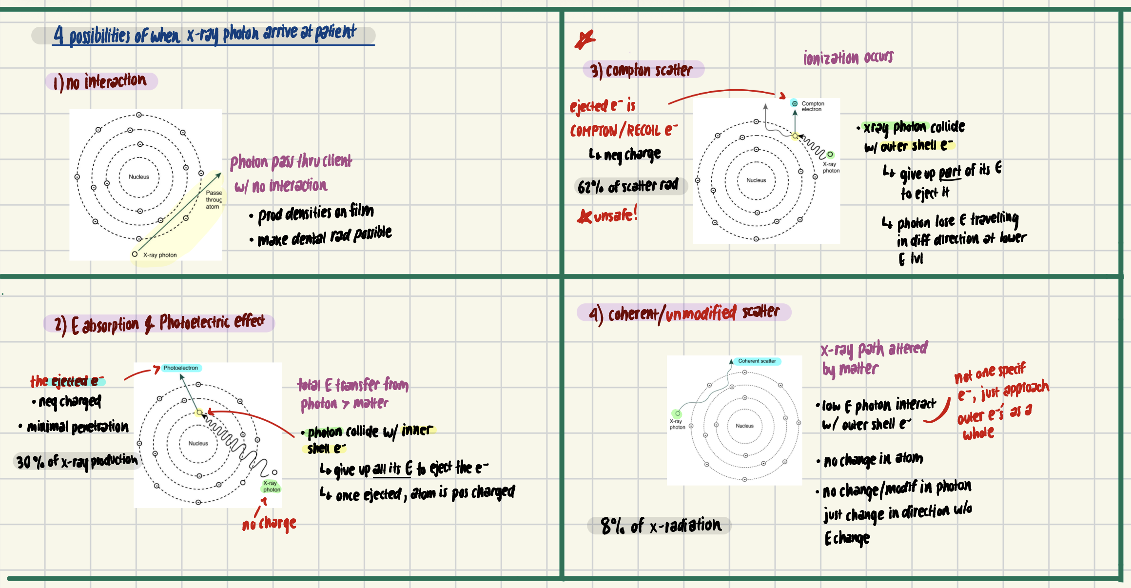
4 possibilities of when xray photon arrive at patient
Rad biology
study of effects of ionizing radiation on living tissue
Dr. William Rollins
father of rad protection (not alara tho)
some xrays dont reach film & are absorbed by patient tissue
when absorb, chem changes occur > biologic damage
absorp/photoelectric effect (completely absorbed by patient)
compton (xray photon cause ionization w various atoms until all E gone
*refer to diagram i made*
2 mechs of rad injury
ionization (when xray strike tissue)
free radical formation (INVOLVE IN DEGENERATIVE DISEASE + CANCER)
cell damage primarily occur thru formation of free radicals
formed when photon ionize water (component of cells)
so what is it
uncharged (neut) atom/molec that exist w/ single unpaired e- in its outermost shell
highly reactive + unstable
Free radicals are highly reactive and unstable because they have an unpaired electron.
react with important cell structures, including DNA.
can cause cell damage, mutations, or cell death.
Why are free radicals dangerous?
DNA damage → Can lead to mutations.
Cell death → Can damage tissues.
Cancer risk → DNA mutations may increase the risk of cancer.
theories of rad injury
most xray pass thru cell little/no damage
but can result from…
direct hit & absorp of photon in cell
DIRECT THEORY
cell damage result when ionizing rad direct hit critical areas within cell
ex: DNA
occur infreq (most pass thru no damage)
indirect absorp of photon by WATER IN CELL + free rad formation
photons absorbed in cell & cause formation of toxins (free rad) - damage cell
damage not from hit
occur FREQ bc cell is 70-80% water
Biological effects from rad (2)
Stochastic (CHANCE)
no threshold (no completely safe rad dose)
linear relationship (as dose inc, harm inc)
severity of effect NOT dose related
ex: genetic mutations like tumor/leukemia
Nonstochastic
somatic (phys) effects that have a threshold (certain dose needed for effect)
higher dose > more severe (but not nec linear)
large dose need for serious issue
ex: erythema, brun, hair loss, cataract form, infertility
Sequence of Rad Injury
Latent Period
time between exposure to ionizing rad / observable clinical sign
depend on total rad dose received & amt of receiving time
more rad receive = faster dose rate = shorter the latent period
ex: guy who radiated hand
Injury Period
follow latent, variety of injury result in….
cell death, cell fxn change, cessation of mitotic activity (or abnormality)
Recovery Period (if not then perm damage)
not all cellular rad injuries perm
most damage caused by low level rad repaired within body cells
can have cumulative/additive effects if unrepaired
ex: cancer, catarct formation, birth defects (if no recovery)
5 contrib factors to rad injury
total dose (type/amt/ energy lvl/ duration)
more famage occur when large quant of rad absorbed
dose rate = rate at which rad occurs & absorp take place
dose rate = dose/time
more damage if high dose bc rapid delivery dont allow time for cellular damage repair
area exposed = larger = greater injury
localized better than full body rad
cell sensitivity
faster if cell rapid dividing type or young type
radiosensitive = small lymphocte, bone marrow, reprod, young bone
radioresistant = muscle, nerve, mature bone
age = child + old more suscept
in old = thinner body tissue
Rad effects
1. Short- and long-term effects (not applic dentistry)
2. Somatic and genetic effects**** (focus on these left out rest)
3. Radiation effects on cells
4. Radiation effects on tissues and organs
Somatic & genetic effects
biologic effect of rad classified into 2
somatic cells = all cells EXCEPT reprod
genetic cells = REPROD

these cells more sensitive to rad
ones that div freq more sensitive to rad
immature/not highly specialized
ones w higher metabolism
MOST SENSITIVE = SMALL LYMPHOCYTE***
amt of exposure depend on 4 things
film speed = F speed film instead of D redue by 60%
collimation = rectangle better than round (60-70%)
technique = long cone paralleling
exposure = high KvP better

A new patient in your office is told that she is in need of a
full series of x-rays. She is concerned about the risks with
taking so many x-rays. The patient asks the dental radiographer
for more background information regarding radiation exposure.
What type of information can the dental radiographer
provide the patient?
led apron
thyroid collar
alara
dig xray
PID
led collimator
expose rate very low
A new patient presents in your office. The front desk have
looked into their insurance coverage and tells you to do
an FMS on that client and a Panoramic because their
insurance covers it.
no baseline on him (as dh) bone lvl & sus caries
age? med hist?
Voltage
KvP
reg penetrating power of xray beam
control e- speed from cathode>anode
IF INC, OTHER FACTORS STAY CONSTANT
for proper density + cpntrast = inc kvp/dec exposure time
mainly control contrast
high kvp = more E = short wavelength
low kvp = less E = longer wavelength
kV
control quality (wL & E of beam)
control density (darkness)
control contrast (grey shades)
time
interval in which xrays produced
measured in impulses (60/s)
if we dec kvp, we can inc time FOR PROPER DENS/CONT
mA
# e- or current flowing thru cathode fil
1) Distance from source of rad > patient’s skin
TARGET – SURFACE DISTANCE
2) Distance from source of radiation > tooth
TARGET – OBJECT DISTANCE
3) Distance from source of radiation to the film
TARGET – FILM DISTANCE

1. High kVp → LOW contrast
X-rays have more penetrating power.
Produces more shades of grey.
Less difference between black and white.
Called long-scale contrast.
2. Low kVp → HIGH contrast
X-rays have less penetrating power.
Produces fewer shades of grey.
Greater difference between black and white.
Called short-scale contrast.