Lesson 2.1: Embryology and dental eruption
 Call Kai
 Learn
 Practice Test
 Spaced Repetition
 Match
 Flashcards
 Knowt Play
1/44

There's no tags or description
Looks like no tags are added yet.
45 Terms
What is embryology?
The study of the progressive growth and differentiation that takes place during the early stages of embryonic development
What is Odontogenesis?
Tooth formation and development, which is a continuous and complex biological process that occurs as part of embryogenesis of cranial and maxillofacial complex
What does odontogenesis involve and when does it begin
It involves ectoderm, mesoderm and cells from neural crest.
It begins between the 4th-6th week of intraeuterine life
What are the stages of odontogenesis?
Dental lamina, thickening, initiation, dental bud, cap and bell.

Where do the dental tissues originate from?
Enamel organ: comes from oral ectoderm and produces enamel
Dental papilla: comes from mesenchyme and produces dentin and dental pulp
Dental sac: comes from mesenchyme and gives rise to cementum and pdl
What are the 2 main phases of odontogenesis?
Morphogenesis and Hystogenesis which converge at bell stage
Morphogenesis
Consists of the development and formation of the coronary and radicular patterns as a result of the division, displacement and organization of the different parts involved in the process.
It begins at the sixth week and the first manifestation is the differentiation of the dental lamina from the ectoderm that lines the primitive oral cavity.
Hystogenesis
Consists of the formation of the different types of dental tissues: enamel, dentin and pulp, in those previously formed patterns.
The reciprocal induction phenoemenon
Ectomesenchymal cells that are derived from the neural crest epithelial cells (ectoderm) migrate into cephalic region and acquire a mesenchymal phenotype. These cells will give rise to dentin-pulp complex
How are ectomesenchymal cells formed?
Epithelial cells of neural crest with ectodermal origin begin to divide by mitosis and occupie some space and then migrate towards mesoderm so that they come in contact with the mesoderm and form ectomesenchymal cells.
Later the dental lamina is formed

How many thickenings of the oral epithelium are formed in each maxillary bone?
10 in each = 20
During 5th month of pregnancy 32 germs of permanent teeth are formed
Structures of Dental organ

Vestibular and dental lamina
Vestibular lamina
Located towards exterior
Cells proliferate with the ectomesenchyme, rapidly increase their volume and then degenerate forming that forms the vestibular sulcus between cheeka dn tooth zone
Dental lamina
Cells proliferate rapidly increase their volume an intense and localized manner
8th week of intrauterine life: 10 epithelial growths are formed within the ectomesenchyma of each jaw
5th month of gestation: 32 germs of permanent dentition
What induces the proliferation of the buccal epithelium to form the vestibular and dental lamina?
Underlying ectomesenchyme
Primordium / tooth germ
Formed by aggregation of differentiating cells that will form the future tooth
Onset of first permanent molar appears as early as the 4th month intrauterine life
2nd and 3rd molar begin to develop after birth with 4-5 years
Which key signaling molecules are involved in dental development ?
Msx: Muscle segment homeobox
Lef1: Lymphoid cell enhancer factor
Pax9: Paired box gene
BMP: Bone Morphogenetic Protein
FGF: Fibroblast growth factor
Msx: Muscle segment homeobox
Expressed where epithelium-mesenchyme interaction is necessary
Necessary for beginning of incisor formation and the budding and enamel formation stage
Alterations in these genes produce hypodontia and cleft palata
Lef1: Lymphoid cell enhancer factor
Important for the formation of the cap stage
Pax9: Paired box gene
Expressed in mesenchyme and is essential for mophogenesis
Alterations in this gene cause severe oligodontia
BMP: Bone Morphogenetic Protein
Components of osteo-induction
Expressed during early morphogenesis
BMP4 is main signalling component capable of inducing odontogenesis
Induce expression of Msx genes
FGF: Fibroblast growth factor
Mediate growth and differentiation of developing cells
Stimulate epithelial and mesenchymal divison and proliferation
Induce Mx1 expression
Expressed by developmental epithelial cells in dental Germs when epithelial-mesenchymal interactions regulate odontogenic morphogenic
Other transcription factors and signalling molecules such as p63, TNF and ectodyplasin (EDA)
Involved in formation and growth of the germ
Defects cause ectodermal dyplasias characterized by lack of teeth (agenesis)
Overactivation of the receptor for EDA leads to additional teeth with aberrant morphology
Extra tooth germs = supernumerary teeth, smaller germs = missing and smaller teeth, Overactivation = larger teeth
Stages of odontogenesis
Initiation. Thickening stage. Formation of the dental lamina.
Bud stage. Appearance of all buds.
Cap stage: formation of the enamel organ
Bell stage. Differentiation and histogenesis.
Secretory stage. Follicle stage. Enamel and dentin secretion

Bud stage (proliferation stage)
First epithelial growth occurring within the ectomesenchyma of the jaws, that arises as a result of mitotic divison of stemcells from basal layer of epithelium
Future enamel organs that will give rise to only ectodermal tissue of tooth the enamel
Differentiation of dental lamina with local round proliferations that originate from it in each arch at different positions corresponding to the position of the primary teeth (10 mandible / 10 maxilla)
Beginning of dental organ called dental follicles
What are the 3 types of cells in the bud stage?
Cylindrical cells in the periphery
Polygonal cells with very narrow intercellular spaces inside
Cells of underlying ectomesenchyme are condensed below the lining around the bud

Transition between cap and bud stage
Pax9 and Msx1 are necessary for transitions between different satges
Marks the beginning of morphological differences between tooth germs that give rise to different types of teeth
Cap stage
Bud undergoes an invagination due to unequal proliferation and originates a concavity forming the cap
Ectomesenchymal cells condense and capillaries appear under the influence of the proliferative epithelium forming the dental papilla which form the dentin-pulp complex
Mesenchymal tissue surrounding the cap will condense forming the primitive or dental follicle
What are the structures of cap stage?
Enamel organ (ectodermic):
OEE = Peripheral cells arranged along the enamel organ that consists of a single layer of colloid cells
IEE = tall cyclndrical cells, innermost layer that surround the papilla, will transform into a layer of ameloblasts
Stellate reticulum
Dental papilla (Ectomesenchyme) = Will form pulp
Dental sac (Ectomesenchyme)

Transition between cap and bell stage
Enamel knot that stimulates cusp formation
Receptors for enamel knot signals are located in the epithelial cells surrounding the enamel knot
Organized center that orchestrates morphogenesis
FGF4 and Slit-1 are the best molecular markers for enamel knot formation because they are the only genes that have been observed in both primary and secondary knots
Bell stage
Occurs between 14-18th weeks of intrauterine life, when invagination of IEE deepens giving the structure the appereance of a bell
Structural and histological modifications take place in the enamel organ, dental papilla and dental sac
Early and late beöö stage
Early bell stage structure
Enamel organ
OEE: Cells change from cuboidal to flattened
Stellate reticulum
Stratum intermedium: between stellate reticulum and IEE, 4-5 rows of flat cells with elongated central nuclei and poorly developed organelles
IEE / Preameloblasts: Cells of IEE differentiate into preameloblasts, including differentiation of dental papilla cells to preodontoblasts (Once odontoblasts are formed they transform into young ameloblasts)
Dental papilla / future pulpar cavity = Differentiation of odontoblasts from dental papilla, which will synthesize dentin at the cuspal level
Dental sac
Internal: cellulovascular
External: Abundant collagen fibers

What happens in late bell stage?
Preameloblasts change polarity and turn into young ameloblasts allowing a secretory function of enamel proteins
They will remain inactive until the odontoblasts secrete the first layer of dentin → Predentin
Preodontoblasts turned into Odontoblasts

Follicular (apposition) stage
Begins in areas of future cusps or incisal edges with the formation of the enamel matrix over developing dentin layers
Differentiation of young ameloblasts to ameloblasts
Development of crown formation:
Odontoblasts start dentin deposition
Ameloblasts start enamel deposition
Specific signals between enamel organ and dental papilla determine morphology of crown resulting in the shape number and distribution of cusps
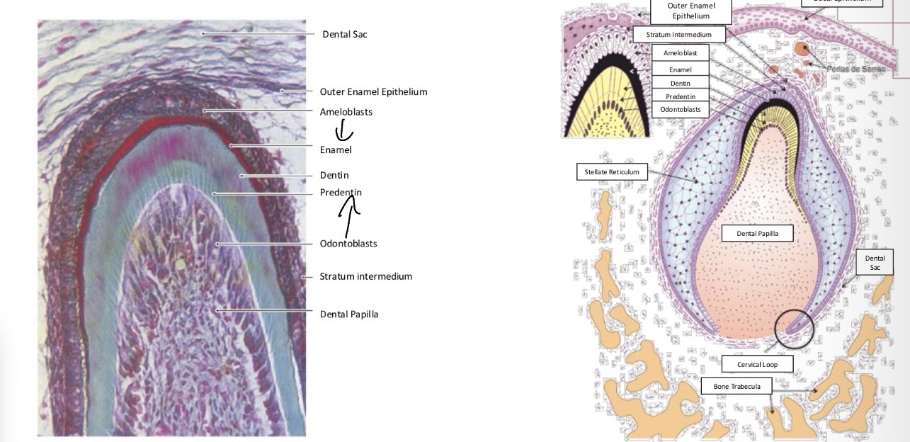
Structures of follicular stage

Anomalies associated with odontogenesis
Defect in migration of neural crest cell
Treacher collins syndrome
Congenital disorder of craniofacial development characterized by bilateral symmetrical otomandibular dysplasia associated with various morphological anomalies of the head and neck.#
Cause by mutations in the TCOF1 gene located on chromosome 5 (dominant)
Most common symptoms: Imcomplete development of malar bones, dental occlusion, ogival palate and sometimes cleft palate, feeding problems in the early years

Dental agenesis
One of most common craniofacial anomalies in human development
Genetically heterogeneous disorder that manifests as the congenital absence of one or more teeth
Hipodontia: Lack of 1-6 teeth
Oligodontia: Lack of more than 6 teeth
Anodontia: complete lack of teeth
Causes of Agenesis
Condition of multifactoral origin influenced by genetic, environmental, pathological and evolutionary factors involved in normal mechanisms of odontogenesis.
PAX9: Necessary for gene expression
MSX1: Encodes transcription factors involved in the different stage of development
What are the genetic anomalies responsible for oligodontia?
Oligodontia due to mutations in the homeobox gene MSX1. It manifests as agenesis of second premolars and third molars. It may be associated with cleft lip and cleft palate.
Oligodontia due to mutations in the PAX9 gene. It results in the absence of most permanent molars, sometimes together with hypodontia in the primary dentition.
Oligodontia due to mutations in the AXIN2 gene. It is characterized by multiple agenesis of permanent teeth, and individuals with these mutations have a significantly higher risk of developing colorectal cancer.
Down syndrome
Agenesis found in 70% of woman and 91% of men with Down syndrome
Maxillary lateral incisors, mandibular central incisors, and second premolars are the most frequently affected teeth
Unilateral or bilateral agenesis of the maxillary lateral incisors occurs in more than 10% of patients.
Non-syndromic dental hypergenesis
Prevalence of supernumerary teeth is 0.2-0.9%
Can appear everywhere in the arches but more frequently in upper
Genetic reason but mutation responsible not identified now
Usually single, can be multiple (Gardner syndrome / Cleidocranial dysplasia)
Hertwigs epithelial root sheath formation
At cervical loop fusion occurs between IEE and OEE of enamel organ giving rise to Hertwigs epithelial root sheath (Without presence of stellate reticulum)
Hertwigs sheath (cuboid cells) is the inducer and modulator of the tooth root. In the papilla it induces the differentiation of root odontoblasts that will form dentin
Hertwigs sheath looses its continuinity and fragments forming the epithelial remains of Malassez which persist in pdl
Sheath epithelium shapes the future dentinocemental junction, by inducing the formation of dentin on the inside and cementum on the outside
Present At eruptive stage
Differentiation of tooth sac cells
Cells penetrate and rest against newly formed root dentin
Mesenchymal cells of the tooth sac surrounding the sheath differentiate into:
Cementoblasts: Cementum
Fibroblasts: PDL
Osteoblasts: Alveolar bone
Which growth and signalling factors are involved in the differentiation of tooth sac cells?
TGF: Transforming Growth Factor
PDGF: Platelet-derived growth factor
FGF: Fribroblast growth factor.
Adhesion Molecules: E-Cadherin

Formation of Uni- or mult-radicular roots
HERS may produce 2-3 tongue like extensions directed toward the axis of the tooth
Once the floor is delimited, they will proliferate independently in each of the roots.
After root formation is completed, the epithelial sheath curves inward forming the diaphragm, which defines the root limit and the new primary apical foramen.
Through the apical foramen, nerves and blood vessels enter and exit the pulp chamber. Some authors state that at this point the dental papilla becomes the dental pulp