LAB MICRO 4
 Call Kai
 Learn
 Practice Test
 Spaced Repetition
 Match
 Flashcards
 Knowt Play
1/44

Earn XP
Description and Tags
anaerboes & parasites
45 Terms
Define different types of isolates based on atmospheric requirements:
Obligate Aerobe: only aerobic growth; oxygen required
Facultative Anaerobe: both aerobic & anaerobic growth; greater growth in presence of oxygen
Obligate Anaerobe: only anaerobic growth; cannot grow in the presence of oxygen (toxic)
Aerotolerant Anaerobe: anaerobic growth; but continues in presence of oxygen
Microaerophilic: aerobic growth; oxygen required in low concentration
Capnophilic: aerobic growth; CO2 required in high concentration

What clinical clues are associated with anaerobes?
Infectious process next to mucosal surfaces with normal anaerobic flora in GI, female genital tract, & oral cavity
Foul odor or gas
Tissue necrosis & abscess formation
Failure to respond to antibiotics that are not active against anaerobic bacteria
Specimen collection requirements for anaerobes
Avoid contamination by anaerobic normal flora of mucosal surfaces
Sterile body fluids only (blood, pleural & peritoneal fluids, aspirates)
Anaerobic transport media – agar gel with buffered mineral salts & oxygen free
Room temp 15-20 minutes or longer if in anaerobic vial
DO NOT INCUBATE, may cause overgrowth
DO NOT REFRIGERATE, cold increases oxygen diffusion
Anaerobic growth methods
anaerboic chamber
anaerobic jars & sealed boxes
anoxomat
oxyplate
anaerobic indicators
Describe Anaerobic Chamber
materials introduced through small doors in airlock while operator works through arm ports in airtight sleeves
85% N2, 10% H2, 5% CO2 as compressed gases
Describe Anaerobic Jars & Sealed Boxes:
Water mixed with chemical packet containing sodium bicarbonate & sodium borohydride, hydrogen & CO2 generated
Hydrogen & atmospheric oxygen in jar react with palladium catalyst & combine to form water, removing oxygen
Atmosphere contains: <5% oxygen, need 80-90% N2, 5% hydrogen, 5-10% CO2
Anaerobic indicator placed in jar to monitor atmospheric conditions
Methylene Blue = blue → colorless
Resazurin = pink → colorless
Describe Anoxomat
jar evacuation & replacement system uses compressed gas to create anaerobic atmosphere
80% N2, 10% hydrogen, 10% CO2
Palladium pouch = removes any O2, forming water
Describe OxyPlate
each petri dish becomes anaerobic chamber
Culture medium contains enzyme (oxyrase) which combines oxygen with hydrogen to remove oxygen as water is formed
Describe anaerobic indicators
Methylene Blue = blue → colorless
Resazurin = pink → colorless
Clostridium perfringens: place inoculated plate in anaerobic environment, growth = anaerobic conditions maintained
Why do we use anaerobic culture media?
Contains hemin, vitamin K to enhance anaerobic growth – selective most important because anaerobic infections are polymicrobial (infection caused by 2 or more species)
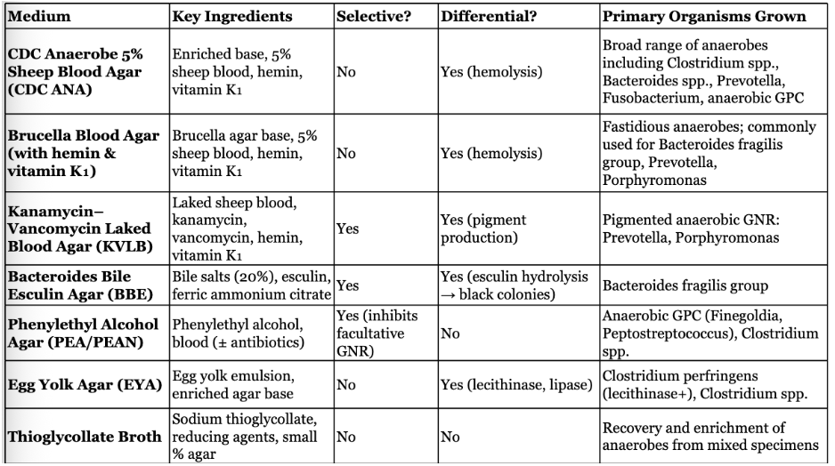
Anaerobic Blood Culture (CDC):
Anaerobic Blood Culture (CDC): enriched (5% sheep blood, hemin, K1), differential based on hemolysis
general purpose medium for cultivation of all anaerobic bacteria
Bacteroides Bile Esculin Agar (BBE)
Bacteroides Bile Esculin Agar (BBE): enriched (bile salts 20%, esculin, ferric ammonium citrate), selective, & differential (esculin hydrolysis = black colonies & iron)
Isolation & presumptive ID of bacteroides fragilis based on bile tolerance & esculin hydrolysis
Kanamycin-Vancomycin Laked Sheep Blood Agar (KVL)
Kanamycin-Vancomycin Laked Sheep Blood Agar (KVL): enriched (laked sheep blood, kanamycin, vancomycin, hemin, K1), selective, differential (pigment)
Isolation of anaerobic bacteria bacteroides & prevotella
Prevents growth of facultative gram neg & gram pos bacteria
Lysed blood enhances pigment formation
Phenylethyl Alcohol Agar (PEA)
Phenylethyl Alcohol Agar (PEA): enriched (antibiotics), selective (inhibits gneg rods)
Isolation of most anaerobic organisms (gpos & gneg) from mixed cultures
Inhibits facultative anaerobic gram neg bacteria like enterobacterales
Colistin Nalidixic Acid Agar (CNA)
Colistin Nalidixic Acid Agar (CNA): inhibits gram neg organisms & grows most gpos anaerobic & facultative anaerobic bacteria
Egg Yolk Agar
Egg Yolk Agar: detect proteolytic enzymes (lipase & lecithinase) produced by clostridium
Lecithinase = opaque zone around colony
Lipase = blue green sheen on agar surface around colony
Broths with Reducing Agents
Broths with Reducing Agents: thioglycollate & cooked meat
Oxygen tension decreases toward bottom of tube allowing growth of anaerobic isolates without incubation in anaerobic conditions
Describe aerotolerance testing
Bacteria must first be demonstrated to be an anaerobe
Inoculate onto anaerobic BAP & increased CO2 CAP
Growth only on anaerobic BAP = obligate anaerobe
Growth on aerobic CAP, but better on anaerobic BAP = aerotolerant anaerobes
Anaerobic GNB
Bacteroides sp – GNCB
Prevoltella sp – GNCB
Porphyromonas sp – GNCB
Fusobacterium sp – GNR with tapered ends
Definitive ID with MALDI-TOF or MOLECULAR

Bacteroides fragilis
GNCB
BBE = brown
20% bile tolerant
Bile esculin + (can hydrolyze esculin)
KVL = growth, vancomycin & kanamycin & colistin RESISTANT
Indole -
Prevotella
GNBC
BBE = NO GROWTH, inhibited by bile
KVL = GROWTH, vanco/kana/colistin RESISTANT
Indole -
Black colonies
Porphyromonas
GNBC
BBE = NOW GROWTH
KVL = NO GROWTH, vancomycin SENSITIVE
Indole +
Black colonies
Fusobacterium sp
GN fusiform, bread crumb colonies
F. nucleatum: common, cause brain abscess
F. necrophorum: serious, lemierre’s
BBE: NO GROWTH
Inhibited by 20% bile
KVL: NO GROWTH
Vancomycin RESISTANT
Kanamycin, colistin SENSITIVE
Most common are indole +
Anaerobic GNC
Veillonella: most common infections in mouth & urogenital
Nitrate reduction +
Glucose fermentation -
Acidaminococcus: most common infections in GI
Nitrate reduction -
Glucose fermentation -
Megasphaera: most common infections in GI
Nitrate reduction -
Glucose fermentation +
Anaerobic GPB
C. perfringes
C. tetani
C. botulinum
C. difficile
C. septica
C. perfringes
Clostridium perfringens: Gas gangrene (myonecrosis), post-abortion sepsis, food poisoning, histotoxin & enterotoxin
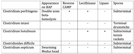
Double zone beta hemolysis
Boxcar shaped cells
Setup on EYA
Lecithiase +
Lipase -
Subterminal spores
Reverse CAMP +
Nagler test (anti-lecithinase) +
Tributyrin test -
C. tetani
Clostridium tetani: tetanus, neurotoxin
Terminal drumstick spores
Lecithinase -
Lipase -
C. botulinum
Clostridium botulinum: botulism, neurotoxin
Subterminal oval spores
Motile with peritrichous flagella
Lecithinase +
Lipase +
C. difficile
Clostridioides difficile: antibiotic associated diarrhea, hard to culture
Yellow ground glass colonies on CCFA with horse stable odor
Commercial antigen kits for rapid detection of toxin (enterotoxin & cytotoxin)
C. septicum
Clostridium septica: NF of GI tract suggests colon cancer in blood culture
Virulence = histotoxins
Swarming medusa head colonies
Lecithinase & lipase -
Cutibacterium acne
Cutibacterium acne:
GP pleomorphic rods – anaerobic diphtheroid
Cat +
Indole +
NF of skin, most commonly isolated anaerobic GPB
Opportunistic pathogen – recovered from shoulder joints
Chart for Clostridium
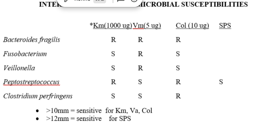
describe the AST used for anaerobic ID
Kirby-bauer disc setup for ID
Disc antimicrobial concentrations utilized for anaerobic ID ONLY, not interchangeable for routine organisms
Definitive ID of anaerobic organisms
Biochemical test batteries
Metabolic end product analysis using gas liquid chromatography
Cell wall fatty acid profiling by gas liquid chromatography
MALDI-TOF
Molecular testing
Why might we collect parasite stool samples?
Based on patient signs & symptoms, clinical history, & travel to endemic areas
Cysts found in formed stool
Trophs found in watery stool
Where do we find parasitic organisms?
GI tract is most common site
CSF – acanthamoeba sp
Eye – acanthomoeba spp, loa loa
Skin – onchocerca volvulus
Blood – plasmodium sp, babesia sp.
Fresh fecal samples in clean sterile wide mouth containers
Stool collection vials – clean vial with no preservatives or some preservatives
Liquid samples examined within 20 minutes of passage if not in preservative, formed within 24 hours
What type of analysis do we do for O & P (ova & parasite) examination?
Fresh for quick wet mount – saline & 1% iodine
Preservatives – increase recovery of many that degrade quickly
Formalin
PVA (polyvinyl alcohol)
SAF (sodium acetate-acetic acid formalin)
Schaudinn’s Fluid (PVA & mercuric acid)
Macroscopic Analysis – visual determination of eggs, worms, proglottids, color
Microscopic –
wet mount (motile trophozoites, cysts, helminth eggs)
Trichrome stain – ID of protozoan trophozoites or cysts
Modified Acid Fast stain – oocytes of cryptosporidium
What symptoms do we see in parasitic infections?
Diarrheal disease (bloody, watery, purulent, bowel obstruction, belching, steatorrhea)
Eosinophilia
Skin rash, itching
Hepatosplenomegaly, lymphadenopathy
Jaundice
UTI symptoms
Inflammation of lungs, cough
Fever, chills, night sweats, malaise, muscle pain
What kind of treatments may interfere with parasite testing?
Mineral oil
Bismuth
Antibiotics (tetracycline) – wait 2 weeks before sample collection
Barium – wait 5-10 days
Antimalarial agents
What are the standard recommendations for parasite stool samples?
3 specimens (2 before laxative, 1 after)
Amebiasis – 6, detects 90% of infections (cost is issue)
Post treatment collection to confirm clearance (3-4 weeks protozoa, 5-6 wks taenia)
Determine specimen collection # based on lab testing performed (ELISA, microscopy, fresh vs concentrated vs permanent smear, pooling)
Samples submitted within 10 day period, NEVER same day OR 1 per day 3 consecutive days
Pros & Cons of fresh stool
Pros:
no chemical preservatives & low cost
Motile trophoxoites
Perform all testing – ELISA, stains, direct examination, concentrations, permanent stains, acid fast stains
Cons:
Time to receipt & processing can result in organism degradation
Pros & Cons of preserved stool
Pros:
Easier diagnosis as preservations of organisms morphology
Delivery time not critical & home collection encouraged
Perform most testing – concentrations, permanent stains, ELISA, acid fast
Cons:
Cost of vials & disposal of mercuric chloride preservatives
If not mixed, organisms may not be preserved