Cardiovascular System II
 Call Kai
 Learn
 Practice Test
 Spaced Repetition
 Match
 Flashcards
 Knowt Play
1/23

There's no tags or description
Looks like no tags are added yet.
24 Terms
Cardiovascular System II
READING GUIDE Bold terms = memorize • Arrows = cause → effect • Notes = common mistakes / advanced details
Formula quick reference
Formula | What it measures |
CO = HR × SV | Cardiac output |
SV = EDV − ESV | Stroke volume |
EF = (SV ÷ EDV) × 100 | Ejection fraction |
PP = SBP − DBP | Pulse pressure |
MAP ≈ DBP + PP/3 | Mean arterial pressure |
Flow = ΔP ÷ R | Pressure–flow relation |
Topics: cardiac output; stroke volume; preload, afterload, and contractility; blood pressure; vascular resistance; capillary exchange; BP regulation; blood components; RBCs and WBCs; oxygen and CO₂ transport; hemostasis; blood groups; special circulations.
Abbreviations: HR = heart rate; SV = stroke volume; CO = cardiac output; EDV = end-diastolic volume; ESV = end-systolic volume; EF = ejection fraction; LV/RV = left/right ventricle; RA/LA = right/left atrium; BP = blood pressure; SBP/DBP = systolic/diastolic blood pressure; MAP = mean arterial pressure; PP = pulse pressure; SVR = systemic vascular resistance; CVP = central venous pressure; Hb = hemoglobin; Hct = hematocrit; RBC/WBC = red/white blood cell.
A. Cardiac Output & Heart Function
. Cardiac Output (CO)
CO = amount of blood ONE ventricle pumps per minute.
FORMULA CO = HR x SV
• HR: beats/min.
• SV: mL/beat.
• CO: mL/min or L/min.
Typical resting references (approximate): HR 60–100 bpm; SV 55–100 mL/beat; CO 4–8 L/min; average resting CO about 5 L/min.
Example: HR 75 bpm x SV 70 mL/beat = 5,250 mL/min = 5.25 L/min.
Important:
• RV and LV normally pump about the same volume per minute over time.
• Do NOT add right-heart and left-heart CO together.
• CO changes with exercise, body size, metabolic demand, and cardiovascular health.
2. Stroke Volume (SV)
SV = amount of blood ejected by ONE ventricle in ONE beat.
FORMULA SV = EDV - ESV
• EDV: blood in ventricle after filling (end diastole).
• ESV: blood remaining after ejection (end systole).
Example: EDV 120 mL → contraction → ESV 50 mL → SV = 70 mL.
Ventricles do NOT normally empty completely.
Three factors affect SV: PRELOAD (filling/stretch), CONTRACTILITY (squeeze strength), AFTERLOAD (load opposing ejection).
3. Ejection Fraction (EF)
EF = percentage of EDV ejected during systole.
FORMULA EF = (SV / EDV) x 100
Example: EDV 120, ESV 50 → SV 70 → EF = (70/120) x 100 = 58.3%.
• Typical normal LV EF: about 55–70%.
• Reduced EF can indicate reduced systolic pumping.
• Preserved EF does NOT guarantee normal filling or CO; heart failure can occur with preserved EF.
DISTINCTION: EF = percent; SV = volume/beat; CO = volume/min.
4. Preload — Ventricular Filling
Preload = stretch of ventricular muscle at end diastole, just before contraction.
Often estimated from EDV and filling pressure, but NOT identical to EDV.
FLOW:
Increased venous return → increased EDV → increased myocardial stretch → usually stronger contraction → increased SV.
FRANK–STARLING LAW: Within normal limits, more filling/stretch makes the ventricle contract more forcefully, increasing SV. Mechanisms include sarcomere mechanics and calcium sensitivity.
Preload increases with:
• More venous return.
• Higher blood volume.
• Longer filling time when other conditions allow.
Preload decreases with:
• Dehydration or hemorrhage.
• Reduced venous return.
• Very fast HR (shorter diastole).
Advanced:
• Preload technically refers to end-diastolic muscle stretch, not just blood volume.
• Too much stretch or a failing ventricle may NOT produce a further rise in SV.
• Excessive preload can worsen congestion in heart failure.
5. Afterload — Resistance to Ejection
Afterload = load/pressure the ventricle must overcome to eject blood.
• LV afterload: strongly affected by aortic pressure, arterial resistance, wall stress, and outflow obstruction.
• RV afterload: strongly affected by pulmonary arterial pressure/resistance and RV outflow obstruction.
FLOW (if preload and contractility are unchanged):
Increased arterial pressure / valve narrowing → increased afterload → ventricle works harder → increased ESV → decreased SV.
Increases with:
• Hypertension or aortic stenosis (LV).
• Pulmonary hypertension or pulmonary valve stenosis (RV).
stenosis: abnormal narrowing of a blood vessel, spinal canal, or other tubular structure in the bod
Decreases with:
• Lower arterial resistance/pressure or appropriate vasodilation.
TEAS TRAP: Afterload is the load resisting ejection, NOT the force of contraction.
6. Contractility — Intrinsic Squeeze Strength
Contractility = inherent force-generation ability at a given preload and afterload.
FLOW: Increased contractility → stronger contraction → decreased ESV → increased SV → increased CO.
Can increase with:
• Sympathetic stimulation.
• Epinephrine/norepinephrine acting on beta-1 receptors.
• Increased available intracellular Ca²⁺.
• Positive inotropic medications; some hormones.
Can decrease with:
• Myocardial ischemia, severe hypoxia, severe acidosis.
• Certain negative inotropic drugs.
• Significant electrolyte abnormalities.
Inotropy = contractile strength: positive inotrope increases it; negative inotrope decreases it.
inotrpoic: any substance or medication that alters the force or energy of muscular contractions
Cardiac Ca²⁺ entry triggers additional Ca²⁺ release from sarcoplasmic reticulum.
→ ca+ needed for movement of muscle (actin/tripin/myosin) Ca+ a binding agent
TRAP: Increased force due to Frank–Starling preload is NOT the same as increased intrinsic contractility.
7. The Three Stroke-Volume Factors
PRELOAD ↑ (more filling/stretch) → SV usually ↑.
CONTRACTILITY ↑ (stronger squeeze) → SV usually ↑.
AFTERLOAD ↑ (harder to eject) → SV usually ↓.
Assume other factors stay constant and the ventricle is functioning within physiological limits.
MEMORY: Preload = FILL; Contractility = SQUEEZE; Afterload = PUSH AGAINST.
8. Heart Rate vs. Cardiac Output
At unchanged SV: increased HR → increased CO.
At VERY high HR:
High HR → shortened diastole → reduced filling/EDV → reduced SV → CO may decrease.
Chronotropy = effect on HR (positive = faster; negative = slower).
Dromotropy = effect on conduction speed, especially AV node (positive = faster; negative = slower).
EXERCISE:
Sympathetic activity ↑ + vagal effect ↓ → HR ↑ and contractility ↑.
Skeletal muscle pump + respiratory pump → venous return ↑.
Usually SV ↑ + HR ↑ → CO ↑ → more oxygen delivered to active muscle.
Important: extreme HR may limit filling despite sympathetic stimulation.
9. Conditions That Change CO
CO = HR x SV.
• Exercise: increased HR, contractility, venous return → CO usually rises.
• Hemorrhage: blood volume ↓ → preload ↓ → SV and CO may fall.
• Dehydration: venous return ↓ → CO may fall.
• Severe ischemia: contractility ↓ → SV/CO ↓.
• High afterload: SV/CO may fall.
• Severe bradycardia: CO may fall.
• Extreme tachycardia: filling time ↓ → SV/CO may fall.
CARDIAC RESERVE = ability to increase CO above resting output as demand rises.
Compensation can temporarily preserve CO despite underlying disease.
B. Blood Pressure & Vessel Physiology
10. Blood Pressure (BP)
BP = force of circulating blood against vessel walls.
Example: 120/80 mmHg.
• SBP 120: peak arterial pressure during ventricular systole.
• DBP 80: lowest arterial pressure during ventricular diastole.
Terms:
• PP (pulse pressure) = SBP - DBP.
• MAP = average arterial pressure across one cardiac cycle.
• Perfusion = blood delivery to tissues.
120/80 is an example, NOT a universal target.
11. Pulse Pressure (PP)
FORMULA PP = SBP - DBP.
Example: 120/80 → PP = 40 mmHg.
PP affected by stroke volume, arterial stiffness/compliance, and ejection characteristics.
• SV ↑ → PP often ↑.
• Stiffer arteries (compliance ↓) → PP often ↑.
• Major blood loss can narrow PP.
12. Mean Arterial Pressure (MAP)
MAP = average arterial pressure driving systemic tissue blood flow.
Resting approximation:
MAP ≈ DBP + (SBP - DBP)/3
OR MAP ≈ (SBP + 2 x DBP)/3.
Example: BP 120/80 → MAP = 80 + 40/3 ≈ 93 mmHg.
WHY: Normally, more of the cardiac cycle occurs in diastole.
Advanced: This estimate becomes less accurate at extremely high HR or with unusual pressure waveforms.
13. CO, Resistance, and Pressure
APPROXIMATE: MAP ≈ CO x SVR.
MORE COMPLETE: MAP - CVP = CO x SVR.
CO = cardiac output; SVR = systemic vascular resistance; CVP = central venous pressure (usually small compared with MAP).
If other factors constant:
• CO ↑ or SVR ↑ → MAP tends to ↑.
• CO ↓ or SVR ↓ → MAP tends to ↓.
IMPORTANT: BP can look adequate despite poor tissue perfusion; vasoconstriction may preserve MAP during hemorrhage while CO falls.
14. Blood Flow and Pressure Gradients
Blood flows from HIGHER to LOWER pressure.
FORMULA Flow (Q) = pressure difference (ΔP) / resistance (R).
At fixed ΔP:
• Resistance ↑ → flow ↓.
• Resistance ↓ → flow ↑.
A resistance increase can raise pressure UPSTREAM while reducing flow through the affected pathway. The overall response depends on the full circulation.
15. Vascular Resistance
Resistance = opposition to blood flow.
Affected by:
• Vessel radius (most important rapidly adjustable factor).
• Blood viscosity.
• Vessel length.
• Vessel shape/branching/irregularities.
For ideal laminar flow in a rigid tube:
R = 8ηL / (πr^4).
So R is proportional to 1/(radius^4).
If radius is cut in half → resistance rises 16x; at unchanged ΔP, flow falls to 1/16.
Advanced: Real vessels are elastic, branched, and pulsatile; this is a useful approximation.
16. Vasoconstriction vs. Vasodilation
VASOCONSTRICTION: radius ↓ → resistance ↑ → local flow ↓ (if pressure difference unchanged).
VASODILATION: radius ↑ → resistance ↓ → local flow ↑ (if pressure difference unchanged).
Arterioles are key resistance vessels because smooth muscle quickly changes their diameter.
TRAP: All arteries carry blood AWAY from heart; arterioles have a major role in resistance and local distribution.
17. Blood Viscosity
Viscosity = resistance of fluid to flow.
• RBC concentration/hematocrit ↑ → viscosity often ↑ → resistance ↑.
• Severe anemia → viscosity may ↓.
TRAP: Lower viscosity does NOT guarantee better oxygen delivery; anemia can severely impair oxygen transport.
18. Arterial Compliance and Elastic Recoil
Compliance = how easily a vessel expands with pressure.
FORMULA Compliance = change in volume / change in pressure (ΔV/ΔP).
Compliant/elastic arteries: stretch during systole, store energy, recoil during diastole → help maintain blood flow between beats.
Stiff arteries: expand less → often higher systolic BP and pulse pressure.
The aorta is a major elastic artery.
19. BP and Velocity Across the Circulation
PATH:
Aorta/large arteries (high, pulsatile BP)
→ small arteries and arterioles (large pressure drop)
→ capillaries (lower BP; gas/nutrient exchange)
→ venules and veins (low BP; return to heart).
• Highest systemic arterial pressures are near aortic outflow.
• Much resistance/pressure drop occurs across small arteries and arterioles.
• Capillaries have extremely LARGE TOTAL cross-sectional area → LOW average blood velocity → time for exchange.
TRAP: One capillary is tiny, but the sum of all capillary areas is huge.
20. Venous Return
Venous return = systemic venous blood reaching the right atrium.
At steady state: venous return ≈ CO.
Mechanisms:
• Skeletal muscle pump: muscles squeeze veins.
• Venous valves: limit backflow.
• Respiratory pump: pressure changes assist flow toward heart.
• Sympathetic venoconstriction: mobilizes stored blood centrally.
• Sufficient blood volume: maintains filling pressure.
FLOW: leg muscle contraction → vein squeezed → valves favor heartward flow → venous return ↑ → preload ↑.
CVP = pressure near right atrium, related to right-sided filling conditions.
Advanced: CVP alone does NOT reliably determine total blood volume or predict fluid responsiveness.
21. Standing and Orthostatic Compensation
Standing suddenly → gravity pools blood in leg veins → venous return ↓ → preload ↓ → SV ↓ → BP may ↓.
Compensation: lower BP → baroreceptor response → sympathetic output ↑ → HR ↑ and vasoconstriction ↑ → BP supported.
If compensation inadequate → dizziness, weakness, or syncope.
Normal compensation may prevent a dramatic BP fall.
22. Capillary Exchange
Capillaries = major sites of gas, nutrient, waste, and fluid exchange.
1) DIFFUSION: particles move down concentration/partial-pressure gradients (O₂, CO₂).
2) TRANSPORT THROUGH CELLS/OPENINGS: pores, clefts, transcytosis; selected solutes/proteins.
3) BULK FLOW: fluid movement caused by hydrostatic and osmotic pressure forces (filtration/reabsorption).
Other points:
• Lipid-soluble molecules can cross endothelial cell membranes.
• Glucose/ions may use channels, carriers, or intercellular pathways.
• Water responds to osmotic/hydrostatic forces.
• Fenestrated capillaries and sinusoids allow greater passage of selected materials.
23. Hydrostatic vs. Oncotic Pressure
CAPILLARY HYDROSTATIC PRESSURE: pushes fluid OUT of capillary → filtration.
PLASMA ONCOTIC (COLLOID OSMOTIC) PRESSURE: plasma proteins, especially ALBUMIN, oppose outward filtration and favor water retention in blood.
Traditional introductory model:
• Arterial capillary end: hydrostatic pressure often dominates → filtration.
• Venous end: lower hydrostatic pressure; oncotic pressure may favor reabsorption.
Modern refinement: Many systemic capillaries have sustained net filtration; lymphatics return excess fluid. Continuous venous-end reabsorption is NOT universal.
24. Edema
Edema = excess fluid in interstitial tissues.
Main causes:
• Capillary hydrostatic pressure ↑ → more fluid pushed out.
• Plasma albumin ↓ → oncotic force ↓ → edema risk ↑.
water flows from low to high solute concentration of particles
water flows from high to low water concentration
• Capillary permeability ↑ → fluid/proteins leak out.
• Lymphatic obstruction → inadequate drainage.
• Na+/water retention → volume and capillary pressure may ↑.
LOW ALBUMIN: albumin ↓ → oncotic pressure ↓ → net filtration rises/fluid retention in vessels falls → edema.
LEFT HEART FAILURE: LV pump failure → pressure backs up in pulmonary circulation → pulmonary edema may develop.
TRAP: Edema can occur even when total body fluid is normal or high; problem is DISTRIBUTION of fluid.
25. Lymphatic Return
PATH:
Plasma fluid filters into tissues → excess interstitial fluid enters blind-ended lymphatic capillaries → lymphatic vessels and nodes → returns near subclavian veins.
Functions:
• Return fluid and proteins to blood.
• Help prevent edema.
• Immune surveillance.
• Carry dietary fats absorbed through intestinal lacteals.
Circulatory blood stays in a closed, heart-pumped system; lymph begins in tissue lymphatics and drains into veins.
C. Regulation Of Blood Pressure And Circulation
26. Cardiovascular Control Centers
Medulla oblongata (brainstem) = main rapid cardiovascular control center.
Controls HR, contractility, vessel tone, and blood-pressure responses.
SYMPATHETIC: beta-1 in heart → HR ↑ and contractility ↑; alpha-1 in many vessels → vasoconstriction.
PARASYMPATHETIC: vagus nerve → acetylcholine → M2 receptors → SA firing ↓ and AV conduction ↓.
LOCAL SIGNALS: alter vessel tone according to tissue needs.
27. Baroreceptor Reflex (Rapid BP Control)
Baroreceptors = STRETCH sensors in carotid sinus and aortic arch.
BP FALLS:
BP ↓ → arterial stretch ↓ → baroreceptor firing ↓ → medulla increases sympathetic and reduces parasympathetic output → HR ↑, contractility ↑, vasoconstriction ↑ → BP tends to recover.
BP RISES:
BP ↑ → stretch/firing ↑ → sympathetic ↓ and parasympathetic ↑ → HR and contractility tend to decrease; vasoconstriction decreases → BP tends to fall.
Afferent nerves:
• Carotid sinus → glossopharyngeal nerve (CN IX).
• Aortic arch → vagus (CN X).
Negative feedback, seconds–minutes. Baroreceptors can reset during chronic hypertension and do not alone set long-term BP.
28. Chemoreceptors
Peripheral chemoreceptors: CAROTID BODIES and AORTIC BODIES.
Detect ↓ arterial O₂ (especially marked decreases), ↑ CO₂, and ↑ H+ (↓ pH).
Abnormal gases/pH → chemoreceptor signaling → respiratory/cardiovascular centers → increased ventilation and cardiovascular adjustments.
Central chemoreceptors respond especially to CO₂-induced H+ changes in brain fluid/CSF.
TRAP: BARORECEPTORS sense stretch/pressure; CHEMORECEPTORS sense blood chemistry.
29. Renin–Angiotensin–Aldosterone System (RAAS)
Purpose: conserve Na+/water, raise vascular tone, and help maintain BP/volume.
Triggers: renal perfusion pressure ↓; NaCl sensed at macula densa ↓; sympathetic beta-1 signaling to juxtaglomerular cells ↑.
PATHWAY:
Kidney juxtaglomerular cells release RENIN
→ renin converts liver-made ANGIOTENSINOGEN to ANGIOTENSIN I
→ ACE (present widely in endothelium, including lungs) produces ANGIOTENSIN II
→ Ang II causes vasoconstriction + stimulates adrenal ALDOSTERONE
→ kidneys increase Na+ reabsorption, often retaining water
→ blood volume/BP tend to rise.
ANGIOTENSIN II:
• Strong vasoconstrictor → SVR ↑.
• Stimulates aldosterone.
• Promotes thirst and ADH.
• Supports renal Na+ retention.
ALDOSTERONE:
• Made in adrenal cortex, zona glomerulosa.
• Acts on distal nephron to increase Na+ reabsorption and K+ secretion.
• Water may follow retained Na+ when water is available and permeability permits.
TRAP: Renin = enzyme; ACE = converting enzyme; Ang II = vasoconstrictor/hormonal messenger; aldosterone = Na+-retaining hormone.
30. Antidiuretic Hormone (ADH, Vasopressin)
Made in hypothalamus; stored/released from posterior pituitary.
Triggers: plasma osmolality ↑; significant blood volume or pressure ↓.
PATH:
ADH ↑ → aquaporin-2 water channels increase in collecting ducts → water reabsorption ↑ → urine volume ↓ and concentration ↑ → blood water conserved.
Higher ADH can also cause vasoconstriction through V1 receptors.
ADH vs. ALDOSTERONE:
• ADH: mainly retains WATER; collecting-duct aquaporins; posterior pituitary release.
• Aldosterone: mainly increases SODIUM retention (and K+ secretion); adrenal cortex; water often follows.
31. Atrial/Brain Natriuretic Peptides (ANP/BNP)
ANP: released mainly from atria with chamber stretch.
BNP (B-type): released mainly from ventricles during increased wall stress.
PATH:
Blood volume/chamber stretch ↑ → ANP/BNP ↑ → kidney Na+ excretion (natriuresis) ↑ and water excretion (diuresis) ↑ → volume/BP tend to fall.
Also oppose RAAS and promote vascular relaxation.
MEMORY: RAAS = RETAIN salt/water; ANP/BNP = RELEASE salt/water.
32. Other Hormones That Affect Circulation
• Epinephrine: beta-1 increases HR/contractility; vascular effects vary by receptor.
• Norepinephrine: cardiac stimulation and alpha-1 vasoconstriction.
• Thyroid hormone: raises metabolic demand and cardiovascular responsiveness.
• Erythropoietin (EPO): raises RBC production and affects oxygen-carrying capacity.
Advanced: Epinephrine can stimulate beta-2 vasodilation in some skeletal-muscle vascular beds, plus alpha-1 vasoconstriction in others.
33. Local Autoregulation
Autoregulation = tissues adjust their own blood supply.
METABOLIC: increased metabolism → CO₂/H+/adenosine signals ↑ → local arterioles often dilate → flow ↑.
MYOGENIC: vessel pressure/stretch ↑ → smooth-muscle constriction in some vascular beds → stabilizes local perfusion.
Other mediators: nitric oxide (NO) promotes vasodilation; endothelin promotes vasoconstriction.
Systemic sympathetic constriction and local metabolic dilation can occur at the same time in DIFFERENT tissues.
34. Short-Term vs. Long-Term BP Control
FAST (seconds–minutes): baroreceptor reflex, autonomic HR/contractility, arteriole diameter, venous tone.
LONGER (hours–days): renal Na+/water balance; RAAS; aldosterone; ADH; natriuretic peptides.
Nervous system changes pressure QUICKLY; kidneys/hormones set longer-term fluid balance and support BP regulation.
35. Hemorrhage and Hypovolemic Shock
HEMORRHAGE = blood loss.
HYPOVOLEMIC SHOCK = insufficient tissue perfusion due to severe circulating-volume loss.
PATH:
Blood loss → circulating volume ↓ → venous return/preload ↓ → SV ↓ → CO ↓ → tissue perfusion ↓.
Compensation:
• Sympathetic output ↑ → HR/contractility ↑ + peripheral vasoconstriction.
• Renin/Ang II/aldosterone ↑.
• ADH and thirst ↑.
Severe oxygen-delivery failure → anaerobic metabolism ↑ → lactate accumulation/acidosis → cell dysfunction.
BP may appear near normal at FIRST due to compensatory vasoconstriction.
Shock types:
• Hypovolemic: insufficient volume.
• Cardiogenic: pump failure.
• Distributive: loss of normal vascular tone/maldistributed circulation.
• Obstructive: physical obstruction of flow.
TRAP: Shock means inadequate tissue PERFUSION, not merely a low BP number.
D. Blood Composition And Oxygen Transport
36. Functions of Blood
Blood = fluid connective tissue.
TRANSPORT: O₂/CO₂, nutrients, hormones, wastes, heat.
PROTECTION: immune defense and clotting.
REGULATION: pH, temperature, fluid balance.
Typical blood pH: 7.35–7.45. More viscous than water. Adult volume often approximately 4–6 L (varies by size/physiology).
37. Blood Components (After Centrifugation)
TOP = PLASMA (~55%).
THIN MIDDLE = BUFFY COAT (<1%): WBCs + platelets.
BOTTOM = RBCs (~45%; varies).
Formed elements = RBCs + WBCs + platelets.
HEMATOCRIT (Hct) = fraction/percentage of blood occupied by RBCs.
PLASMA contains clotting proteins (including fibrinogen).
SERUM = liquid after clot formation; many clotting factors have been consumed/removed.
38. Plasma Composition
About 90–92% water.
Includes Na+, K+, Ca²⁺, Cl-, HCO3-, glucose, amino acids, lipids, hormones, waste, dissolved gases, proteins.
Important plasma proteins:
• ALBUMIN: plasma oncotic pressure; transport.
• GLOBULINS: antibodies and transport proteins.
• FIBRINOGEN: converted into fibrin during clotting.
Albumin ↓ → oncotic pressure ↓ → edema risk ↑.
39. Red Blood Cells (RBCs / Erythrocytes)
Main job = O₂ transport; also help transport CO₂ and regulate acid-base status.
Features:
• BICONCAVE disc → large surface-area/volume ratio; short diffusion distance.
• FLEXIBLE → pass through narrow capillaries.
• Mature human RBCs: NO nucleus and NO mitochondria.
• Rely on glycolysis for ATP; don't use the oxygen they carry for mitochondrial respiration.
• Packed with hemoglobin.
• Lifespan ≈ 120 days.
40. Hemoglobin (Hb)
Hb = O₂-carrying protein inside RBCs.
1 Hb = 4 globin chains + 4 heme groups; each heme contains Fe2+ and binds ONE O₂.
Therefore one Hb molecule carries up to FOUR O₂ molecules.
In lungs: Hb binds O₂ → oxyhemoglobin.
In tissues: Hb releases O₂ → deoxyhemoglobin.
O₂ binding is reversible.
41. Oxygen Transport
PATH:
Alveoli → O₂ diffuses into lung capillaries → binds Hb inside RBCs → transported to tissues → releases from Hb → diffuses into cells → mitochondria use O₂ for aerobic ATP formation.
O₂ diffuses down its PARTIAL-PRESSURE gradient:
• Alveolar PO₂ > incoming venous blood PO₂ → O₂ enters blood.
• Arterial blood PO₂ > tissue PO₂ → O₂ enters tissues.
Hb releases DIFFERENT amounts of O₂ according to tissue conditions/demand.
Most blood O₂ is bound to Hb; a small amount is dissolved in plasma.
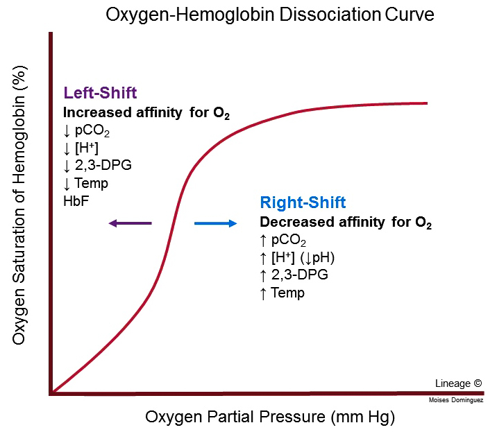
42. Hemoglobin–Oxygen Dissociation Curve
O₂ saturation = % of Hb O₂-binding sites occupied.
Curve is S-shaped because O₂ binding is cooperative. At high PO₂, saturation approaches a plateau.
RIGHT SHIFT → Hb affinity for O₂ ↓ → O₂ release to tissues ↑ at a given PO₂.
Causes: CO₂ ↑, H+ ↑ / pH ↓, temperature ↑, 2,3-BPG ↑.
LEFT SHIFT → Hb affinity for O₂ ↑ → O₂ release ↓ at a given PO₂.
Causes: CO₂ ↓, H+ ↓ / pH ↑, temperature ↓, 2,3-BPG ↓.
BOHR EFFECT: More tissue CO₂/H+ promotes O₂ unloading.
TRAP: Higher Hb affinity can impair tissue oxygen release.
43. O2 Saturation vs. O2 Content vs. O2 Delivery
SATURATION = percentage of Hb sites carrying O₂.
CONTENT = total oxygen in a specific volume of blood.
DELIVERY = oxygen carried to tissues per minute; depends on CO and O₂ content.
Advanced formulas:
Arterial O₂ content: CaO₂ ≈ (1.34 x Hb x SaO₂) + (0.003 x PaO₂).
• Hb in g/dL; SaO₂ expressed as a fraction; PaO₂ in mmHg.
O₂ delivery: DO₂ = CO x CaO₂ x 10 (if CO in L/min and CaO₂ in mL O₂/dL).
Examples:
• ANEMIA: Hb ↓ → oxygen content ↓ despite possibly normal SpO₂.
• HYPOXEMIA: arterial oxygen partial pressure low.
• LOW CO: blood delivered per minute ↓.
• CARBON MONOXIDE: binds Hb, reduces useful O₂ binding and unloading.
TEAS PRIORITY: O₂ delivery depends on BOTH blood flow and oxygen carried per unit blood. Advanced equation constants are supporting material.
44. Carbon Dioxide (CO2) Transport
CO₂ travels from tissues → blood → lungs.
Approximate forms:
• ~70% as bicarbonate (HCO3-).
• ~20–25% bound to Hb and other proteins.
• ~5–10% dissolved.
Chemical reaction (reversible):
CO₂ + H2O <→ H2CO3 <→ H+ + HCO3-.
CARBONIC ANHYDRASE speeds the reaction.
In tissues: CO₂ enters RBC → bicarbonate forms; HCO3- often moves out in exchange for Cl- (CHLORIDE SHIFT).
In lungs: reaction reverses → CO₂ formed → exhaled; chloride shift reverses.
HALDANE EFFECT: Oxygenation of Hb in lungs promotes CO₂/H+ release; deoxygenated Hb in tissues can carry more CO₂/H+.
TRAP: Most CO₂ travels as bicarbonate, NOT bound directly to Hb.
45. RBC Production (Erythropoiesis)
ERYTHROPOIESIS = RBC production in red bone marrow.
PATH:
Tissue oxygen delivery ↓ → kidneys release EPO ↑ → red bone marrow produces more RBCs → oxygen-carrying capacity ↑.
EPO = erythropoietin; produced mainly by kidneys in adults.
Needs: iron for heme, vitamin B12 + folate for DNA synthesis in immature cells, amino acids for globin, functioning marrow.
46. RBC Destruction and Recycling
RBC lifespan ≈ 120 days.
Macrophages in spleen, liver, and marrow remove aged/damaged RBCs.
PATH:
Old RBC → Hb breakdown:
• Globin → amino acids reused.
• Heme → iron recycled + pigment breakdown → bilirubin → liver processing → biliary excretion.
Iron may be stored as ferritin or reused for new Hb.
47. Anemia vs. Polycythemia
ANEMIA = low Hb concentration or RBC mass → oxygen-carrying capacity ↓.
Potential causes: blood loss, iron deficiency, B12/folate deficiency, hemolysis, low EPO, marrow disease.
POLYCYTHEMIA = increased RBC mass/concentration → viscosity ↑, possible clot risk.
TRAP: More RBCs are not automatically beneficial; excessive RBC mass can make blood harder to move.
48. White Blood Cells (WBCs / Leukocytes)
WBCs = nucleated immune cells that defend against infection, tissue damage, and abnormal cells. Made through hematopoiesis; many leave blood vessels by DIAPEDESIS to enter tissues.
FIVE TYPES:
• NEUTROPHILS: phagocytose bacteria; early acute inflammation.
• LYMPHOCYTES: B, T, NK cells; adaptive and innate immune defense.
• MONOCYTES: enter tissues → macrophages/dendritic cells; phagocytosis/antigen presentation.
• EOSINOPHILS: parasites and allergic responses.
• BASOPHILS: histamine and other inflammatory/allergic mediators.
GRANULOCYTES = neutrophils + eosinophils + basophils.
AGRANULOCYTES = lymphocytes + monocytes.
B cells → antibody-secreting plasma cells.
T cells → coordinate/carry out cellular immune responses.
NK cells → kill some infected or abnormal cells (innate immune system).
WBC counts may rise with infection, inflammation,
49. Hematopoiesis
HEMATOPOIESIS = production of formed blood elements in bone marrow.
Stem cell → two broad lineages:
• MYELOID: RBCs, platelets, neutrophils, eosinophils, basophils, monocytes.
• LYMPHOID: B cells, T cells, NK cells.
Regulators: EPO increases RBC production; thrombopoietin (TPO) supports platelet production via megakaryocytes; colony-stimulating factors/interleukins regulate WBC lineages.
50. Platelets (Thrombocytes)
Platelets = small, non-nucleated cell FRAGMENTS derived from bone-marrow MEGAKARYOCYTES.
Jobs: adhere at injury, activate and recruit platelets, make temporary plug, supply surface for clotting, help repair.
Lifespan ≈ 7–10 days.
Platelets are NOT full nucleated cells.
51. Hemostasis — Stopping Bleeding
HEMOSTASIS = controlled stopping of bleeding.
THREE STAGES:
1) VASCULAR SPASM: damaged vessel constricts → blood loss ↓.
2) PLATELET PLUG: platelets adhere, activate, aggregate.
3) COAGULATION: clotting factors create fibrin → stable clot.
PLATELET PLUG:
Vessel injury exposes collagen → von Willebrand factor (vWF) helps platelet adhesion → platelets activate → ADP/thromboxane A2 recruit more platelets → aggregation/temporary plug.
COAGULATION:
Clotting cascade → prothrombin (factor II) becomes THROMBIN (IIa) → thrombin converts FIBRINOGEN (factor I) to FIBRIN → factor XIII stabilizes fibrin → stronger clot traps platelets/blood cells.
Intrinsic and extrinsic pathways feed into a common pathway; in vivo clotting depends on cell surfaces and interacting coagulation factors.
Needed:
• Ca²⁺ (factor IV) supports clotting reactions.
• Vitamin K supports synthesis of functional factors II, VII, IX, X and proteins C/S.
TRAP: Platelet plug forms first; fibrin reinforces/stabilizes it.
52. Clot Breakdown (Fibrinolysis)
FIBRINOLYSIS = breakdown of fibrin during clot resolution.
Plasminogen → activated to PLASMIN → plasmin breaks down fibrin → clot gradually removed.
Natural anticoagulants prevent excessive clot spread.
Terms:
• Thrombus = clot formed inside a vessel/heart.
• Embolus = material traveling in blood that may lodge downstream.
• Thromboembolus = traveling detached thrombus.
• Hemorrhage = excessive bleeding.
• Hemostasis = controlled prevention/stopping of bleeding.
• Fibrinolysis = fibrin breakdown.
Clotting includes positive-feedback amplification, but also regulatory inhibition.
53. ABO Blood Groups
RBC type depends on A and B surface ANTIGENS; plasma contains corresponding ANTIBODIES.
TYPE A: A antigen; anti-B antibodies.
TYPE B: B antigen; anti-A antibodies.
TYPE AB: A + B antigens; neither anti-A nor anti-B naturally.
TYPE O: neither A nor B antigen; anti-A and anti-B antibodies.
ANTIGEN = RBC surface marker.
ANTIBODY = immune protein in plasma that recognizes/binds antigen.
Incompatible RBC transfusion → antibodies bind donor RBCs → agglutination/hemolysis → potentially severe reaction.
54. Rh Blood Group
Rh+ = D antigen present. Rh- = D antigen absent.
Unlike naturally occurring ABO antibodies, anti-D usually forms after exposure to Rh+ RBCs (sensitization).
Pregnancy scenario:
Rh- pregnant patient + exposure to fetal Rh+ RBCs → anti-D may form → in later Rh+ pregnancy, antibodies may damage fetal RBCs.
Rh immune globulin helps PREVENT sensitization when indicated
55. RBC Transfusion Compatibility
Simplified ABO compatibility for PACKED RBCs:
A recipient → A or O RBCs.
B recipient → B or O RBCs.
AB recipient → A, B, AB, or O RBCs.
O recipient → O RBCs only.
Rh- recipients generally need Rh- RBCs when feasible; Rh+ recipients may receive Rh+ or Rh- RBCs.
CLASSIC MEMORIZATION:
O negative = universal RBC donor (ABO/Rh simplified).
AB positive = universal RBC recipient (ABO/Rh simplified).
CAUTION: These labels are NOT a guarantee of clinical compatibility. Blood typing, antibody screen, crossmatching, etc. still matter. Plasma compatibility follows different rules because plasma carries antibodies.
E. Additional Circulations And Integration
56. Hepatic Portal Circulation
PORTAL PATH:
GI tract + spleen + pancreas → HEPATIC PORTAL VEIN → liver sinusoids → HEPATIC VEINS → IVC → right atrium.
Why: absorbed nutrients reach liver for processing/storage; liver also metabolizes many substances/drugs and regulates blood glucose.
TRAP: Portal vein carries blood TO liver; hepatic veins drain blood FROM liver to IVC.
57. Fetal vs. Adult Circulation
Before birth: PLACENTA supplies gas exchange; lungs are not yet primary site.
Fetal shunts:
• DUCTUS VENOSUS: bypasses much liver circulation, directing blood from umbilical vein toward IVC.
• FORAMEN OVALE: opening RA → LA.
• DUCTUS ARTERIOSUS: pulmonary artery/trunk → aorta.
PATH (simplified):
Placenta → UMBILICAL VEIN (O₂-rich) → ductus venosus → IVC → RA → fetal shunts divert much blood away from lungs.
UMBILICAL ARTERIES: relatively O₂-poor blood fetus → placenta.
UMBILICAL VEIN: O₂-rich blood placenta → fetus.
At birth:
Lungs expand → pulmonary resistance falls.
Placental circulation stops → systemic resistance rises.
Foramen ovale closes functionally; ductus arteriosus constricts/closes; ductus venosus closes.
Higher-level supporting material: memorize three shunts + routes first.
58. Common TEAS Traps
• CO is per MINUTE; SV is per BEAT.
• SV = EDV - ESV, not EDV + ESV.
• Preload ↑ is not ALWAYS beneficial in failure.
• Preload-related stretch is NOT intrinsic contractility.
• Afterload ↑ usually reduces SV if preload/contractility fixed.
• Extremely fast HR may lower CO via decreased filling.
• Higher BP does NOT guarantee better tissue perfusion.
• Viscosity contributes to vascular resistance; not identical.
• Capillary velocity is LOW because combined capillary cross-sectional area is huge.
• Hydrostatic pressure pushes fluid OUT; albumin-related oncotic pressure opposes outward filtration.
• Lower BP reduces baroreceptor stretch/firing, NOT increases it.
• ADH generally DECREASES urine volume.
• Aldosterone increases Na+ reabsorption (water may follow).
• ANP/BNP favor Na+/water excretion, opposing aspects of RAAS.
• Most O₂ travels BOUND to hemoglobin.
• Normal SpO₂ alone does NOT establish adequate O₂ delivery; Hb and CO matter.
• Most CO₂ travels as BICARBONATE.
• Mature human RBCs have NO mitochondria.
• Platelets are CELL FRAGMENTS, not full nucleated cells.
• Fibrinogen is soluble precursor; fibrin forms stable clot mesh.
• O-negative universal donor refers to simplified RBC ABO/Rh compatibility, NOT all products/antigens.
59. Formulas to Memorize
1. CO = HR x SV.
2. SV = EDV - ESV.
3. EF = (SV / EDV) x 100%.
4. PP = SBP - DBP.
5. MAP ≈ DBP + (PP / 3).
6. MAP - CVP = CO x SVR (use compatible units).
7. Blood flow Q = ΔP / R.
8. For ideal laminar flow: resistance proportional to 1 / radius^4.
Advanced supporting:
CaO₂ ≈ (1.34 x Hb x SaO₂) + (0.003 x PaO₂).
DO₂ = CO x CaO₂ x 10 (CO L/min; CaO₂ mL/dL).
60. Worked Calculations
EXAMPLE 1 — CO:
HR = 90; SV = 65 mL → CO = 90 x 65 = 5,850 mL/min = 5.85 L/min.
EXAMPLE 2 — SV:
EDV = 150 mL; ESV = 60 mL → SV = 90 mL.
EXAMPLE 3 — EF:
SV = 90 mL; EDV = 150 mL → EF = 90/150 x 100 = 60%.
EXAMPLE 4 — MAP:
BP = 138/78 → PP = 60 → MAP = 78 + 60/3 = 98 mmHg.
EXAMPLE 5 — RESISTANCE:
Radius decreases 2 → 1: R(new)/R(old) = (2/1)^4 = 16 → resistance 16x greater if other factors fixed.
61. Cardiovascular Integration
Heart pumps blood → CO = HR x SV → arterial pressure drives flow → arterioles distribute blood to capillaries → capillaries exchange O₂, CO₂, nutrients, waste → veins return blood → nerves, kidneys, hormones, and local signals regulate circulation.
OXYGEN DELIVERY proportional to CO x blood oxygen content.
Low tissue oxygen may result from:
• Low CO / poor pumping.
• Low Hb (anemia).
• Low blood oxygenation (hypoxemia).
• Impaired local circulation.