BIOL 2311 Exam 3
 Call Kai
 Learn
 Practice Test
 Spaced Repetition
 Match
 Flashcards
 Knowt Play
1/113

There's no tags or description
Looks like no tags are added yet.
114 Terms
List two of the seven criteria listed in your lab manual that are used to name muscles?
location
direction of muscle fibers
relative size
number of origins
location of origin and insertion
shape
action

Identify the type of muscle tissue shown?
cardiac

Identify the indicated layer of connective tissue.
endomysium
A strong, cord-like bundle of connective tissue that connects a muscle to another muscle or to a bone is called:
tendon
The connective tissue sheath that forms around an individual skeletal muscle cell is called:
endomysium
A flat, sheet-like bundle of connective tissue that connects a muscle to another muscle or to a bone is called:
aponeurosis
What do we call muscles that aid the action of other muscles by reducing undesirable or unnecessary movement?
synergists
Which of the seven criteria used for naming a muscle applies to the deltoid muscle?
shape
Which of the seven criteria used for naming a muscle applies to the rectus femoris muscle?
direction of fibers and location
Name one action of the zygomaticus minor muscle.
elevation of the lateral corners of the mouth
Name one action of the vastus medialis muscle.
extension of the lower leg at the knee
Name one action of the infraspinatus muscle.
lateral rotation of the arm at the shoulder
Name one action of the sternocleidomastoid muscle.
rotation of the head
Name one insertion of the platysma muscle.
lower margin of the mandible
Name one insertion of the rectus femoris muscle.
tibial tuberosity
Name one insertion of the external oblique muscle.
linea alba
Name one insertion of the infraspinatus muscle.
greater tubercle of the humerus
Name one origin of the tibialis anterior muscle.
lateral condyle of the tibia
Name one origin of the external oblique muscle.
lower eight ribs
Name one origin of the pectoralis major muscle.
sternum
Name one origin of the sternocleidomastoid muscle.
manubrium of the sternum

Identify the indicated muscle.
rectus abdominis

Identify the indicated muscle.
external oblique abdominal

Identify the indicated muscle.
zygomaticus minor

Identify the indicated muscle.
orbicularis oculi

Identify the indicated muscle.
sternocleidomastoid

Identify the indicated muscle.
buccinator
You locate a broad, fan shaped muscle filling the roughly circular low area on the lateral surface of the cranium. This muscle extends from the margin of this area to the coronoid process of the mandible. The muscle you have located is the:
Temporalis
You locate a deep, triangular muscle on the upper portion of the chest. This muscle extends from the anterior surface of the third, fourth, and fifth ribs to the coracoid process of the scapula. The muscle you have located is the:
Pectoralis minor
You locate a series of muscle segments following the pattern of the ribs along the lateral surface of the chest. These muscles extend from the back to the lateral surface of the chest. The muscle group you have located is the:
Serratus anterior
You locate a muscle on the posterior surface of the neck, extending from the occipital bone and the spinous processes of cervical vertebra 7 and the thoracic vertebrae to the acromion and spine of the scapula and the lateral third of the clavicle. The muscle you have located is the:
Trapezius
You locate a muscle on the anterior surface of the lower leg, just lateral to the visible tibia bone. This muscle extends from the upper part of the lateral surface of the tibia to the medial cuneiform. The muscle you have located is the:
Tibialis anterior
You locate the muscle on the posterior side of the forearm whose distal tendons extend onto the hand towards the second, third, fourth, and fifth digits. This muscle extends from these tendons to the lateral epicondyle of the humerus. The muscle you have located is the:
Extensor digitorum
You locate a muscle at the proximal end of the long, strap-like tendon that runs along the lateral side of the thigh to the knee. The muscle you have located is the:
Tensor fasciae latae
You locate the muscle between the gracilis and the medial side of the gluteus maximus, close to the proximal end of the leg. This muscle extends from the ischial tuberosity to the medial condyle of the femur. The muscle you have located is the:
Adductor magnus

Identify the indicated muscle.
sternocleidomastoid

Identify the indicated muscle.
sternothyroid
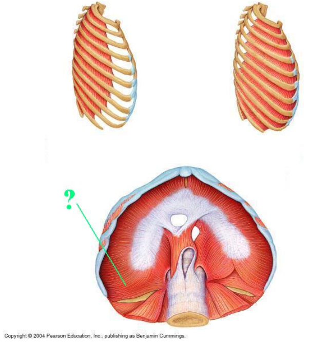
Identify the indicated muscle.
diaphragm
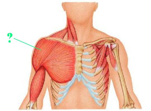
Identify the indicated muscle.
deltoid

Identify the indicated muscle.
pectoralis minor

Identify the indicated muscle.
latissimus dorsi

Identify the indicated muscle.
rhomboid major
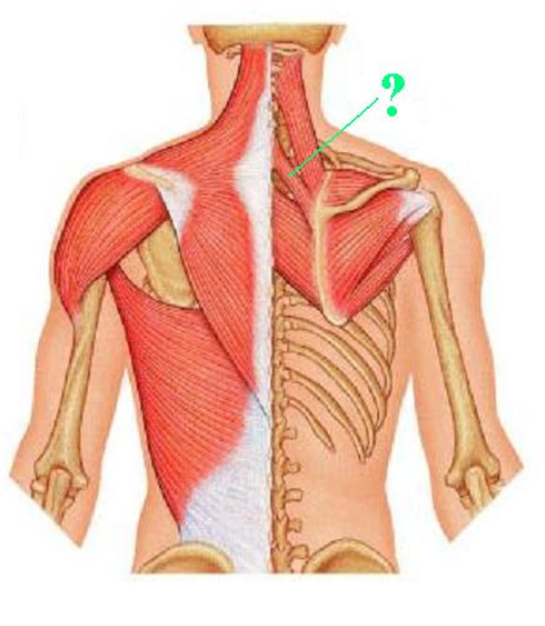
Identify the indicated muscle.
rhomboid minor

Identify the indicated muscle.
extensor digitorum

Identify the indicated muscle.
extensor carpi radialis longus

Identify the indicated muscle.
flexor carpi radialis
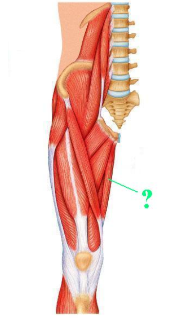
Identify the indicated muscle.
gracilis

Identify the indicated muscle.
iliacus

Identify the indicated muscle.
soleus

Identify the indicated muscle.
semitendinosus
A bundle of skeletal muscle cells within a skeletal muscle is called:
fascicle
The connective tissue sheath that forms around an entire skeletal muscle is called:
epimysium
The connective tissue sheath that forms around a bundle of skeletal muscle cells is called:
perimysium
Define "insertion" as it applies to skeletal muscles.
moveable site of attachment
Which of the seven criteria used for naming a muscle applies to the sternocleidomastoid muscle?
location of origin and insertion
Which of the seven criteria used for naming a muscle applies to the tibialis anterior muscle?
location
Name one action of the rectus femoris muscle.
flexion of the thigh at the hip
Name one action of the external oblique muscle.
flexion and rotation of vertebral column
Name one action of the zygomaticus major muscle.
elevation of the lateral corners of the mouth
Name one insertion of the triceps brachii muscle.
olecranon of ulna
Name one insertion of the semimembranosus muscle.
medial condyle of the tibia
Name one origin of the triceps brachii muscle.
posterior humerus
Name one origin of the zygomaticus minor muscle.
zygomatic bone
Name one origin of the semitendinosus muscle.
ischial tuberosity
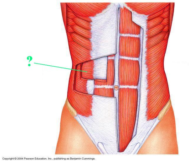
Identify the indicated muscle.
transverse abdominis

Identify the indicated structure.
linea alba

Identify the indicated muscle.
frontalis
epicranius frontal belly

Identify the indicated muscle.
masseter

Identify the indicated muscle.
platysma

Identify the indicated muscle.
orbicularis oris
You locate a large sheet of muscle between the medial margin of the scapula and the midline of the spinal column. This muscle extends from the spinous processes of thoracic vertebrae 2-5 to the medial margin of the scapula below the spine of the scapula. The muscle you have located is the:
Rhomboid major
You locate a muscle that runs along the inferior margin of the posterior side of the scapula. This muscle extends from the medial margin of the scapula to the intertubercular sulcus of the humerus. The muscle you have located is the:
Teres major
You locate this muscle extending from the superior margin of the orbit to the anterior margin of the flat sheet of tendon that covers the top of the cranium. The muscle you have located is the:
Frontalis
You locate a muscle that extends from the tip of the chin to the skin just below the lower lip. The muscle you have located is the:
Mentalis
You locate the large muscle on the lateral side of the anterior surface of the thigh. This muscle extends from the upper part of the femur to the patella. The muscle you have located is the:
Vastus lateralis
You locate a thin, strap-like muscle wrapping around the anterior surface of the thigh from lateral to medial and proximal to distal. This muscle extends from the anterior superior iliac spine to the medial surface of the tibia just below the tibial tuberosity. The muscle you have located is the:
Sartorius
You locate a thin muscle between the triceps brachii and the biceps brachii on the anterior (medial) side of the arm. This muscle extends from the coracoid process of the scapula to the middle of the body of the humerus. The muscle you have located is the:
Coracobrachialis
You locate a large muscle on the posterior surface of the lower leg. This muscle is the most superficial of this muscle group. This muscle extends from the medial and lateral condyles of the femur to the calcaneus via the calcaneal tendon. The muscle you have located is the:
Gastrocnemius

Identify the indicated muscle.
sternothyroid

Identify the indicated muscle.
mylohyoid

Identify the indicated muscle.
pectoralis major

Identify the indicated muscle.
biceps brachii

Identify the indicated muscle.
teres minor

Identify the indicated muscle.
extensor carpi ulnaris

Identify the indicated muscle.
palmaris longus
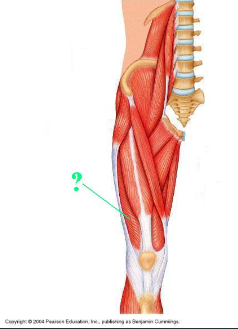
Identify the indicated muscle.
vastus lateralis

Identify the indicated muscle.
gastrocnemius

Identify the indicated muscle.
gluteus medius
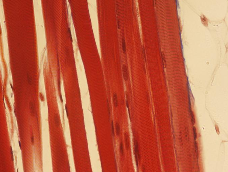
Identify the type of muscle tissue shown?
skeletal
Define "origin" as it applies to skeletal muscles.
immovable site of attachment
Which of the seven criteria used for naming a muscle applies to the trapezius muscle?
shape
Which of the seven criteria used for naming a muscle applies to the transversus abdominis muscle?
direction of fibers and location
Name one action of the orbicularis oculi muscle.
closes the eyelids
Name one action of the rectus abdominis muscle.
Flexion and rotation of the vertebral column
Name one insertion of the masseter muscle.
angle and ramus of the mandible
Name one insertion of the buccinator muscle.
orbicularis oris muscle
Name one origin of the rectus abdominis muscle.
pubic crest and symphysis
Name one origin of the gastrocnemius muscle.
medial and lateral condyles of the femur
Name one origin of the rectus femoris muscle.
anterior inferior iliac spine
Name one origin of the extensor digitorum muscle.
lateral epicondyle of the humerus