protozoa: class zoomastigophora (the flagellates)
 Call Kai
 Learn
 Practice Test
 Spaced Repetition
 Match
 Flashcards
 Knowt Play
1/18

There's no tags or description
Looks like no tags are added yet.
19 Terms
flagellate
single-celled parasite that moves by means of tail-like structure known as flagellum. two morph forms: trophs & cysts. ingestion of infected cysts = initiation of human intestinal infection.
specimen of choice for intestinal (G. intestinalis, C. mesnili, D. fragilis): stool. extraintestinal (T. vaginalis) varies by spp: centrifuged urine, vaginal/urethral discharge, prostatic secretions.
life cycle & pathogenesis of flagellates
trophs emerge in intestine & actively multiply by asexual binary fission. if change in intestinal environment, excystation, when cysts are present. some troph in outside world survive. cysts survive in outside world & continue to develop. human can get diarrhea w/o apparent cause. most intestinal infections asymptomatic. giardia intestinalis only intestinal one considered pathogenic.
axostyle
rod-like support structure in select flagellates
axoneme
intracellular portion of a flagellum
cytostome
rudimentary mouth present in some flagellates
median bodies
comma-shaped structures inside giardia intestinalis. structure associated w/ energy, metabolism, or support.
costa
rod-like structure located at the base of the undulating membrane (fin-like structure)

giardia intestinalis trophozoite
shape: pear-shaped, teardrop.
motility: falling leaf.
appearance: bilaterally symmetrical.
nuclei: 2 ovoid-shaped, each w/ large karyosome. no peripheral chromatin.
flagella: 4 pairs. one pair anterior end, one pair posterior end, two pairs central & extending laterally.
other structures: 2 median bodies, 2 axonemes, sucking disk


giardia intestinalis cyst
shape: ovoid.
nuclei: immature have 2, mature have 4. central karyosomes, no peripheral chromatin.
cytoplasm: retracted from cell wall.
other structures: median bodies, 2 in immature or 4 in fully mature. interior flagellar structures
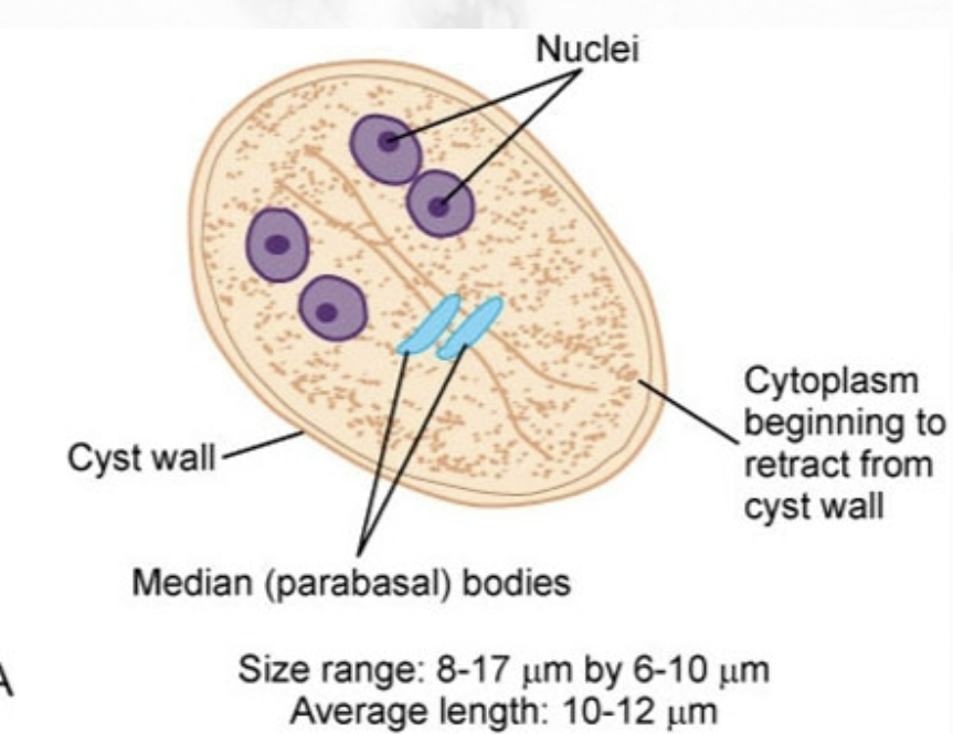
giardia intestinalis (lamblia, duodenalis), giardiasis
can be asymptomatic or cause traveler’s diarrhea. found worldwide in multiple water sources. transmitted via contaminated food/water, hand-to-mouth, unprotected sex, circulating flies & cockroaches. cysts resistent to routine water chlorination. travelers who drink contaminated water in known endemic areas, children, people who practice unprotected sex at risk. several animal hosts.
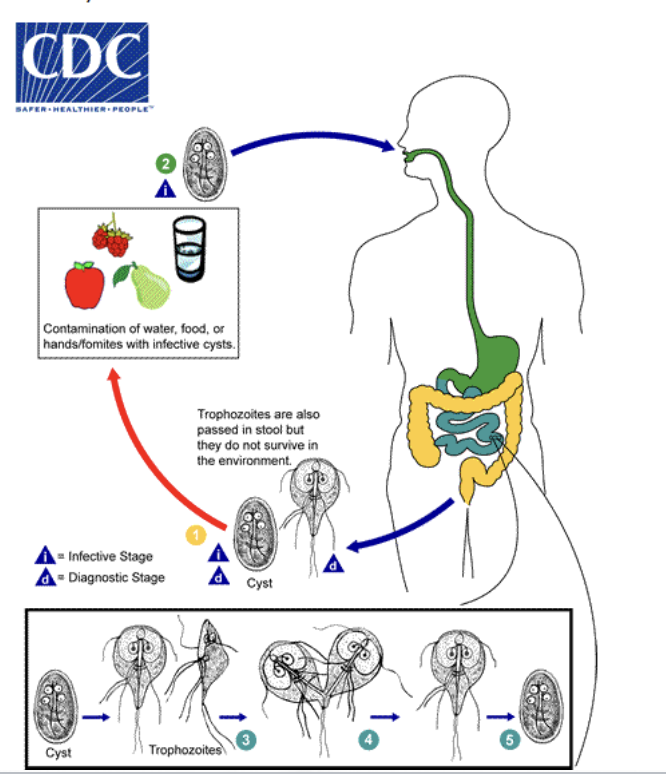
traveler’s diarrhea caused by giardia intestinalis
aka symptomatic intestinal amebiasis. diarrhea, abdominal pain/cramping, weight loss, anorexia, chronic fatigue, flatulence. severe cases: fat soluble vitamin deficiencies, folic acid deficiencies, hypoproteinemia w/ hypogamma-globulinemia, intestinal villi structure changes. individuals with IgG deficiency seem to be susceptible recurring infections. found by stool, small intestine biopsies: ELISA, RTPCR, etc.

chilomastix mesnili trophozoite
shape: pear.
motility: stiff, rotary, directional.
nuclei: 1 w/ small central or eccentric karyosome, no peripheral chromatin.
flagella: 4 total, 3 extend from anterior end & 1 extends posteriorly from cytostome region.
other structures: prominent cytostome extending 1/3 to ½ body length, spiral groove


chilomastix mesnili cyst
shape: lemon w/ clear hyaline knob extending from anterior end.
nuclei: 1 w/ large central karyosome, no peripheral chromatin.
other structures: well-defined cytostome on 1 side of nucleus.

chliomastix mesnili
cosmopolitan, prefers warm climates. transmitted by consuming contaminated food/water or hand-to-mouth contamination. stool (standard methods) for lab diagnosis. usually asymptomatic. prevented by good sanitation & personal hygiene, protection of food from insects


dientamoeba fragilis trophozoite
the only known morphologic form. found in mucosal crypts of large intestine (no surrounding tissue invasion) after human ingestion. can rarely ingest RBCS.
shape: irregularly round.
motility: progressive, broad hyaline pseudopodia.
nuclei: 2, each consists of massed clumps of 4-8 chromatin granules. no peripheral chromatin.
cytoplasm: bacteria-filled vacuoles common

dientamoeba fragilis
life cycle poorly understood. exact mode of transmission unknown: theory that it may be transmitted via select helminth eggs. diagnosed via PCR or detection of trophs in trichrome stained fecal smears (tends to blend in with background), not detectable by concentration methods.
children & individuals in semi-communal or institutional environments at risk. possibly transmitted person-to-person, oral-anal. usually asymptomatic. symptoms can vary if infected.


trichomonas vaginalis trophozoite
hardy by nature. can survive in urine, wet sponges, & damp towels for several hours. survive in water for 40 min.
shape: ovoid, round, or pear.
motility: rapid, jerky.
nuclei: 1, ovoid, nondescript.
flagella: all originating anteriorly, 3-5 extend anteriorly, 1 extending posteriorly.
other features: undulating membrane extending ½ of body length. prominent axostyle that often curves around nucleus, granules may be seen along axostyle.

trichomonas vaginalis
reside on mucosal surface of vagina in women: trophs replicated by longitudinal binary fission, feed on bacteria & leukocytes, & thrive in slightly alkaline or slightly acidic pH (unhealthy). reside in prostate gland region & epithelium of urethra in men. transmitted by sex or passage from mother to child via birth canal.
diagnosed using saline wet preps (urine, discharge, prostatic secretions). InPouch TV culture system (most sensitive): women vaginal swabs, men urine or semen sediment, incubation of up to 3 days.
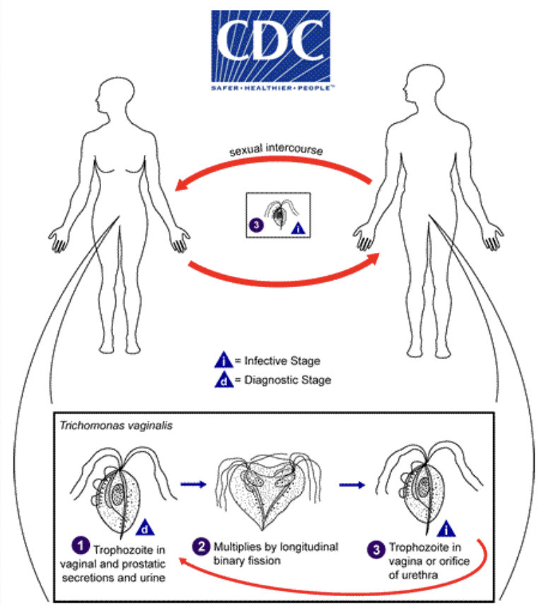
symptoms of trichomonas vaginalis infection
most men are asymptomatic. women are symptomatic. persistent urethritis: enlarged tender prostate, dysuria, nocturia, epididymitis. persistent vaginitis: foul-smelling, greenish-yellow, liquid vaginal discharge; burning, itching, chafing