Exam 2 Complete
 Call Kai
 Learn
 Practice Test
 Spaced Repetition
 Match
 Flashcards
 Knowt Play
1/211

There's no tags or description
Looks like no tags are added yet.
212 Terms
What are the two girdles of the body? What do they allow? Which one is more stable?
Pelvic and shoulder girdle.
Girdles allow connection between the axial and appendicular skeleton.
PG is more stable than the shoulder girdle, with a deep socket, the labrum, and strong ligaments that trade ROM for stability.
What is the PG made up of?
Where do they fuse
PG is made up of the ilium, ischium, and pubis. They all fuse at the acetabulum.
What are the gender differences within the PG?
In females the PG is lighter, thinner, and wider, because females are biologically built for child birth.
Sacroiliac joint
what is its ROM?
What is its function?
Very strong ligamentous arrangements between the sacrum and pelvis, so there is minimal movement.
The function of the joint is to transmit weight from the spine to the femur bones.
What are some gender differences of the SIJ?
In some males, their sacrum is fused to their ilium, so there is very little ROM
However, in females, the SIJ is typically not fused because of childbirth, so there is greater ROM (but still not much)
What are the movements of the sacrum (independent of the pelvis)
Sacral flexion and extension

What is sacral flexion?
when does it occur?
Along the sagittal plane, the sacrum tilts forward.
it occurs when the spine extends

What is sacral extension?
when does it occur?
Along the sagittal plane, the sacrum tilts backwards
occurs when the spine flexes.
What are the movements of the pelvis?
Anterior and posterior tilt
Right and left lateral tilt
Left and right rotation

Pelvis: What is an anterior tilt? What is a posterior tilt?
Along the sagittal plane.
Anterior tilt: ASIS and the iliac crest tilt forward
hip flexion and lumbar spine extension
Posterior tilt: ASIS and the iliac crest tilt backwards.
hip extension and lumbar spine flexion

P: What is a right lateral tilt? What is a left lateral tilt?
Frontal plane.
Right lateral tilt: Right side drops and left side rises
Left lateral tilt: Left side drops and right side rises

P: What is left rotation of the pelvis? Right rotation?
Along the transverse plane.
Left rotation: Pelvis is twisted to the left
Right rotation: Pelvis is twisted to the right
What is the relationship of the pelvis to the trunk and lower extremities?
It’s the link between them. It simultaneously needs to contribute to the motion of the trunk and lower extremities and stability.
Acts as a base of support and contributes to movements.
What is it called when movements are initiated by the pelvis?
Primary movement
Primary movements of pelvis and what is causes in → Spinal Joints / Trunk and Hip Joints
Anterior Tilt → Hyperextension → Flexion
Posterior Tilt → Hyperflexion→ Extension
Left Lateral Tilt → Right lateral flexion → Abduction at L & Adduction at R
Right Lateral Tilt → Left lateral flexion → Abduction at R & Adduction at L
Left Rotation → Right Rotation → Slight lateral rotation (R) & Slight medial rotation (L)
Right Rotation → Left Rotation → Slight medial rotation (R) & Slight lateral rotation (L)
Open chain vs Closed chain movements.
Open: Proximal segment is fixed and distal segment moves.
Closed: Distal segment is fixed and proximal segment moves.

Hip joint
Scientific name
Joint Classification
Cartilage
Acetabulum femoral joint
Ball-and-Socket Joint
Head is covered with articular cartilage except the fovea capitis
Acetabulum is covered in articular and fibrocartilage.

Fibrocartilage in hip joint.
Acetabulum is lined with articular cartilage and fibrocartilage (acetabulum labrum). It follows (most) of the rim of the acetabulum, adding depth inwards and outwards. It functions to provide additional cushion for high impact activities of femoral head.
What are the ligamentous reinforcements of the SIJ
Transverse acetabular, teres femoris (ligamentum teres), Iliofemoral (Y ligament), pubofemoral, and ischiofemoral ligament.
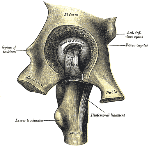
Transverse acetabular ligament
what does it complete
Made up of
Reinforces
Completes the ring of the acetabulum labrum, bridging the acetabular notch.
Made up of stronger material than fibrocartilage
Reinforces the inferior portion of the acetabulum, ensuring the femur does not drop in space during high-impact activities.
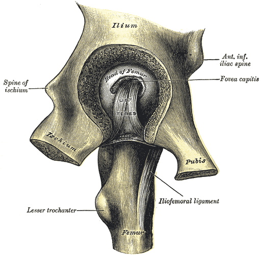
Teres femoris (ligamentum teres)
Inserts
Reinforces
Runs from the fovea capitis of the femoral head to the acetabular notch (inferior portion of the acetabulum); carries a small blood vessel to the head.
Internal reinforcement

Iliofemoral (Y) ligament
Reinforces what during what movement…
The anterior and strongest ligament of the hip.
Ensures the head of the femur does not come outwards anteriorly, which occurs during hip extension and external rotation.
limits extension

Pubofemoral ligament
ensures..
prevents..
Anterior/inferior.
Ensures the head of the femur does not drop downwards, which occurs during abduction.
Prevents excessive abduction (and extension).

Ischiofemoral ligament
Ensures..
Limits…
Strong posterior, triangular ligament
Ensures the head does not come out posteriorly.
Limits internal rotation and adduction (particularly in a flexed position)

What is the angle of inclination of femoral neck?
Is the angle being created by line trajectory from greater trochanter to acetabulum, and down the shaft.

Coxa Valga
What is it
Reduces the effectiveness of…
Decreases and increases stress where on the femur?
When the head-neck alignment angle is >125 (more longitudinal)
lengthens limb
Reduces the effectiveness of abductors
moment arm is shorter
Decreases bending neck stress but increases load on the head
more likely to break head than neck

What is Coxa Vara
Increases the effectiveness of..
Decreases and increases stress where on the femur?
When the head-neck alignment angle is <125
shortens limb
Increases the effectiveness of hip abductors
moment arm is longer
Increased bending force on neck and decreased load on head
Femoral neck in the transverse plane
Anteversion, excessive anteversion, and retroversion
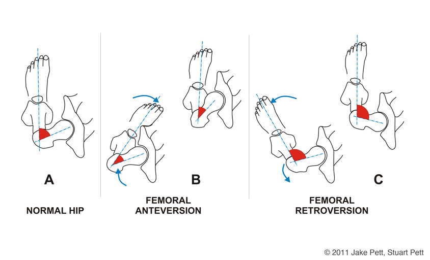
What is anteversion?
The head and neck of the femur is rotated 12-14 degrees anteriorly (normal).
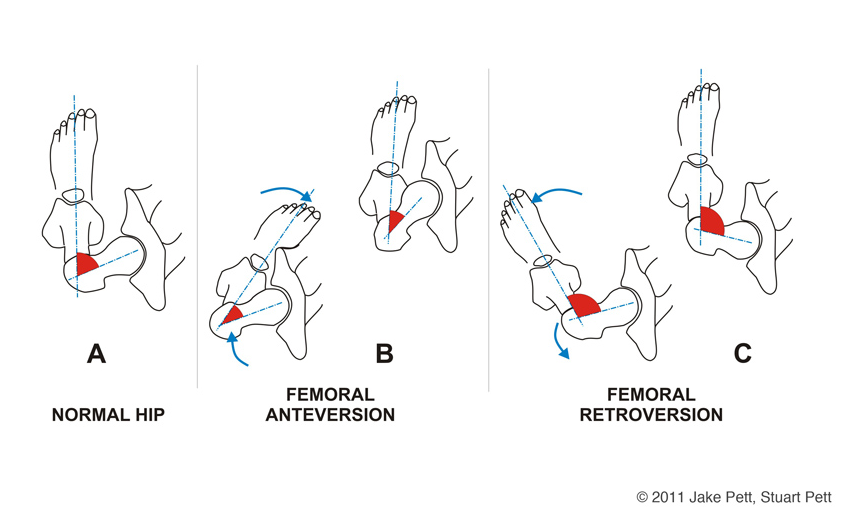
Excessive anteversion
What is it?
What happens to the head of the femur when the person stands straight?
How does the person walk?
The head and neck of the femur is rotated more than 12-14 degrees anteriorly.
If the person were to stand with their foot and knee straight, the head of the femur becomes uncovered anteriorly. When the head is jutted out the hip joint does not function, so…
The person internally rotates their thigh for the head to be in the acetabulum, causing an internally rotated gait.
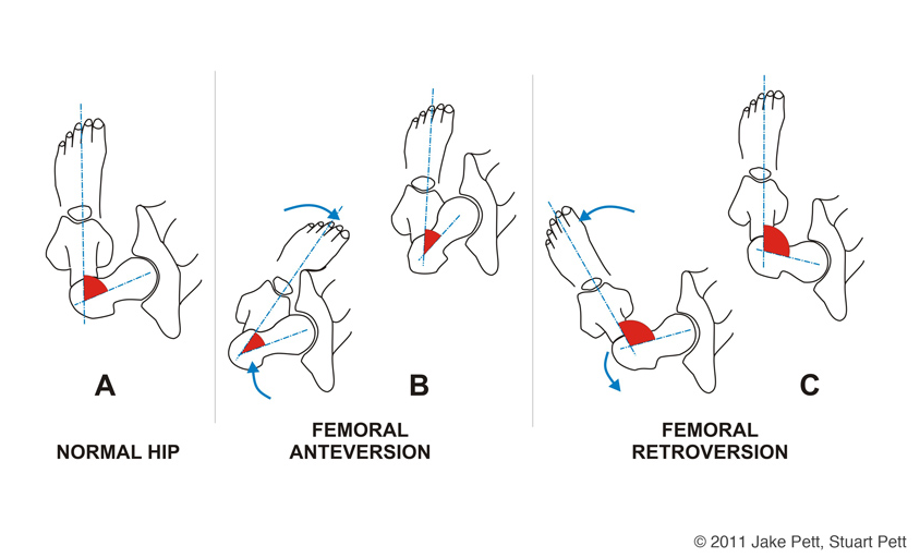
What does excessive anteversion cause?
Patellar-femoral problems as the torsional load goes into knee.
Forces the person into lordosis: increase in curvature of lumbar spine.
Person walks more pronated, as there is to much weight on medial side of foot.
Increases Q-angle
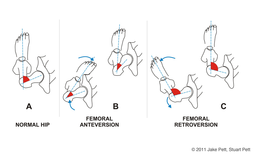
Retroversion
What is it?
What happens to the head of the femur when the person stands straight?
How does the person walk?
The head and neck of the femur is rotated less than 12-14 degrees posteriorly.
If the person were to stand with their foot and knee straight, the head of the femur becomes uncovered posteriorly. When the head is jutted out the hip joint does not function, so…
The person externally rotates their thigh for the head to be in the acetabulum, causing an externally rotated gait.
Retroversion causes
Supination, as more weight is on the lateral side of the foot
Decreases the Q angle

What is the Q-angle?
How does the Q-angle change during excessive anteversion and retroversion.
A line from the ASIS to the patellar midpoint and a vertical line from the patellar midpoint. The angle between the two vectors is the midpoint.
Excessive anteversion: ASIS vector is pushed out, increasing the Q-angle
Retroversion: ASIS vector is pushed closer, decreasing the Q-angle
When looking at hip and pelvic movements we first want to ID…
1) The direction of movement
2) position of the body in relation to gravity (contraction type)
3) ID the movement of the segment
proximal: pelvis
distal: femur
How is the segment moving when… Hip flexor muscles are moving concentrically
Thigh is moving towards the trunk.
How is the segment moving when… Hip extensor muscles are used eccentrically
Moving pelvis on top of stable thigh
How is the segment moving when…Hip extensor muscles are used concentrically in a closed chain movement.
Rising from a squat
How is the segment moving when…Hip extensors are used concentrically in a open chain movement
Raising the thigh up towards the trunk posteriorly
How is the segment moving when… Hip flexors are used concentrically in a closed-chain movement
Doing a sit up
What is their line of pull
Iliopsoas
Pectineus
Rectus Femoris
Sartorius
Anterior line of pull, primarily hip flexion
What is their line of pull
Adductor brevis
Adductor longus
Adductor magnus
Gracilis
Medial line of pull, primarily hip adduction.
What is their line of pull
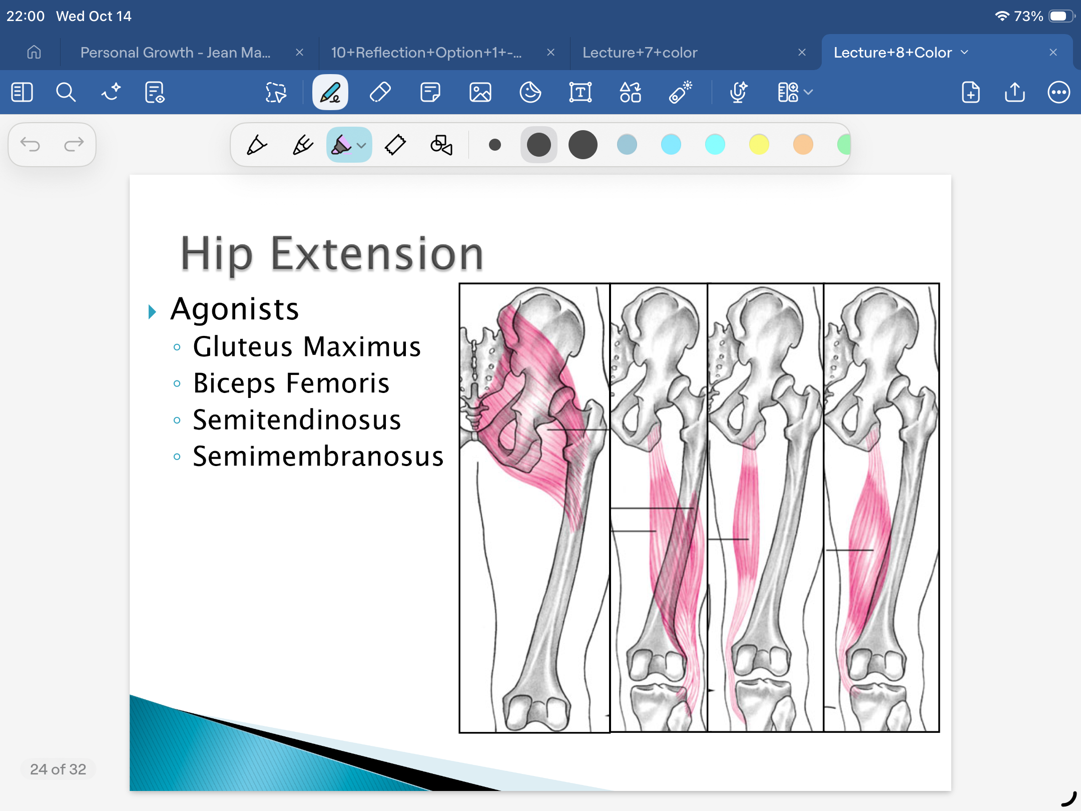
Gluteus maximus
Biceps femoris
Semitendinosus
Semimembranosus
6 Deep External Rotators
Posterior line of pull, mainly hip extension
What is their line of pull
Gluteus medius
Gluteus minimus
Tensor fasciae latae
Lateral line of pull, mainly hip abduction.
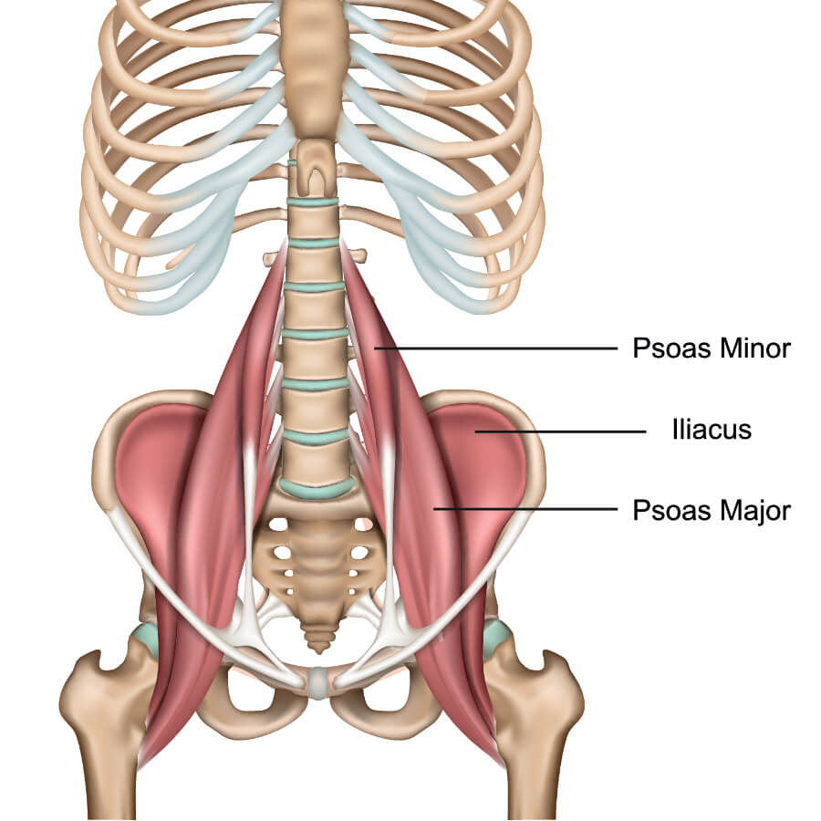
Iliopsoas Muscle
Composed of
LoP
Actions
Composed of three different muscles
Anterior LoP
A: Flexion of hip
A: External rotation
Iliopsoas inserts on LT and twists it towards the front.

Sartorius
What type of muscle is it?
OI
LoP?
Actions
Bi-articular muscle
O: ASIS
I: Proximal anterior medial surface of tibia
Anterior LoP
A: Hip abduction
as O is slightly lateral to hip joint center
A: External rotator
brings medial I to lateral O
A: Knee flexion
has a posterior LoP as it wraps posteriorly around the knee before inserting on the tibia

Rectus Femoris
What type of muscle is it?
OI
LoP?
Actions
Bi-articular muscle
O: ASIS
I: Tibial tuberosity via patellar tendon
Anterior LoP
A: Hip flexion
A: Knee extension

Tensor Fascia Latae
What type of muscle is it?
OI
LoP?
Actions
TFL is a small muscle with a long I, it is not bi-articular, because the iliotibial tract (IT band) is not an active mover, it does not have the ability to contract.
O: Iliac crest, posterior to ASIS
I: Iliotibial tract
Lateral LoP
A: Hip abductor
A: Hip flexor
A: Internally rotates hip as it flexes it (tendency to)

Gluteus Maximus
LoP?
Actions
LoP is separated by upper and lower fibers
Action of all fibers: Hip extension and external rotation.
Action of upper fibers: Abduction
Action of lower fibers: Adduction

Gluteus Medius
OI
LoP?
Actions
Lateral LoP
O: Gluteal surface of ilium
I: lateral aspect of greater trochanter
All fibers: hip abduction
Anterior fibers action: internal rotation
Posterior fibers action: external rotation

Gluteus minimus
OI
LoP?
Actions
O: Gluteal surface of ilium
I: Anterior aspect of greater trochanter
Lateral LoP
A: abduction of hip
A: internal rotation as femur abducts
many of its fibers are towards the anterior of pelvis
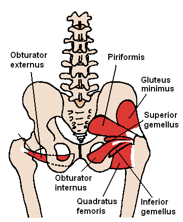
Six Deep External Rotator Muscles
LoP?
Actions
Lateral LoP
A: external rotation of hip

Semitendinosus
OI
LoP?
Actions
O: Ischial tuberosity
I: Proximal anterior medial surface of tibia
Medial and posterior LoP
A: Hip extensor
A: Knee flexor
A: Internal rotator of hip and knee

Semimembranosus
OI
LoP?
Actions
O: Ischial tuberosity
I: medial condyle of tibia
Medial and posterior LoP
A: Hip extensor
A: Knee flexor
A: Internal rotator of hip and knee

Biceps Femoris
OI
LoP?
Actions
O: Ischial tuberosity
I: Lateral condyle of tibia and head of fibula
Lateral and posterior LoP
A: Extension of hip
A: Flexion of knee
A: External rotation of hip and knee

Adductor brevis
OI
LoP?
Actions
O: anterior inferior pubic ramus
I: linea aspera
Medial and lateral LoP
A: adduction of hip
A: external rotation as it adducts at hip
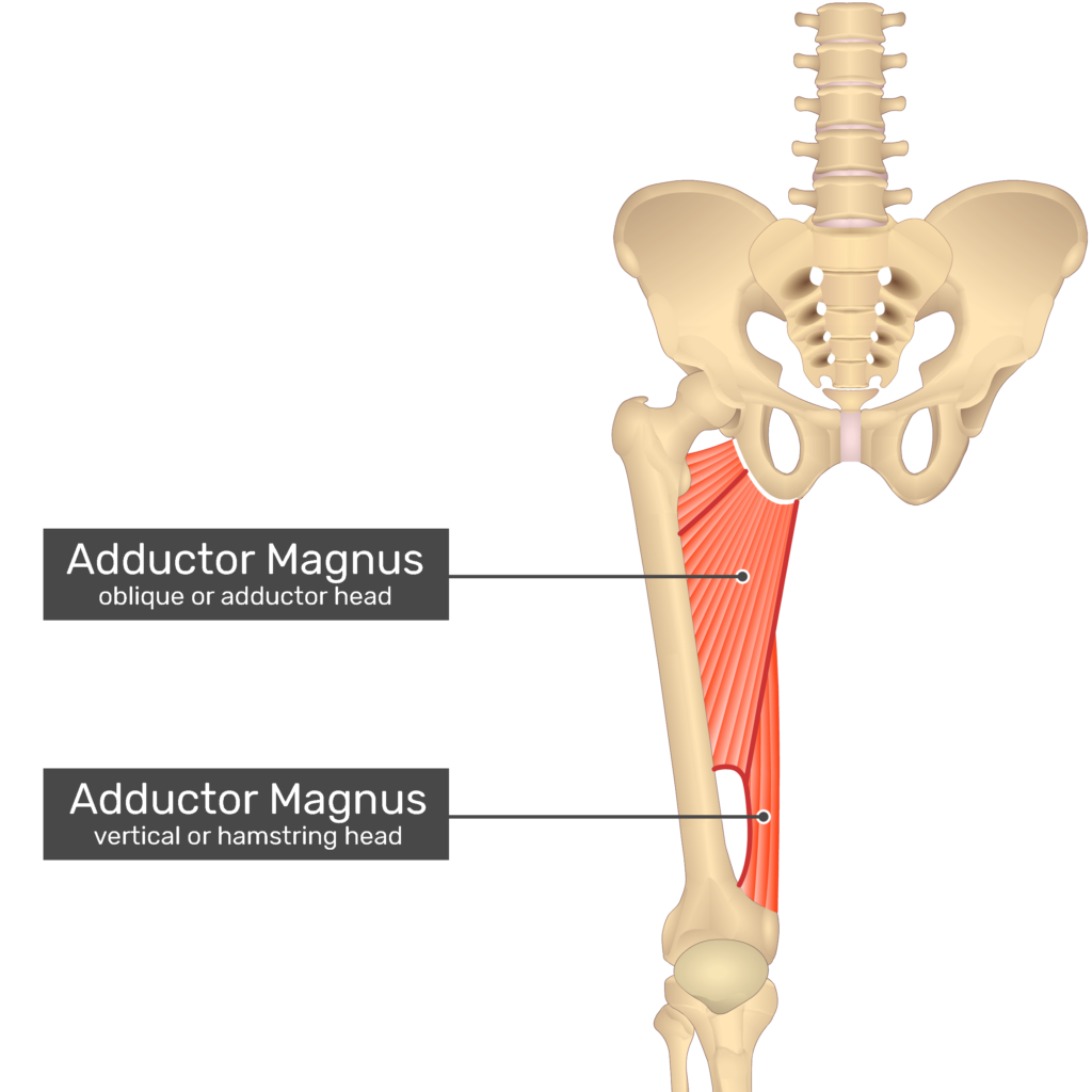
Adductor magnus
OI
LoP?
Actions
O: ramus of pubis and ischium, and ischial tuberosity
I: Entire linea aspera
Medial and lateral LoP
A: adduction of hip
A: external rotation of hip as it adducts

Adductor longus
OI
LoP?
Actions
O: anterior pubis, below crest
I: Medial third of linea aspera
Medial and anterior LoP
A: adduction at hip
A: assists in hip flexion
A: (medial rotator)
because of its I

Pectineus muscle
OI
LoP?
Actions
O: Above pubic crest
I: Lesser trochanter to linea aspera
Lateral, anterior, and medial LoP
A: Hip flexion
A: Adduction of hip
A: external rotation of hip

Gracilis
OI
LoP?
Actions
O: anterior medial edge of descending ramus of pubis
I: anterior medial surface of tibia
Posterior and medial line of pull
A: Weak flexion of knee
goes behind knee before inserting anteriorly on tibia
A: adduction of hip
A: internal rotation of hip
What is the agonist muscle group based on?
Based on anatomical position, and it changes with changes to joint angle
Hip flexor agonists

Hip extensors agonist
Hip abductors agonists

Hip adductors agonist

Hip internal rotators agonist

Hip external rotators agonist

What is hip muscle strength?
How a muscle group can be made stronger by increasing their MA by increasing their moment arm and L/T relationship
Hip muscle strength extension
How
Why
Example
Starting with hip flexed close to 90 degrees makes the agonist muscle group more effective at hip extension.
because it increases the moment arm and L/T relationship closer to optimum
Ex) With the hip flexed, the ischium is pushed outward; therefore, the posterior muscles are moved farther from the hip joint, increasing its moment arm and the L/T relationship.
Hip muscle strength flexion
How
Why
Example
Having hip flexors start slightly extended lengthens them anteriorly, increasing efficiency in flexion
Iliopsoas
diminishes with trunk flexion - L/T is short
increases with slight hip extension - L/T is longer
Hip muscle strength flexion
Rectus femoris
As a bi-articular muscle, by flexing the knee (lengthening it), it is stronger at the hip.
Hip muscle strength abductors
How
Why
Example
Maximum strength near neutral joint angle (anatomical position).
There is a built-in pulley system as the abductors wrap around the greater trochanter. It naturally lengthens them slightly, bringing them closer to an optimum L/T relationship and increasing the moment arm.
Hip muscle strength adductors
how
Greater starting with slight abduction, as it increases the L/T relationship.
Hip muscle strength internal and external rotators
Generally, starting with the hip joint flexed, the muscle agonist in both groups increases the moment arm and L/T relationship.
Both internal and external rotators are stronger when seated (flexed) rather than supine (neutral)
The knee joint is…
The largest and most complex joint in the body, however it is primarily considered a hinge joint.
The joint is formed by…
Enlarged femoral condyles are received by the medial and lateral surface (condyles) of the tibia.
The tibia is
The fibula is
The patella is
The tibia is the medial weight bearing bone of the shank.
The fibula is the lateral bone of the shank. It does not have a function at the knee, it serves as an area where structures insert.
Does NOT articulate with femur or patella
The patella is a sesamoid (floating) bone, embedded in the quadriceps/patellar tendon.
What is the knee joint proper?
arguments
The tibiofemoral joint is classified as a hinge joint.
Argued to be referred to as a trochoginglymus or condyloid.
What is the patellofemoral joint?
It is the gliding joint created between the patella and femur (condyles).
What structures provide stability and what type?
Ligaments provide static stability for joint (noncontractile)
Muscles provide dynamic stability for joint (contractile)
Menisci are fibrocartilage pads that attach to and deepen the tibial surface, where the femoral condyles sit, enhancing stability and absorbing shock.
What are some features and structure of menisci?
Thick border that tapers downwards and inwards, thinning out.
They are held in place by small ligaments, allowing the menisci to move slightly.
less likely to tear during flexion and extension
The medial meniscus is larger and has a more open C appearance, because the medial femoral condyle is larger and it is the weight-bearing side.
The lateral meniscus is smaller and has a closed C configuration.
Injury to menisci
common?
how do they occur?
Meniscus tears are very common and occur in various ways.
Most often due to compression and shear forces while changing in direction with the knee flexed or extended.
Define: Blow
A blow is named by where it lands, and it injures the opposite side.
Anterior and Posterior Cruciate Ligaments
Cross
Functions (2)
Cross within the knee between the femur and the tibia
F: Gives anterior and posterior stability to the tibia
tibia does not move too forward or back in respect to the femur.
F: Gives rotary stability.
stops tibia from internally and externally rotating
ACL
Prevents…
Injury
Common? Cause?
Prevents the tibia from sliding forward on the femur (hyperextension).
Is one of the most common, serious injuries to the knee.
It’s often due to noncontact forces, either from vigorously contracting the quads, planting with a straight leg, cutting (rotary forces), pivoting, or landing with the knee caving in.
PCL
Prevents
Injury
Common?
Causes
Prevents the tibia from sliding backwards.
Not often injured by noncontact forces, as we can flex the knee to a large degree.
Most likely injured in contact sports due to an anterior blow on the tibia, slamming into the knee anteriorly, or the playing surface.
Fibular (lateral) collateral ligament
Injury
Common?
Causes
Resists varus forces
Not often injured, as it is less weight-bearing
However, it can be injured in a contact sport if a medial blow is received, which blows open the lateral side.
Tibial (medial) collateral ligament
Maintains
Injury
common?
causes
Maintains medial stability by resisting valgus forces (knock knee) or preventing the knee from being abducted.
It is bigger and more commonly injured than the LCL in contact and noncontact sports.
Abnormal valgus forces (falling on the medial side of the knee)
Mechanisms of a teammate or opponent falling on the lateral aspect of the knee or leg. So, a lateral blow opening the medial side of the knee joint.
Where is the synovial cavity?
Capsule is behind the patella and in between the femur and tibia.
What is the function of the fat pads surrounding the knee joint?
Shock absorption, cushioning, and separating structures which reduces friction.
What is Plica?
They are?
What may happen to them?
Folds on fat that are small sacks of synovial fluid.
They are an anatomical variant
May be irritated or inflamed with injuries or overuse of knee, causing it to swell greatly.
What are bursae?
Small capsules of synovial fluid.
They function as shock absorbers and reduce friction.
When does the knee lock? How does the knee lock? What is the name for it?
When the knee reaches neutrality/full extension/ neutral angle, the knee is locked into place for stability.
Known as the screws home mechanism, for proper alignment the femoral condyles need to be received by the menisci of the tibia.
For this to occur, the tibia externally rotates approximately 10 degrees to be “screwed home” into its neutral alignment.
How does the knee unlock?
To unlock the knee (flexion), the tibia needs to internally rotate approximately 10 degrees so the menisci and femoral condyles are disarticulated enough to flex.
The popliteus starts the unlocking.
Why does the locking and unlocking of knee happen?
Why the external rotation (locking) and internal rotation (unlocking) of the knee happens is because the lateral meniscus is smaller than the medial meniscus. So there needs to be a twist to lock and unlock the medial side into and out of alignment.
Femur and tibia during open and closed chain movements.
Femur moves in closed chain movements
Tibia moves in open chain movements
when extended, the tibia is externally rotated with respect to the femur
when flexed, the tibia is internally rotated with respect to the femur
What are the movements of the knee and their degrees.
Flexion: occurs to about 140 degrees
Extension: extends to 180 degrees
hyperextension: 10 degrees or less
When the knee is unlocked at 30 degrees or more (flexed)
internal rotation can occur to about 30 degrees
external rotation can occur to about 45 degrees
What is the Q-angle?
Line from the tibial tuberosity through the midpoint of the patella.
ASIS to the midpoint of the patella.
angle between the two vectors forms the Q-angle.
What is the LoP of the vastus lateralis in relation to the other quad muscles?
LoP of the vastus lateralis is more lateral than the rest of the quads which have a more medial LoP.
The Q-angle is women tend to be…, which causes……
In women, the Q-angle tends to be larger (20 degrees or less) because the pelvis is wider, which causes the angle to be more lateral overall.
Causes more stress on knee joint structures as a lateral LoP comes onto it. Especially on the ACL, because a larger Q-angle goes with more knee valgus, loading it as the ACL functions to prevent twisting.
Menisci as well