Lab exam review 2 - Mitosis

0.0(0)
Studied by 0 people
call kaiCall Kai
learnLearn
examPractice Test
spaced repetitionSpaced Repetition
heart puzzleMatch
flashcardsFlashcards
GameKnowt Play
Card Sorting

1/97

encourage image

There's no tags or description

Looks like no tags are added yet.

98 Terms

1
New cards

Mitosis

A type of nuclear division in eukaryotic cells that divides replicated DNA into two equal, genetically identical sets of chromosomes. It produces two daughter cells with the exact same chromosome number and genetic content as the parent cell

  • The precise process by which a single somatic cell divides to produce two genetically identical daughter cells is called mitosis


2
New cards

Intro to mitosis

In cells, growth and development are fundamental to life. While many organisms start as a single cell, that cell will divide trillions of times to allow for growth, tissue repair, organ regeneration, and asexual reproduction.


3
New cards

Multicellular organisms use cell division for:

  • Growth

  • Maintenance

  • Repair injured cells and tissues

  • Development


4
New cards

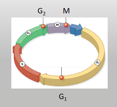
Interphase

Between periods of active division, cells spend most of their life cycle in this phase. During interphase, the nucleus appears uniform under a standard microscope as the cell performs its everyday metabolic functions. Though it looks inactive, the cell is actively increasing in size, synthesizing new organelles, and replicating its DNA

Interphase occurs in 3 stages

  • G1 phase (first gap)

  • S phase

  • G2 phase (second gap)

  • During interphase, DNA exists in a loose, uncoiled state called chromatin


<p><span style="background-color: transparent;">Between periods of active division, cells spend most of their life cycle in this phase. During interphase, the nucleus appears uniform under a standard microscope as the cell performs its everyday metabolic functions. Though it looks inactive, the cell is actively increasing in size, synthesizing new organelles, and replicating its DNA</span></p><p><span style="background-color: transparent;">Interphase occurs in 3 stages</span></p><ul><li><p>G1 phase (first gap)</p></li><li><p>S phase</p></li><li><p>G2 phase (second gap)</p></li><li><p><span style="background-color: transparent;">During interphase, DNA exists in a loose, uncoiled state called <strong>chromatin</strong></span></p></li></ul><p></p>
5
New cards

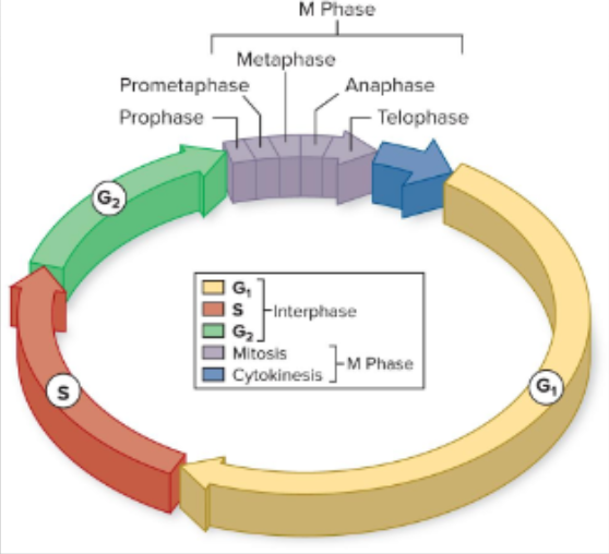
G1 phase (Gap 1 or first growth phase)

Cell grows and performs normal functions

  • the first of three sub-phases of interphase in the eukaryotic cell cycle, occurring right after M phase (mitosis and cytokinesis) and right before S phase (DNA synthesis)

  • During G1, a newly formed cell undergoes intense growth and metabolic activity to recover from the previous division and prepare its internal machinery for copying its genome

<p><span>Cell grows and performs normal functions</span></p><ul><li><p>the first of three sub-phases of <strong>interphase</strong> in the eukaryotic cell cycle, occurring right after M phase (mitosis and cytokinesis) and right before S phase (DNA synthesis)</p></li><li><p>During G1, a newly formed cell undergoes intense growth and metabolic activity to recover from the previous division and prepare its internal machinery for copying its genome</p></li></ul><p></p>
6
New cards

S Phase

The middle stage of Interphase in the eukaryotic cell cycle where the cell replicates its entire genome (nuclear DNA) and duplicates its centrosomes

  • DNA synthesis occurs

  • (Synthesis phase) of interphase, the cell synthesizes a complete copy of its nuclear DNA and duplicates its centrosomes

  • Before a cell can divide, it must undergo DNA replication to duplicate its genetic material

  • chromosomes are replicated during the S phase (synthesis phase) of interphase. Each single chromosome is copied to form two identical copies called sister chromatids, which stay joined together at a region called the centromere


<p>The middle stage of Interphase in the eukaryotic cell cycle where the cell replicates its entire genome (nuclear DNA) and duplicates its centrosomes</p><ul><li><p>DNA synthesis occurs</p></li><li><p>(Synthesis phase) of interphase, the cell synthesizes a complete copy of its nuclear DNA and duplicates its centrosomes</p></li><li><p><span style="background-color: transparent;">Before a cell can divide, it must undergo DNA replication to duplicate its genetic material</span></p></li><li><p><span>chromosomes are replicated during the </span>S phase (synthesis phase) of interphase. Each single chromosome is copied to form two identical copies called sister chromatids, which stay joined together at a region called the centromere</p></li></ul><p></p>
7
New cards

G2 Phase

(Gap 2 phase) is the third and final subphase of interphase in the eukaryotic cell cycle, occurring directly after DNA replication in the S phase and right before mitosis (M phase)

Cell gets ready for mitosis:

  • Makes proteins needed for mitosis

  • Produces new organelles

  • Checks DNA damage


<p>(Gap 2 phase) is the third and final subphase of interphase in the eukaryotic cell cycle, occurring directly after DNA replication in the S phase and right before mitosis (M phase)</p><p>Cell gets ready for mitosis:</p><ul><li><p>Makes proteins needed for mitosis</p></li><li><p>Produces new organelles</p></li><li><p>Checks DNA damage</p></li></ul><p></p>
8
New cards

Chromatin

The complex of DNA and protein (specifically histone proteins) found inside the eukaryotic nucleus. It allows long strands of DNA to pack tightly so they can fit inside a tiny cell nucleus

  • During interphase, DNA exists in a loose, uncoiled state called chromatin

  • a complex of DNA, RNA, and proteins—primarily spool-like proteins called histones—found inside the nucleus of eukaryotic cells


<p>The complex of DNA and protein (specifically histone proteins) found inside the eukaryotic nucleus. It allows long strands of DNA to pack tightly so they can fit inside a tiny cell nucleus</p><ul><li><p><span style="background-color: transparent;">During interphase, DNA exists in a loose, uncoiled state called <strong>chromatin</strong></span></p></li><li><p><span>a complex of DNA, RNA, and proteins—primarily spool-like proteins called </span><strong>histones</strong><span>—found inside the nucleus of eukaryotic cells</span></p></li></ul><p></p>
9
New cards

What state is DNA in during interphase?

During interphase, DNA exists in a loose, uncoiled state called chromatin

  • During interphase, DNA exists as chromatin—a loose, uncondensed complex of DNA wrapped around histone proteins


<p><span style="background-color: transparent;">During interphase, DNA exists in a loose, uncoiled state called <strong>chromatin</strong></span></p><ul><li><p><span>During interphase, DNA exists as </span><strong>chromatin</strong>—a loose, uncondensed complex of DNA wrapped around histone proteins</p></li></ul><p></p>
10
New cards

Centromere

The specialized, constricted region of a duplicated chromosome where two sister chromatids remain tightly attached after DNA replication

  • It also serves as the primary assembly site for the kinetochore, a protein structure where spindle fibers (microtubules) attach during mitosis and meiosis

  • the region where two sister chromatids stay connected after DNA replicates in S phase, and it's the attachment point that spindle fibers grab to pull chromosomes apart during mitosis and meiosis.


<p>The specialized, constricted region of a duplicated chromosome where two sister chromatids remain tightly attached after DNA replication</p><ul><li><p>It also serves as the primary assembly site for the kinetochore, a protein structure where spindle fibers (microtubules) attach during mitosis and meiosis</p></li><li><p>the region where two sister chromatids stay connected after DNA replicates in S phase, and it's the attachment point that spindle fibers grab to pull chromosomes apart during mitosis and meiosis.</p><p><br></p></li></ul><p></p>
11
New cards

Chromosomes

A thread-like, gene-carrying structure made of a single long deoxyribonucleic acid (DNA) molecule tightly coiled and wrapped around associated histone proteins

  • A duplicated chromosome consists of two identical sister chromatids joined together at a region called the centromere

  • Each individual chromatid features a distinct short arm and a long arm.


<p>A thread-like, gene-carrying structure made of a single long deoxyribonucleic acid (DNA) molecule tightly coiled and wrapped around associated histone proteins</p><ul><li><p><span style="background-color: transparent;">A duplicated chromosome consists of two identical sister chromatids joined together at a region called the <strong>centromere</strong></span></p></li><li><p><span style="background-color: transparent;">Each individual chromatid features a distinct short arm and a long arm.</span></p><p><br></p></li></ul><p></p>
12
New cards

Chromatin vs. Chromosome

During normal cell life (interphase), DNA is loosely structured and decondensed as chromatin. Right before cell division (mitosis or meiosis), chromatin condenses tightly into visible chromosomes

Chromatin and chromosomes are both made of DNA wrapped around histone proteins, but they represent different physical states of genetic material in the cell

  • State: A loose, open, thread-like complex of DNA and proteins.

  • When it appears: Present during interphase (the normal, non-dividing phase of the cell cycle).

Chromosome

  • State: A tightly coiled, condensed, distinct rod- or X-shaped structure.

  • When it appears: Formed just before and visible during mitosis or meiosis (cell division).


13
New cards

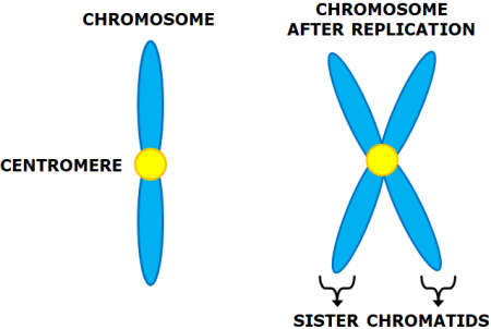
Chromatid

One of the two identical halves of a duplicated chromosome formed during the S phase of interphase

  • Sister Chromatids: two identical copies of a single chromosome connected at a specialized region called the centromere. They remain attached until anaphase (in mitosis or meiosis II), when they are pulled apart to become individual chromosomes

  • Sister Chromatids: two identical copies of a chromosome made by DNA replication, held together by cohesin proteins and connected at the centromere

  • Each individual chromatid features a distinct short arm and a long arm


<p>One of the two identical halves of a duplicated chromosome formed during the S phase of interphase</p><ul><li><p>Sister Chromatids: two identical copies of a single chromosome connected at a specialized region called the centromere. They remain attached until anaphase (in mitosis or meiosis II), when they are pulled apart to become individual chromosomes</p></li><li><p>Sister Chromatids: two identical copies of a chromosome made by DNA replication, held together by cohesin proteins and connected at the centromere</p></li><li><p><span style="background-color: transparent;">Each individual chromatid features a distinct short arm and a long arm</span></p><p><br></p></li></ul><p></p>
14
New cards

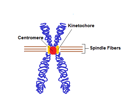
Kinetochore

A specialized protein structure that assembles on the centromere of each sister chromatid during cell division

  • Its primary role is to serve as the physical anchoring site for spindle microtubules, allowing the cell to move, align, and accurately separate chromosomes

  • a large, disc-shaped protein complex that assembles on the centromere of a chromosome during cell division (mitosis and meiosis). It acts as the anchor point where spindle microtubules attach to pull sister chromatids apart

<p>A <strong>specialized protein structure</strong> that assembles on the <strong>centromere</strong> of each sister chromatid during cell division</p><ul><li><p>Its primary role is to serve as the physical anchoring site for <strong>spindle microtubules</strong>, allowing the cell to move, align, and accurately separate chromosomes</p></li><li><p>a large, disc-shaped protein complex that assembles on the centromere of a chromosome during cell division<span> (mitosis and meiosis). It acts as the anchor point where spindle microtubules attach to pull sister chromatids apart</span></p></li></ul><p></p>
15
New cards

Cohesin

A ring-shaped protein complex that holds replicated sister chromatids together from S-phase of interphase until anaphase of mitosis or meiosis, ensuring proper chromosome segregation

  • a multi-protein ring-shaped complex that holds newly replicated sister chromatids together from DNA replication until anaphase in mitosis


<p>A ring-shaped protein complex that holds replicated sister chromatids together from S-phase of interphase until anaphase of mitosis or meiosis, ensuring proper chromosome segregation</p><ul><li><p>a multi-protein ring-shaped complex that holds newly replicated sister chromatids together from DNA replication until anaphase in mitosis</p></li></ul><p></p>
16
New cards

Centrioles

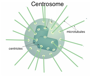
Barrel-shaped, cylindrical organelles composed of microtubules that serve as the structural backbone for cell division and motility in animal cells

  • cylindrical, barrel-shaped organelles made of nine triplets of microtubules. Located inside the centrosome, they organize the mitotic spindle that attaches to chromosomes and separates sister chromatids into daughter cells during mitosis in animal cells

<p>Barrel-shaped, cylindrical organelles composed of microtubules that serve as the structural backbone for cell division and motility in animal cells</p><ul><li><p>cylindrical, barrel-shaped organelles made of nine triplets of microtubules. Located inside the centrosome, they organize the mitotic spindle that attaches to chromosomes and separates sister chromatids into daughter cells during mitosis in animal cells</p></li></ul><p></p>
17
New cards

Centrosome

The primary microtubule-organizing center (MTOC) of an animal cell. Located near the nucleus, it consists of a pair of centrioles surrounded by a protein matrix. It replicates during interphase (G2 phase) and moves to opposite poles during mitosis to form the spindle fibers that separate chromosomes

  • Centrioles are found inside centrosomes. A typical animal cell centrosome contains a pair of barrel-shaped centrioles (a mother and a daughter centriole) positioned at right angles to each other, all embedded within a protein cloud


<p>The primary microtubule-organizing center (MTOC) of an animal cell. Located near the nucleus, it consists of a pair of centrioles surrounded by a protein matrix. It replicates during interphase (G2 phase) and moves to opposite poles during mitosis to form the spindle fibers that separate chromosomes</p><ul><li><p><span>Centrioles are found inside centrosomes</span>. A typical animal cell centrosome contains a pair of barrel-shaped centrioles (a mother and a daughter centriole) positioned at right angles to each other, all embedded within a protein cloud </p></li></ul><p></p>
18
New cards

Centrosome

The primary microtubule-organizing center (MTOC) in animal cells. It organizes the mitotic spindle fibers that attach to and pull replicated chromosomes apart during cell division

  • Located near the nucleus, it consists of a pair of centrioles surrounded by a protein matrix

  • It nucleates and organizes microtubules, which form the mitotic spindle during cell division to capture and separate chromosomes into daughter cells

  • an organelle that serves as the main microtubule-organizing center (MTOC) in animal cells


<p>The primary microtubule-organizing center (MTOC) in animal cells. It organizes the mitotic spindle fibers that attach to and pull replicated chromosomes apart during cell division</p><ul><li><p>Located near the nucleus, it consists of a pair of centrioles surrounded by a protein matrix</p></li><li><p><span>It nucleates and organizes <strong>microtubules</strong>, which form the <strong>mitotic spindle</strong> during cell division to capture and separate chromosomes into daughter cells</span></p></li><li><p>an organelle that serves as the main <strong>microtubule-organizing center (MTOC)</strong> in animal cells</p></li></ul><p></p>
19
New cards

Spindle fibers

Dynamic bundles of microtubules that form the mitotic or meiotic spindle, originating from centrosomes to organize, align, and separate chromosomes during eukaryotic cell division (mitosis and meiosis)

  • Composed of polymers of tubulin protein dimers (microtubules) organized by the centrosome or microtubule-organizing center (MTOC)


<p>Dynamic bundles of microtubules that form the mitotic or meiotic spindle, originating from centrosomes to organize, align, and separate chromosomes during eukaryotic cell division (mitosis and meiosis)</p><ul><li><p>Composed of polymers of <strong>tubulin protein</strong> dimers (microtubules) organized by the centrosome or microtubule-organizing center (MTOC)</p></li></ul><p></p>
20
New cards

Kinetochore microtubules

Spindle fibers that bind directly to the kinetochore—a protein complex located at the centromere of a chromosome. During mitosis and meiosis, these microtubules pull sister chromatids or homologous chromosomes toward opposite poles of the cell

  • Structure: They are protein filaments made of tubulin that grow outward from centrosomes at opposite poles of the cell

  • They anchor to the kinetochore, a multi-protein complex located at the centromere of each sister chromatid or homologous chromosome

  • Function in Anaphase: During anaphase, these microtubules shorten via depolymerization (disassembling at the kinetochore end), which physically pulls the sister chromatids or homologous chromosomes apart toward opposite poles of the cell


<p>Spindle fibers that bind directly to the <strong>kinetochore</strong>—a protein complex located at the centromere of a chromosome. During mitosis and meiosis, these microtubules pull sister chromatids or homologous chromosomes toward opposite poles of the cell</p><ul><li><p><strong>Structure:</strong> They are protein filaments made of tubulin that grow outward from centrosomes at opposite poles of the cell</p></li><li><p>They anchor to the <strong>kinetochore</strong>, a multi-protein complex located at the <strong>centromere</strong> of each sister chromatid or homologous chromosome</p></li><li><p><strong>Function in Anaphase:</strong> During anaphase, these microtubules shorten via <strong>depolymerization</strong> (disassembling at the kinetochore end), which physically pulls the sister chromatids or homologous chromosomes apart toward opposite poles of the cell</p></li></ul><p></p>
21
New cards

Centromere

The specialized, constricted region of a chromosome where two sister chromatids remain tightly attached after DNA replication. It acts as the assembly site for the kinetochore, a protein complex that binds spindle fibers (microtubules) to pull chromosomes apart during mitosis and meiosis

  • On the chromosome (constricted region)

  • Connects sister chromatids and attaches spindle fibers

  • Kinetochore: on the centromere surface; protein plaque that physically binds microtubules

  • the region holding two sister chromatids together after DNA replication, and it's where spindle fibers pull during mitosis and meiosis


<p>The specialized, constricted region of a chromosome where two sister chromatids remain tightly attached after DNA replication. It acts as the assembly site for the kinetochore, a protein complex that binds spindle fibers (microtubules) to pull chromosomes apart during mitosis and meiosis</p><ul><li><p>On the chromosome (constricted region)</p></li><li><p>Connects sister chromatids and attaches spindle fibers</p></li><li><p>Kinetochore: on the centromere surface; protein plaque that physically binds microtubules</p></li><li><p><strong><em>the region holding two sister chromatids together</em></strong><span> after DNA replication, and it's where spindle fibers pull during mitosis and meiosis</span></p></li></ul><p></p>
22
New cards

Cohesin

A ring-shaped protein complex that holds duplicated sister chromatids together after DNA replication

  • It acts as a molecular "glue," ensuring that the genetic copies remain physically tethered until they are ready to be separated


<p><span>A ring-shaped protein complex that holds duplicated sister chromatids together after DNA replication</span></p><ul><li><p><span>It acts as a molecular "glue," ensuring that the genetic copies remain physically tethered until they are ready to be separated</span></p></li></ul><p></p>
23
New cards

Kinetochore

A specialized protein structure that assembles on the centromere of a chromosome during cell division

  • Its primary function is to link the chromosome to the spindle microtubules, allowing the mitotic or meiotic spindle to pull the sister chromatids or homologous chromosomes apart

  • The centromere is the region of DNA where sister chromatids are held closest together

  • The kinetochore is the protein complex built on top of that DNA region

  • a complex of proteins associated with the centromere of a chromosome during cell division, to which the microtubules of the spindle attach


<p>A specialized protein structure that assembles on the centromere of a chromosome during cell division</p><ul><li><p>Its primary function is to link the chromosome to the spindle microtubules, allowing the mitotic or meiotic spindle to pull the sister chromatids or homologous chromosomes apart</p></li><li><p><span>The centromere is the region of DNA where sister chromatids are held closest together</span></p></li><li><p><span>The kinetochore is the protein complex built on top of that DNA region</span></p></li><li><p>a complex of proteins associated with the centromere of a chromosome during cell division<span>, to which the microtubules of the spindle attach</span></p></li></ul><p></p>
24
New cards

Spindle fibers vs. Kinetochore micotubules

Spindle fibers are the general term for all the microtubule-based structures that form the mitotic spindle to move and separate chromosomes.

  • Kinetochore microtubules are a specific subtype of spindle fiber that physically bind to the protein kinetochores on chromosome centromeres


<p><span>Spindle fibers</span> are the general term for all the microtubule-based structures that form the mitotic spindle to move and separate chromosomes.</p><ul><li><p>Kinetochore microtubules are a specific <em>subtype</em> of spindle fiber that physically bind to the protein kinetochores on chromosome centromeres</p></li></ul><p></p>
25
New cards

How many chromosomes do human somatic cells contain?

Human somatic cells contain 46 chromosomes (ordered in 23 pairs)

  • Following DNA replication, the cell still has 46 chromosomes, but each chromosome is now duplicated and contains twice the original amount of DNA (92 chromatids total)

  • Human somatic cells contain 46 chromosomes, which is the diploid (\(2n\)) number. These are organized into 23 homologous pairs, consisting of 22 pairs of autosomes and 1 pair of sex chromosomes (XX or XY)


26
New cards

Somatic cells

Any body cells in a multicellular organism other than reproductive gametes (sperm and egg cells). They are defined as diploid (2n) cells containing two full sets of chromosomes (one from each parent) that divide via mitosis to support growth, repair, and tissue maintenance

  • any cells in an organism other than reproductive cells. These cells make up most of our body tissues and organs, such as skin, muscle, and bone.


27
New cards

Diploid

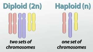
A diploid cell contains two complete sets of chromosomes (2n), with one set inherited from each parent. In humans, diploid somatic cells have 46 chromosomes arranged in 23 homologous pairs.

  • diploid cells divide by mitosis to maintain chromosome number or undergo meiosis to form haploid gametes

  • Mitosis: A 2n diploid cell divides into two identical 2n diploid daughter cells

  • Diploid is represented as 2n (twice the haploid n) number found in gametes

  • a cell that contains two complete sets of chromosomes (2n), with one set inherited from each biological parent. In humans, diploid cells have 46 chromosomes arranged in 23 homologous pairs (22 pairs of autosomes and 1 pair of sex chromosomes)

  • Cells that have two sets of chromosomes

<p>A diploid cell contains two complete sets of chromosomes (2n), with one set inherited from each parent. In humans, diploid somatic cells have 46 chromosomes arranged in 23 homologous pairs.</p><ul><li><p>diploid cells divide by mitosis to maintain chromosome number or undergo meiosis to form haploid gametes</p></li><li><p><strong>Mitosis:</strong> A 2n diploid cell divides into two identical 2n diploid daughter cells</p></li><li><p>Diploid is represented as 2n (twice the haploid n) number found in gametes</p></li><li><p>a cell that contains two complete sets of chromosomes (2n), with one set inherited from each biological parent. In humans, diploid cells have 46 chromosomes arranged in 23 homologous pairs (22 pairs of autosomes and 1 pair of sex chromosomes)</p></li><li><p><span style="font-family: Calibri, sans-serif;">Cells that have two sets of chromosomes</span></p></li></ul><p></p>
28
New cards

Diploid (2n)

A cell that contains two complete sets of chromosomes (2n), with one set inherited from each biological parent. In humans, diploid cells have 46 chromosomes arranged in 23 homologous pairs (22 pairs of autosomes and 1 pair of sex chromosomes)

  • Mitosis: A 2n diploid cell divides into two identical 2n diploid daughter cells

  • n = 23


<p>A cell that contains two complete sets of chromosomes (2n), with one set inherited from each biological parent. In humans, diploid cells have 46 chromosomes arranged in 23 homologous pairs (22 pairs of autosomes and 1 pair of sex chromosomes)</p><ul><li><p><strong>Mitosis:</strong> A 2n diploid cell divides into two identical 2n diploid daughter cells</p></li><li><p>n = 23</p></li></ul><p></p>
29
New cards

Haploid

A cell that contains a single set of unpaired chromosomes, designated by the symbol n. Examples include human gametes (sperm and egg cells), which have n = 23 chromosomes, compared to normal diploid (2n) somatic body cells which have 46

  • Represented as n (or 1n), meaning one complete set of chromosomes

  • Created via meiosis, a specialized type of cell division that reduces a diploid (2n) parent cell down to haploid (n) daughter cells


<p>A cell that contains a single set of unpaired chromosomes, designated by the symbol <strong>n</strong>. Examples include human gametes (sperm and egg cells), which have <strong>n = 23</strong> chromosomes, compared to normal diploid (<strong>2n</strong>) somatic body cells which have <strong>46</strong></p><ul><li><p>Represented as <strong>n</strong> (or <strong>1n</strong>), meaning one complete set of chromosomes</p></li><li><p>Created via <strong>meiosis</strong>, a specialized type of cell division that reduces a diploid (<strong>2n</strong>) parent cell down to haploid (<strong>n</strong>) daughter cells</p></li></ul><p></p>
30
New cards

Haploid

A haploid cell contains a single set of unpaired chromosomes, designated as n. Haploid cells include gametes (sperm and egg cells) and plant spores. They are produced through meiosis, which cuts the chromosome number of a diploid (2n) parent cell in half

  • a cell that contains a single set of unpaired chromosomes, denoted as n (or 1n). For example, human gametes (sperm and egg cells) are haploid and contain 23 total chromosomes, compared to diploid (2n) somatic cells which have 46

  • Cells that have only one set of chromosomes are termed haploid (n).  Sex cells, or gametes, are haploid


<p>A haploid cell contains a single set of unpaired chromosomes, designated as n. Haploid cells include gametes (sperm and egg cells) and plant spores. They are produced through meiosis, which cuts the chromosome number of a diploid (2n) parent cell in half</p><ul><li><p>a cell that contains a single set of unpaired chromosomes, denoted as n (or 1n). For example, human gametes (sperm and egg cells) are haploid and contain 23 total chromosomes, compared to diploid (2n) somatic cells which have 46</p></li><li><p><span>Cells that have only one set of chromosomes are termed <strong>haploid</strong> (n).&nbsp; Sex cells, or gametes, are haploid</span></p></li></ul><p></p>
31
New cards

Chromosome notation

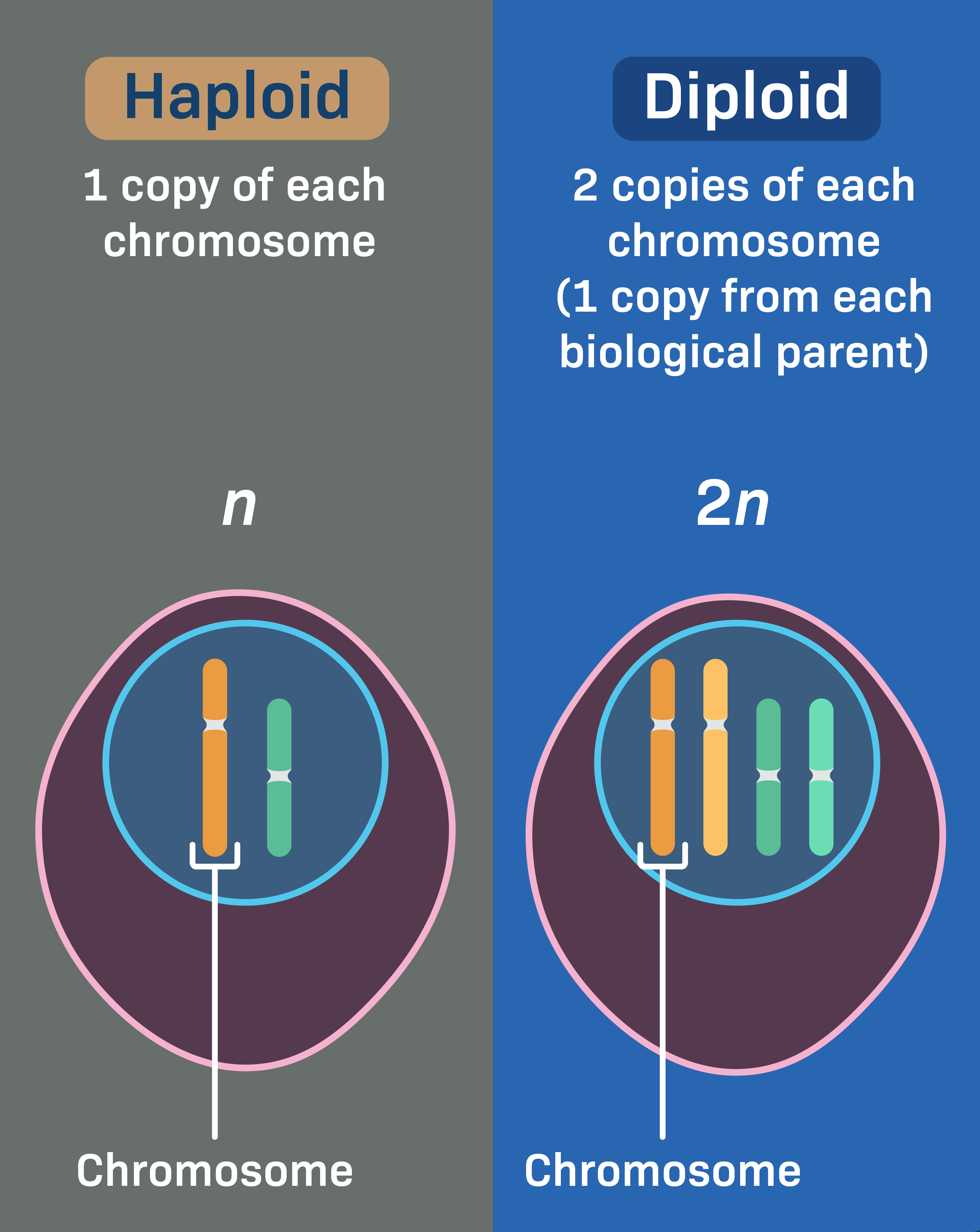
  • n represents the haploid number (one complete set of chromosomes).

  • 2n represents the diploid number (two sets of homologous chromosomes, one from each parent).

  • In humans, n = 23 (gametes) and 2n = 46 (somatic body cells)

  • Cells that have only one set of chromosomes are termed haploid (n).  Sex cells, or gametes, are haploid

  • Cells that have two sets of chromosomes are termed diploid (2n).  Somatic (body) cells are often diploid

<ul><li><p><strong><em>n</em></strong> represents the <strong>haploid number</strong> (one complete set of chromosomes).</p></li><li><p><strong><em>2n</em></strong> represents the <strong>diploid number</strong> (two sets of homologous chromosomes, one from each parent).</p></li><li><p>In humans, n = 23 (gametes) and 2n = 46 (somatic body cells)</p></li><li><p><span>Cells that have only one set of chromosomes are termed <strong>haploid</strong> (n).&nbsp; Sex cells, or gametes, are haploid</span></p></li><li><p><span style="font-family: Calibri, sans-serif;">Cells that have two sets of chromosomes are termed <strong>diploid (2n).</strong>&nbsp; Somatic (body) cells are often diploid</span></p></li></ul><p></p>
32
New cards

Mitosis

A type of nuclear division in eukaryotic cells where duplicated chromosomes are separated into two identical nuclei, resulting in two daughter cells with the exact same chromosome number and genetic makeup as the parent cell

<p>A type of nuclear division in eukaryotic cells where duplicated chromosomes are separated into two identical nuclei, resulting in two daughter cells with the exact same chromosome number and genetic makeup as the parent cell</p>
33
New cards

Homologous chromosomes

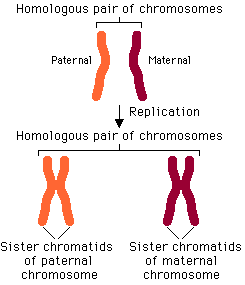
Matched pairs of chromosomes in diploid cells—one inherited from the mother and one from the father—that share the same length, shape, centromere position, and gene loci, though they may carry different versions (alleles) of those genes

  • a matched pair of chromosomes—one inherited from the mother and one from the father—that share the same length, centromere position, and gene loci, though they may carry different alleles

<p>Matched pairs of chromosomes in diploid cells—one inherited from the mother and one from the father—that share the same length, shape, centromere position, and gene loci, though they may carry different versions (<strong>alleles</strong>) of those genes</p><ul><li><p>a matched pair of chromosomes—one inherited from the mother and one from the father—that share the same length, centromere position, and gene loci, though they may carry different alleles</p></li></ul><p></p>
34
New cards

Gametes

Gametes are haploid (1n) reproductive sex cells (sperm in males, eggs or ova in females) that carry a single set of chromosomes. They transmit genetic information from parents to offspring during sexual reproduction and fuse during fertilization to restore the diploid (2n) state

  • gametes do not have paired chromosomes

  • Haploid cells have 23 chromosomes because they contain a single, unpaired set of chromosomes (n = 23), which is half the normal number found in regular human body cells


<p>Gametes are haploid (1n) reproductive sex cells (sperm in males, eggs or ova in females) that carry a single set of chromosomes. They transmit genetic information from parents to offspring during sexual reproduction and fuse during fertilization to restore the diploid (2n) state</p><ul><li><p><strong>gametes</strong> do not have paired chromosomes</p></li><li><p><span>Haploid cells have 23 chromosomes because </span>they contain a single, unpaired set of chromosomes (n = 23), which is half the normal number found in regular human body cells</p></li></ul><p></p>
35
New cards

Prophase and prometaphase

  • Chromatin condenses into duplicated chromosomes

  • The centrosomes move to opposite poles and organize microtubules into spindle fibers

  • The nuclear envelope breaks down. In animal cells, each centrosome contains a pair of centrioles

  • spindle fibers begin to form during prophase


<ul><li><p><span style="background-color: transparent;">Chromatin condenses into duplicated chromosomes</span></p></li></ul><ul><li><p><span style="background-color: transparent;">The centrosomes move to opposite poles and organize microtubules into spindle fibers</span></p></li><li><p><span style="background-color: transparent;">The nuclear envelope breaks down. In animal cells, each centrosome contains a pair of centrioles</span></p></li><li><p><span>spindle fibers begin to form during </span><strong>prophase</strong><br></p></li></ul><p></p>
36
New cards

Metaphase

  • Chromosomes align at the cell’s equator

  • Spindle fibers attach to each chromosome at the centromere

  • a stage of cell division (mitosis and meiosis) where condensed chromosomes align along the imaginary center plane of the cell—known as the metaphase plate or equatorial plane—while spindle fibers attach to their kinetochores

  • M-Phase Checkpoint (Spindle Checkpoint)


<ul><li><p><span style="background-color: transparent;">Chromosomes align at the cell’s equator</span></p></li><li><p><span style="background-color: transparent;">Spindle fibers attach to each chromosome at the centromere</span></p></li><li><p>a stage of cell division (mitosis and meiosis) where condensed chromosomes align along the imaginary center plane of the cell—known as the <strong>metaphase plate</strong> or equatorial plane—while spindle fibers attach to their kinetochores</p></li><li><p>M-Phase Checkpoint (Spindle Checkpoint)</p></li></ul><p></p>
37
New cards

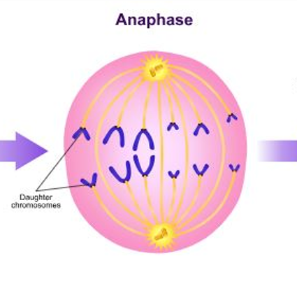
Anaphase

Spindle fibers separate the sister chromatids and pull the daughter chromosomes toward opposite poles

  • the stage of cell division where spindle fibers pull chromosomes toward opposite poles of the cell. In mitosis and meiosis II, sister chromatids separate into individual chromosomes

  • the stage of mitosis where sister chromatids separate at their centromeres and move toward opposite poles of the cell

<p><span style="background-color: transparent;">Spindle fibers separate the sister chromatids and pull the daughter chromosomes toward opposite poles</span></p><ul><li><p>the stage of cell division where spindle fibers pull chromosomes toward opposite poles of the cell. In mitosis and meiosis II, sister chromatids separate into individual chromosomes</p></li><li><p>the stage of mitosis where <strong>sister chromatids</strong> separate at their centromeres and move toward opposite poles of the cell</p></li></ul><p></p>
38
New cards

Telophase

  • Chromosomes uncoil into chromatin

  • spindle fibers disassemble

  • the nuclear envelopes and nucleoli reform

  • the final stage of mitosis where two new nuclear envelopes form around the separated sets of chromosomes at opposite poles of the cell, the chromosomes decondense into chromatin, nucleoli reappear, and the mitotic spindle breaks down, directly preceding cytokinesis


<ul><li><p><span style="background-color: transparent;">Chromosomes uncoil into chromatin</span></p></li><li><p><span style="background-color: transparent;">spindle fibers disassemble</span></p></li><li><p><span style="background-color: transparent;">the nuclear envelopes and nucleoli reform</span></p></li><li><p>the final stage of mitosis where two new nuclear envelopes form around the separated sets of chromosomes at opposite poles of the cell, the chromosomes decondense into chromatin, nucleoli reappear, and the mitotic spindle breaks down, directly preceding cytokinesis</p></li></ul><p></p>
39
New cards

Cytokinesis

The physical division of the cytoplasm that follows nuclear division (mitosis or meiosis), splitting a single eukaryotic cell into two distinct, genetically identical daughter cells

  • Cytokinesis in Animal Cells: The plasma membrane pinches inward, forming a cleavage furrow that divides the cytoplasm.

  • Cytokinesis in Plant Cells: Polysaccharide-containing vesicles gather at the cell’s equator and form a cell plate. Each daughter cell then builds a new cell wall along the plate.


40
New cards

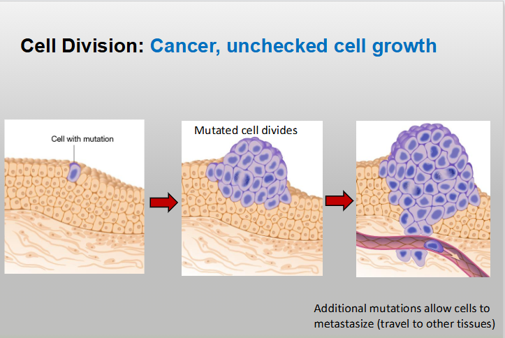
Cancer

A disease caused by uncontrolled cell division that happens when cell cycle checkpoints and regulatory proteins fail to halt the cycle, typically due to accumulated DNA mutations

  • A diease of uncontrolled cell division caused by the failure of cell cycle checkpoints and regulatory proteins

<p>A disease caused by <strong>uncontrolled cell division</strong> that happens when cell cycle checkpoints and regulatory proteins fail to halt the cycle, typically due to accumulated DNA mutations</p><ul><li><p>A diease of <strong>uncontrolled cell division</strong> caused by the failure of cell cycle checkpoints and regulatory proteins</p></li></ul><p></p>
41
New cards

Chromosomes

A cellular structure composed of a single, continuous double-stranded molecule of DNA wrapped around specialized proteins called histones

  • a thread-like, gene-carrying structure found in the nucleus of eukaryotic cells, consisting of a single long molecule of DNA tightly wound and coiled around proteins called histones


<p>A cellular structure composed of a single, continuous double-stranded molecule of DNA wrapped around specialized proteins called histones</p><ul><li><p>a thread-like, gene-carrying structure found in the nucleus of eukaryotic cells, consisting of a single long molecule of DNA tightly wound and coiled around proteins called <strong>histones</strong></p></li></ul><p></p>
42
New cards

Homologous chromosomes

A matching pair of chromosomes—one inherited from the mother (maternal) and one inherited from the father (paternal)—that are similar in length, centromere position, and gene sequence

  • matched pairs of chromosomes—one inherited from the mother and one from the father—that have the same length, shape, and gene sequence


<p>A matching pair of chromosomes—one inherited from the mother (maternal) and one inherited from the father (paternal)—that are similar in length, centromere position, and gene sequence</p><ul><li><p><span>matched pairs of chromosomes—one inherited from the mother and one from the father—that have the same length, shape, and gene sequence</span></p></li></ul><p></p>
43
New cards

Sister chromatids

Two identical copies of a single replicated chromosome that are joined together at a region called the centromere

  • They form during the S phase (Synthesis phase) of interphase when a chromosome makes an exact copy of its DNA

  • They are held tightly together by protein complexes called cohesins

  • Each chromatid contains a protein structure called a kinetochore at the centromere where spindle fibers attach

  • As long as they stay attached at the centromere, they count as one chromosome (though duplicated). Once separated, each is counted as an independent chromosome


<p>Two identical copies of a single replicated chromosome that are joined together at a region called the <strong>centromere</strong></p><ul><li><p>They form during the <strong>S phase (Synthesis phase)</strong> of interphase when a chromosome makes an exact copy of its DNA</p></li><li><p><span>They are held tightly together by protein complexes called <strong>cohesins</strong></span></p></li><li><p><span>Each chromatid contains a protein structure called a <strong>kinetochore</strong> at the centromere where spindle fibers attach</span></p></li><li><p><span>As long as they stay attached at the centromere, they count as <strong>one</strong> chromosome (though duplicated). Once separated, each is counted as an independent chromosome</span></p></li></ul><p></p>
44
New cards

Cohesin

A ring-shaped protein complex that holds replicated sister chromatids together from the time they are duplicated in S phase until they separate during mitosis or meiosis

<p>A ring-shaped protein complex that holds replicated sister chromatids together from the time they are duplicated in S phase until they separate during mitosis or meiosis</p>
45
New cards

Kinetochore

A specialized protein complex that assembles on the centromere of a chromosome during cell division

  • Its primary function is to anchor the chromosome to the spindle apparatus so that sister chromatids (or homologous chromosomes) can be accurately separated into daughter cells

  • a specialized protein structure that assembles at the centromere of each sister chromatid during cell division (mitosis and meiosis)


<p>A specialized protein complex that assembles on the centromere of a chromosome during cell division</p><ul><li><p>Its primary function is to anchor the chromosome to the spindle apparatus so that sister chromatids (or homologous chromosomes) can be accurately separated into daughter cells</p></li><li><p>a specialized protein structure that assembles at the centromere of each sister chromatid during cell division (mitosis and meiosis)</p></li></ul><p></p>
46
New cards

Types of cells

  • Somatic (diploid): 2 sets of chromosomes, diploid (2n)

  • Gametes (haploid): egg and sperm cells, half the number of chromosomes: haploid (1n)


47
New cards

Heterologous chromosomes

(often called non-homologous chromosomes) are chromosomes that belong to different pairs and do not share the same gene sequences, size, shape, or centromere positions

  • In humans X and Y chromosomes are not homologous
    (these are heterologous)

  • chromosomes that belong to different pairs and contain different gene sequences, lengths, and centromere positions


<p>(often called non-homologous chromosomes) are chromosomes that belong to different pairs and do not share the same gene sequences, size, shape, or centromere positions</p><ul><li><p><span>In humans X and Y chromosomes are not homologous<br>(these are heterologous)</span></p></li><li><p>chromosomes that belong to different pairs and contain <strong>different gene sequences</strong>, lengths, and centromere positions</p></li></ul><p></p>
48
New cards

Nucleosome

The basic structural unit of chromatin. It is made up of a coil of DNA wound around a histone core

  • The fundamental, repeating structural unit of chromatin (the complex of DNA and protein found inside a cell's nucleus). It looks like a small bead on a string under a microscope

  • The histone-DNA complex is called nucleosome

  • multiple nucleosomes make up a chromatin fiber


<p>The basic structural unit of chromatin. It is made up of a coil of DNA wound around a histone core</p><ul><li><p>The fundamental, repeating structural unit of chromatin<span> (the complex of DNA and protein found inside a cell's nucleus). It looks like a small bead on a string under a microscope</span></p></li></ul><ul><li><p><span>The histone-DNA complex is called nucleosome</span></p></li><li><p>multiple nucleosomes make up a chromatin fiber</p></li></ul><p></p>
49
New cards

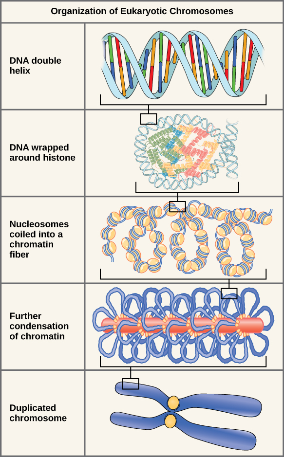
Chromatin

The complex of DNA wrapped around histone proteins inside the nucleus of eukaryotic cells

  • During most of the cell cycle (interphase), DNA exists as diffuse, stringy chromatin. Right before cell division (mitosis/meiosis), chromatin coils and condenses tightly into visible, distinct chromosomes

  • Nucleosomes form the building blocks that make up chromatin

  • Chromatin: nucleosomes coiled into a fiber

<p>The complex of DNA wrapped around histone proteins inside the nucleus of eukaryotic cells</p><ul><li><p>During most of the cell cycle (interphase), DNA exists as diffuse, stringy chromatin. Right before cell division (mitosis/meiosis), chromatin coils and condenses tightly into visible, distinct chromosomes</p></li><li><p>Nucleosomes form the building blocks that make up chromatin</p></li><li><p>Chromatin: nucleosomes coiled into a fiber</p></li></ul><p></p>
50
New cards

Cell cycle

The ordered sequence of growth and division that a eukaryotic cell undergoes to produce two genetically identical daughter cells

  • A series of events in the life of every cell


<p>The ordered sequence of growth and division that a eukaryotic cell undergoes to produce two genetically identical daughter cells</p><ul><li><p>A series of events in the life of every cell</p></li></ul><p></p>
51
New cards

The cell cycle has 2 major phases

  • Interphase (blue) is the cell’s normal growth and preparation for cell division

  • Mitotic phase (red): the DNA and cytoplasm are split and the cell divides


<ul><li><p><span>Interphase (blue) is the cell’s normal growth and preparation for cell division</span></p></li><li><p><span>Mitotic phase (red): the DNA and cytoplasm are split and the cell divides</span></p></li></ul><p></p>
52
New cards

Apoptosis

The process of programmed cell death (cellular suicide) in which a cell systematically breaks itself down in a controlled, orderly manner

  • Serves as a crucial safety control. When cell cycle checkpoints (like the G1 checkpoint) detect unrepairable DNA damage, the cell undergoes apoptosis instead of dividing uncontrollably (which leads to cancer)

  • Apoptosis (programmed cell death) can occur at any phase of interphase (G₁, S, or G₂), depending on the cell type and the nature of the cellular stress or trigger, though it is most frequently executed or decided upon during the G₁/S or G₂M transition checkpoints


<p>The process of programmed cell death (cellular suicide) in which a cell systematically breaks itself down in a controlled, orderly manner</p><ul><li><p>Serves as a crucial safety control. When cell cycle checkpoints (like the G1 checkpoint) detect unrepairable DNA damage, the cell undergoes apoptosis instead of dividing uncontrollably (which leads to cancer)</p></li><li><p><span>Apoptosis (programmed cell death) can occur at </span>any phase of interphase (G₁, S, or G₂), depending on the cell type and the nature of the cellular stress or trigger, though it is most frequently executed or decided upon during the G₁/S or G₂M transition checkpoints</p></li></ul><p></p>
53
New cards

Interphase

The longest part of the eukaryotic cell cycle (accounting for about 90% of the cell's life) during which the cell grows, performs its normal metabolic functions, duplicates its organelles, and replicates its DNA in preparation for cell division

Interphase occurs in 3 stages

  • G1 phase (first gap)

  • S phase

  • G2 phase (second gap)

  • A cell in interphase looks large and intact, with a clearly visible, spherical nucleus surrounded by a nuclear envelope. Under a microscope, the genetic material inside the nucleus does not look like distinct chromosomes; instead, it appears as a loose, grainy, or stringy mass called chromatin


<p>The longest part of the eukaryotic cell cycle (accounting for about 90% of the cell's life) during which the cell grows, performs its normal metabolic functions, duplicates its organelles, and replicates its DNA in preparation for cell division</p><p>Interphase occurs in 3 stages</p><ul><li><p>G1 phase (first gap)</p></li><li><p>S phase</p></li><li><p>G2 phase (second gap)</p></li><li><p><span>A cell in interphase looks </span>large and intact, with a clearly visible, spherical nucleus surrounded by a nuclear envelope. Under a microscope, the genetic material inside the nucleus does not look like distinct chromosomes; instead, it appears as a loose, grainy, or stringy mass called chromatin</p></li></ul><p></p>
54
New cards

S phase

The period of interphase in the eukaryotic cell cycle where the cell replicates its entire genome

  • the period during interphase in the cell cycle when the cell makes a complete copy of all its nuclear DNA


<p>The period of <span><strong>interphase</strong></span> in the eukaryotic cell cycle where the cell replicates its entire genome</p><ul><li><p>the period during <strong>interphase</strong> in the cell cycle when the cell makes a complete copy of all its nuclear DNA</p></li></ul><p></p>
55
New cards

G1 phase

(Gap 1 or First Growth phase) is the first stage of interphase in the eukaryotic cell cycle, occurring right after mitosis and cytokinesis and before the S (synthesis) phase

  • Cell grows and performs normal functions

  • Cellular Growth: The cell physically increases in size, volume, and mass

  • Protein and Organelle Synthesis: The cell makes new proteins, accumulates ATP and nutrients, and duplicates non-nuclear organelles (such as mitochondria and ribosomes)

  • Normal Metabolic Activity: The cell carries out its specialized everyday functions

  • Preperation for DNA replication


<p>(Gap 1 or First Growth phase) is the first stage of interphase in the eukaryotic cell cycle, occurring right after mitosis and cytokinesis and before the S (synthesis) phase</p><ul><li><p>Cell grows and performs normal functions</p></li><li><p><strong>Cellular Growth:</strong> The cell physically increases in size, volume, and mass</p></li><li><p><span><strong>Protein and Organelle Synthesis:</strong> The cell makes new proteins, accumulates ATP and nutrients, and duplicates non-nuclear organelles (such as mitochondria and ribosomes)</span></p></li><li><p><span><strong>Normal Metabolic Activity:</strong> The cell carries out its specialized everyday functions</span></p></li><li><p>Preperation for DNA replication</p></li></ul><p></p>
56
New cards

G2 phase

Cell gets ready for mitosis

  • Makes proteins needed for mitosis

  • Produces new organelles

  • Checks DNA damage


<p>Cell gets ready for mitosis</p><ul><li><p><span>Makes proteins needed for mitosis</span></p></li><li><p><span>Produces new organelles</span></p></li><li><p><span>Checks DNA damage</span></p></li></ul><p></p>
57
New cards

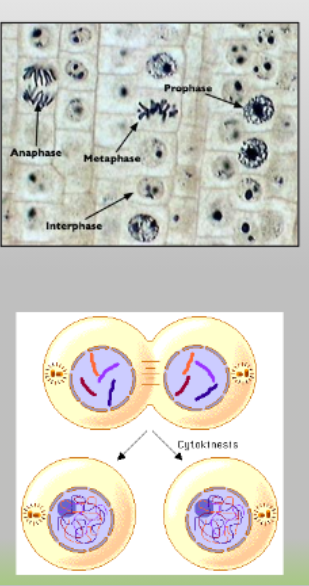
The mitotic phase involves several steps

  • Mitosis is the first step

  • The second step is called cytokinesis, when the cytoplasm and the organelles are divided into two new cells

  • The mitotic (M) phase consists of two main processes: mitosis (nuclear division) and cytokinesis (cytoplasm division), producing two genetically identical diploid daughter cells

<ul><li><p><span>Mitosis is the first step</span></p></li><li><p><span>The second step is called cytokinesis, when the cytoplasm and the organelles are divided into two new cells</span></p></li><li><p><span>The mitotic (M) phase consists of two main processes: </span>mitosis (nuclear division) and cytokinesis (cytoplasm division), producing two genetically identical diploid daughter cells</p></li></ul><p></p>
58
New cards

Prophase

  • Chromosomes condense (the tight coiling and compacting of loose chromatin fibers into short, distinct, rod-shaped chromosomes during the start of cell division)

  • The nucleus membrane breaks down

  • The organelles move toward edges of the cell

  • Centrosomes move to the poles of the cell

  • The spindle microtubules form


<ul><li><p>Chromosomes condense (the tight coiling and compacting of loose chromatin fibers into short, distinct, rod-shaped chromosomes during the start of cell division)</p></li><li><p>The nucleus membrane breaks down</p></li><li><p>The organelles move toward edges of the cell</p></li><li><p>Centrosomes move to the poles of the cell</p></li><li><p>The spindle microtubules form</p></li></ul><p></p>
59
New cards

Prophase

  • Chromosomes condense (the tight coiling and compacting of loose chromatin fibers into short, distinct, rod-shaped chromosomes during the start of cell division)

  • The nucleus membrane breaks down

  • The organelles move toward edges of the cell

  • Centrosomes move to the poles of the cell

  • The spindle microtubules form


<ul><li><p>Chromosomes condense (the tight coiling and compacting of loose chromatin fibers into short, distinct, rod-shaped chromosomes during the start of cell division)</p></li><li><p>The nucleus membrane breaks down</p></li><li><p>The organelles move toward edges of the cell</p></li><li><p>Centrosomes move to the poles of the cell</p></li><li><p>The spindle microtubules form</p></li></ul><p></p>
60
New cards

Prophase: spindle microtubules form

Spindle fibers are made of microtubules, which are long, hollow protein cylinders

  • Spindle fibers (also known as the mitotic or meiotic spindle) are built entirely out of long, dynamic polymers of protein called microtubules

  • they grow outward from the centrosomes or spindle pole bodies located at opposite ends of the cell during cell division (mitosis and meiosis)

  • Kinetochore microtubules: attach directly to the chromosomes to pull them apart


<p><span>Spindle fibers are made of </span>microtubules, which are long, hollow protein cylinders</p><ul><li><p>Spindle fibers (also known as the mitotic or meiotic spindle) are built entirely out of long, dynamic polymers of protein called <strong>microtubules</strong></p></li></ul><ul><li><p>they grow outward from the centrosomes or spindle pole bodies located at opposite ends of the cell during cell division (mitosis and meiosis)</p></li><li><p><strong>Kinetochore microtubules:</strong> attach directly to the chromosomes to pull them apart</p></li></ul><p></p>
61
New cards

Prophase - kinetochore forms

Sister chromatids form a protein called kinetochore in the centromere region which attaches the chromatids to the spindle fibers during prophase

  • the kinetochore assembles during prophase (and finishes forming by prometaphase), right as the nuclear envelope breaks down


<p><span>Sister chromatids form a protein called kinetochore in the centromere region which attaches the chromatids to the spindle fibers during prophase</span></p><ul><li><p><span>the kinetochore assembles </span>during prophase (and finishes forming by prometaphase), right as the nuclear envelope breaks down</p></li></ul><p></p>
62
New cards

Metaphase

The stage of mitosis where the cell’s condensed, duplicated chromosomes align single-file along the metaphase plate (the equator or middle plane of the cell)

  • Chromosomes line up in the middle (metaphase plate)

  • Sister chromatids remain attached

  • Spindle Attachment: Kinetochore microtubules from the mitotic spindle attach to the protein complexes (kinetochores) at the centromere of each sister chromatid


<p>The stage of mitosis where the cell’s condensed, duplicated chromosomes align single-file along the <strong>metaphase plate</strong> (the equator or middle plane of the cell)</p><ul><li><p><span>Chromosomes line up in the middle (metaphase plate)</span></p></li><li><p><span>Sister chromatids remain attached</span></p></li><li><p><strong>Spindle Attachment:</strong> Kinetochore microtubules from the mitotic spindle attach to the protein complexes (kinetochores) at the centromere of each sister chromatid</p></li></ul><p></p>
63
New cards

Metaphase

The stage of mitosis where the cell’s condensed, duplicated chromosomes align single-file along the metaphase plate (the equator or middle plane of the cell)

  • Chromosomes line up in the middle (metaphase plate)

  • Sister chromatids remain attached

  • Spindle Attachment: Kinetochore microtubules from the mitotic spindle attach to the protein complexes (kinetochores) at the centromere of each sister chromatid


<p>The stage of mitosis where the cell’s condensed, duplicated chromosomes align single-file along the <strong>metaphase plate</strong> (the equator or middle plane of the cell)</p><ul><li><p>Chromosomes line up in the middle (metaphase plate)</p></li><li><p>Sister chromatids remain attached</p></li><li><p><strong>Spindle Attachment:</strong> Kinetochore microtubules from the mitotic spindle attach to the protein complexes (kinetochores) at the centromere of each sister chromatid</p></li></ul><p></p>
64
New cards

Anaphase

The stage of mitosis where sister chromatids separate at the centromere and are pulled by spindle fibers toward opposite poles (ends) of the cell

  • Cohesin proteins break down allowing the chromatids to separate

  • Sister chromatids move in opposite directions toward the centrosomes to which their microtubules are attached

  • The cell elongates


<p>The stage of mitosis where sister chromatids separate at the centromere and are pulled by spindle fibers toward opposite poles (ends) of the cell</p><ul><li><p><span>Cohesin proteins break down allowing the chromatids to separate</span></p></li><li><p><span>Sister chromatids move in opposite directions toward the centrosomes to which their microtubules are attached</span></p></li><li><p><span>The cell elongates</span></p></li></ul><p></p>
65
New cards

Anaphase

The stage of mitosis where sister chromatids separate at their centromeres and are pulled by spindle fibers toward opposite poles of the cell

  • Cohesin proteins break down allowing the chromatids to separate

  • Sister chromatids move in opposite directions toward the centrosomes to which their microtubules are attached

  • The cell elongates


66
New cards

During which stage does cohesin break down?

Cohesin breaks down during anaphase of mitosis

  • Cohesin proteins break down allowing the chromatids to separate

  • a ring-shaped multi-protein complex that holds replicated sister chromatids together from the time they are synthesized during DNA replication (S phase) until they separate


67
New cards

Telophase

The final stage of mitosis, during which the genetic material has reached opposite poles of the cell and two new nuclei begin to form. It essentially reverses the structural changes that occurred during prophase and prometaphase

  • Chromosomes reach poles and become less dense

  • Spindles break into tubulin monomers

  • Nuclear membrane forms around chromosomes


<p>The final stage of mitosis, during which the genetic material has reached opposite poles of the cell and two new nuclei begin to form. It essentially reverses the structural changes that occurred during prophase and prometaphase</p><ul><li><p><span>Chromosomes reach poles and become less dense</span></p></li><li><p><span>Spindles break into tubulin monomers</span></p></li><li><p><span>Nuclear membrane forms around chromosomes</span></p></li></ul><p></p>
68
New cards

Telophase

The final phase of mitosis where two new nuclear envelopes form around the separated sets of chromosomes at opposite poles of the cell, and the chromosomes begin to decondense back into chromatin

  • Chromosomes reach poles and become less dense

  • Spindles break into tubulin monomers

  • Nuclear membrane forms around chromosomes


<p>The final phase of mitosis where two new nuclear envelopes form around the separated sets of chromosomes at opposite poles of the cell, and the chromosomes begin to decondense back into chromatin</p><ul><li><p>Chromosomes reach poles and become less dense</p></li><li><p>Spindles break into tubulin monomers</p></li><li><p>Nuclear membrane forms around chromosomes</p></li></ul><p></p>
69
New cards

Cytokinesis is different between plants and animals

  • Animal: forms cleavage furrow

  • Plants in cytokinesis form a cell plate


<ul><li><p><span>Animal: forms cleavage furrow</span></p></li><li><p>Plants in cytokinesis form a cell plate</p></li></ul><p></p>
70
New cards

Cytokinesis

The physical process of cell division that splits the cytoplasm, organelles, and plasma membrane of a parent cell into two distinct daughter cells

  • the physical division of the cytoplasm that splits a single eukaryotic cell into two distinct daughter cells. It occurs at the end of cell division, usually starting during anaphase or telophase right after nuclear division (mitosis or meiosis) is complete

  • Animal: forms cleavage furrow

  • Plants in cytokinesis form a cell plate
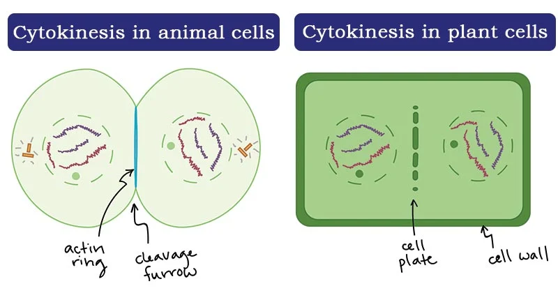

<p>The physical process of cell division that splits the cytoplasm, organelles, and plasma membrane of a parent cell into two distinct daughter cells</p><ul><li><p><mark>the physical division of the cytoplasm that splits a single eukaryotic cell into two distinct daughter cells</mark>. It occurs at the end of cell division, usually starting during anaphase or telophase right after nuclear division (mitosis or meiosis) is complete</p></li></ul><ul><li><p>Animal: forms cleavage furrow</p></li><li><p>Plants in cytokinesis form a cell plate</p></li></ul><p></p>
71
New cards

Cytokinesis in animal cells

The physical division of the cytoplasm that splits a single eukaryotic cell into two separate, genetically identical daughter cells

  • Unlike plant cells (which must build a rigid cell wall), animal cells have a flexible plasma membrane that pinches inward. This inward groove is called a cleavage furrow

  • Cleavage Furrow: a shallow groove or indentation that appears on the surface of an animal cell as the contractile ring tightens (acting like a drawstring) and pulls the plasma membrane inward


<p>The physical division of the cytoplasm that splits a single eukaryotic cell into two separate, genetically identical daughter cells</p><ul><li><p>Unlike plant cells (which must build a rigid cell wall), animal cells have a flexible plasma membrane that pinches inward. This inward groove is called a <strong>cleavage furrow</strong></p></li><li><p><strong>Cleavage Furrow:</strong> a shallow groove or indentation that appears on the surface of an animal cell as the contractile ring tightens (acting like a drawstring) and pulls the plasma membrane inward</p></li></ul><p></p>
72
New cards

Cleavage furrow

A shallow groove or indentation that forms in the plasma membrane of an animal cell during cytokinesis (the final stage of cell division)

  • It is driven by a contractile ring composed of microfilaments (specifically actin) and myosin protein fibers located just beneath the plasma membrane

  • As the actin and myosin filaments slide past each other and the ring contracts, it squeezes the equator of the cell inward, acting like a tight drawstring until the parent cell pinches completely into two separate daughter cells
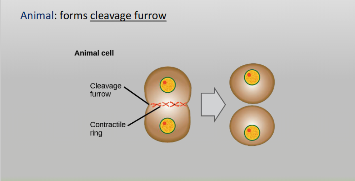

<p>A shallow groove or indentation that forms in the plasma membrane of an animal cell during <strong>cytokinesis</strong> (the final stage of cell division)</p><ul><li><p>It is driven by a <strong>contractile ring</strong> composed of <strong>microfilaments</strong> (specifically <strong>actin</strong>) and <strong>myosin</strong> protein fibers located just beneath the plasma membrane</p></li><li><p>As the actin and myosin filaments slide past each other and the ring contracts, it squeezes the equator of the cell inward, acting like a tight drawstring until the parent cell pinches completely into two separate daughter cells</p></li></ul><p></p>
73
New cards

Cytokinesis in plant cells

The physical division of the cytoplasm and plasma membrane that occurs after mitosis (typically starting in telophase)

  • Unlike animal cells—which pinch inward using a contractile ring—plant cells cannot pinch inward because they possess a rigid, structural cell wall. Instead, they build a new cell wall from the inside out

  • Plant: forms cell plate

  • plant cells form a cell plate during cytokinesis that grows outward and eventually becomes the new cell wall


<p>The physical division of the cytoplasm and plasma membrane that occurs after mitosis (typically starting in telophase)</p><ul><li><p>Unlike animal cells—which pinch inward using a contractile ring—plant cells <strong>cannot pinch inward</strong> because they possess a rigid, structural cell wall. Instead, they build a new cell wall from the inside out</p></li><li><p><span>Plant: forms cell plate</span></p></li><li><p>plant cells form a cell plate during cytokinesis that grows outward and eventually becomes the new cell wall</p></li></ul><p></p>
74
New cards

Cell plate

A disk-like structure that forms in the middle of a dividing plant cell during cytokinesis (the division of the cytoplasm)

  • It fuses with the plasma membrane and develops into a new cell wall, physically separating the two new genetically identical daughter cells

  • plant cells form a cell plate during cytokinesis that grows outward and eventually becomes the new cell wall

  • Plants: form a cell plate


<p>A disk-like structure that forms in the middle of a dividing plant cell during cytokinesis (the division of the cytoplasm)</p><ul><li><p>It fuses with the plasma membrane and develops into a new <strong>cell wall</strong>, physically separating the two new genetically identical daughter cells</p></li><li><p>plant cells form a cell plate during cytokinesis that grows outward and eventually becomes the new cell wall</p></li><li><p>Plants: form a cell plate</p></li></ul><p></p>
75
New cards

Prometaphase

The transitional phase of mitosis that occurs between prophase and metaphase

  • Chromosomes continue to condense

  • Kinetochores appear at the centromeres

  • Mitotic spindle microtubules attach to kinetochores

  • Centrosomes move toward opposite poles

  • Kinetochores appear during prometaphase (the second phase of mitosis), though some assembly begins late in prophase
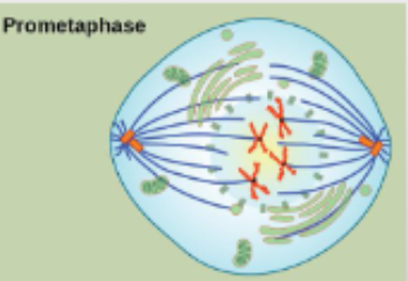

<p>The transitional phase of mitosis that occurs between prophase and metaphase</p><ul><li><p>Chromosomes continue to condense</p></li><li><p>Kinetochores appear at the centromeres</p></li><li><p>Mitotic spindle microtubules attach to kinetochores</p></li><li><p>Centrosomes move toward opposite poles</p></li><li><p><span>Kinetochores appear during </span>prometaphase (the second phase of mitosis), though some assembly begins late in prophase</p></li></ul><p></p>
76
New cards

Prometaphase

The transitional phase of mitosis that occurs between prophase and metaphase

  • Chromosomes continue to condense

  • Kinetochores appear at the centromeres

  • Mitotic spindle microtubules attach to kinetochores

  • Centrosomes move toward opposite poles


<p>The transitional phase of mitosis that occurs between prophase and metaphase</p><ul><li><p>Chromosomes continue to condense</p></li><li><p>Kinetochores appear at the centromeres </p></li><li><p>Mitotic spindle microtubules attach to kinetochores </p></li><li><p>Centrosomes move toward opposite poles</p></li></ul><p></p>
77
New cards

Chromosomes condense

The process of tightly winding and packaging loose, thread-like chromatin (DNA wrapped around histone proteins) into compact, distinct, rod-shaped chromosomes

  • Prophase (of mitosis and meiosis I/II): Chromatin begins to condense and becomes visible under a light microscope

  • Telophase: The reverse happens (decondensation), where chromosomes unwind back into loose chromatin so the cell can access genes for daily functions


<p>The process of tightly winding and packaging loose, thread-like chromatin (DNA wrapped around histone proteins) into compact, distinct, rod-shaped chromosomes</p><ul><li><p><span><strong>Prophase</strong> (of mitosis and meiosis I/II): Chromatin begins to condense and becomes visible under a light microscope</span></p></li><li><p><span><strong>Telophase</strong>: The reverse happens (<em>decondensation</em>), where chromosomes unwind back into loose chromatin so the cell can access genes for daily functions</span></p></li></ul><p></p>
78
New cards

Chromosomes condense

The process of tightly winding and packaging loose, thread-like chromatin (DNA wrapped around histone proteins) into compact, distinct, rod-shaped chromosomes

  • Prophase (of mitosis and meiosis I/II): Chromatin begins to condense and becomes visible under a light microscope

  • Telophase: The reverse happens (decondensation), where chromosomes unwind back into loose chromatin so the cell can access genes for daily functions


<p>The process of tightly winding and packaging loose, thread-like chromatin (DNA wrapped around histone proteins) into compact, distinct, rod-shaped chromosomes</p><ul><li><p><strong>Prophase</strong> (of mitosis and meiosis I/II): Chromatin begins to condense and becomes visible under a light microscope</p></li><li><p><strong>Telophase</strong>: The reverse happens (<em>decondensation</em>), where chromosomes unwind back into loose chromatin so the cell can access genes for daily functions</p></li></ul><p></p>
79
New cards

Chromosomes decondense

The tightly packed, thread-like structures of DNA and proteins (chromosomes) uncoil and relax back into a loose, diffuse form known as chromatin

  • Loose, uncoiled, and looks like spaghetti strands

  • Interphase & Telophase


<p>The tightly packed, thread-like structures of DNA and proteins (chromosomes) <strong>uncoil and relax</strong> back into a loose, diffuse form known as <strong>chromatin</strong></p><ul><li><p>Loose, uncoiled, and looks like spaghetti strands</p></li><li><p>Interphase &amp; Telophase</p></li></ul><p></p>
80
New cards

End product of mitosis

The end product of mitosis (along with cytokinesis) is two genetically identical diploid (2n) daughter cells

  • Cell Type: Somatic (body) cells

  • Chromosome Number: Retains the same diploid (2n) chromosome number as the parent cell (e.g., (2n = 46) in humans)

  • Genetic Content: The daughter cells are exact genetic clones of each other and of the original parent cell

  • Purpose: Growth, tissue repair, regeneration, and asexual reproduction


<p><span>The end product of mitosis (along with cytokinesis) is </span><strong>two genetically identical diploid (2n) daughter cells</strong></p><ul><li><p><span><strong>Cell Type:</strong> Somatic (body) cells</span></p></li><li><p><span><strong>Chromosome Number:</strong> Retains the same diploid (2n) chromosome number as the parent cell (e.g., (2n = 46) in humans)</span></p></li><li><p><span><strong>Genetic Content:</strong> The daughter cells are exact genetic clones of each other and of the original parent cell</span></p></li><li><p><strong>Purpose:</strong> Growth, tissue repair, regeneration, and asexual reproduction</p></li></ul><p></p>
81
New cards

End product of mitosis

The end product of mitosis (along with cytokinesis) is two genetically identical diploid (2n) daughter cells

  • Cell Type: Somatic (body) cells

  • Chromosome Number: Retains the same diploid (2n) chromosome number as the parent cell (e.g., (2n = 46) in humans)

  • Genetic Content: The daughter cells are exact genetic clones of each other and of the original parent cell

  • Purpose: Growth, tissue repair, regeneration, and asexual reproduction


<p>The end product of mitosis (along with cytokinesis) is <strong>two genetically identical diploid (2n) daughter cells</strong></p><ul><li><p><strong>Cell Type:</strong> Somatic (body) cells</p></li><li><p><strong>Chromosome Number:</strong> Retains the same diploid (2n) chromosome number as the parent cell (e.g., (2n = 46) in humans)</p></li><li><p><strong>Genetic Content:</strong> The daughter cells are exact genetic clones of each other and of the original parent cell</p></li><li><p><strong>Purpose:</strong> Growth, tissue repair, regeneration, and asexual reproduction</p></li></ul><p></p>
82
New cards

Mitosis in cells

A. Interphase

B. Prophase

C. Metaphase

D. Anaphase

E. Telophase

F. Cytokinesis
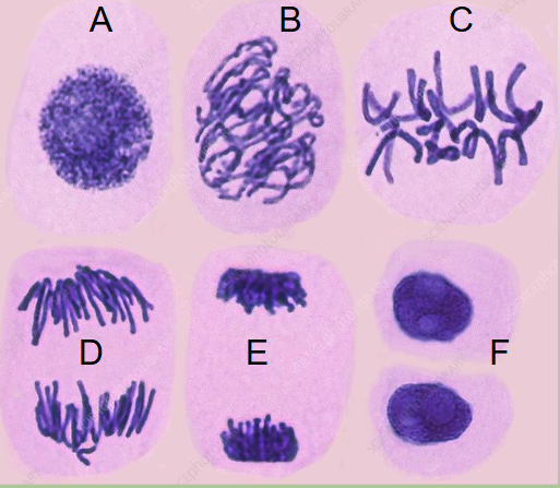
<p>A. Interphase</p><p>B. Prophase</p><p>C. Metaphase</p><p>D. Anaphase</p><p>E. Telophase</p><p>F. Cytokinesis</p>
83
New cards

Functions of cell division

Multicellular organisms use cell division for:

  • Growth

  • Maintenance

  • Repair injured cells and tissues

  • Development

  • Single-celled organisms use cell division to reproduce


84
New cards

Functions of cell division

Multicellular organisms use cell division for:

  • Growth

  • Maintenance

  • Repair injured cells and tissues

  • Development

  • Single-celled organisms use cell division to reproduce


85
New cards

Mitosis in cells

  1. Interphase

  2. Prophase

  3. Metaphase

  4. Anaphase

  5. Telophase

  6. Cytokinesis


<ol><li><p>Interphase</p></li><li><p>Prophase</p></li><li><p>Metaphase</p></li><li><p>Anaphase</p></li><li><p>Telophase</p></li><li><p>Cytokinesis</p></li></ol><p></p>
86
New cards

Cells internal checkpoints regulate their growth

Quality-control stopping points that evaluate a cell’s internal and external conditions before allowing it to advance to the next phase of division

  • G1 checkpoint

  • G2 checkpoint

  • M checkpoint

  • New cell must be identical to the original: fix errors


<p>Quality-control stopping points that evaluate a cell’s internal and external conditions before allowing it to advance to the next phase of division</p><ul><li><p><span>G1 checkpoint</span></p></li><li><p><span>G2 checkpoint</span></p></li><li><p><span>M checkpoint</span></p></li><li><p><span>New cell must be identical to the original: fix errors</span></p></li></ul><p></p>
87
New cards

Regulation at internal checkpoints

Mistakes that affect cell function (wrong number or mutated chromosomes) are regulated at 3 checkpoints:

  1. At end of G1

  2. At G2 to Mitosis transition

  3. In metaphase of mitosis


<p><span>Mistakes that affect cell function (wrong number or mutated chromosomes) are regulated at 3 checkpoints:</span></p><ol><li><p><span>At end of G1</span></p></li><li><p><span>At G2 to Mitosis transition</span></p></li><li><p><span>In metaphase of mitosis</span></p></li></ol><p></p>
88
New cards

Control of the cell cycle

External signals can stimulate or inhibit the cell cycle

  • Death of nearby cells (such as a wound or cut in skin)

  • Release of growth hormones

  • Cell crowding

  • Internal factors also regulate progress of the cell cycle


<p><span>External signals can stimulate or inhibit the cell cycle</span></p><ul><li><p><span>Death of nearby cells (such as a wound or cut in skin)</span></p></li><li><p><span>Release of growth hormones</span></p></li><li><p><span>Cell crowding</span></p></li><li><p><span>Internal factors also regulate progress of the cell cycle</span></p></li></ul><p></p>
89
New cards

Control of the cell cycle

External signals can stimulate or inhibit the cell cycle

  • Death of nearby cells (such as a wound or cut in skin)

  • Release of growth hormones

  • Cell crowding

  • Internal factors also regulate progress of the cell cycle


<p>External signals can stimulate or inhibit the cell cycle</p><ul><li><p>Death of nearby cells (such as a wound or cut in skin)</p></li><li><p>Release of growth hormones</p></li><li><p>Cell crowding</p></li><li><p>Internal factors also regulate progress of the cell cycle</p></li></ul><p></p>
90
New cards

G1 checkpoint

Determines whether all conditions are favorable for cell division

  • External influences, adequate cell reserves and size are important

  • There is a check for DNA damage

  • A cell that does not meet all the requirements does not enter the S phase


<p><span>Determines whether all conditions are favorable for cell division</span></p><ul><li><p><span>External influences, adequate cell reserves and size are important</span></p></li><li><p><span>There is a check for DNA damage</span></p></li><li><p><span> A cell that does not meet all the requirements does not enter the S phase</span></p></li></ul><p></p>
91
New cards

G1 checkpoint

Determines whether all conditions are favorable for cell division

  • External influences, adequate cell reserves and size are important

  • There is a check for DNA damage

  • A cell that does not meet all the requirements does not enter the S phase


<p>Determines whether all conditions are favorable for cell division</p><ul><li><p>External influences, adequate cell reserves and size are important</p></li><li><p>There is a check for DNA damage</p></li><li><p> A cell that does not meet all the requirements does not enter the S phase</p></li></ul><p></p>
92
New cards

G1 checkpoint options

G1 Checkpoint determines whether all conditions are favorable for cell division

The cell has 2 options if conditions aren’t met:

  • Stop the cycle and try to fix theproblem

  • Enter G0 (becomes dormant) and wait for signs that
    conditions are better


<p><span>G1 Checkpoint determines whether all conditions are favorable for cell division</span></p><p><span>The cell has 2 options if conditions aren’t met:</span></p><ul><li><p><span>Stop the cycle and try to fix theproblem</span></p></li><li><p><span>Enter G0 (becomes dormant) and wait for signs that<br>conditions are better</span></p></li></ul><p></p>
93
New cards

G1 checkpoint options

G1 Checkpoint determines whether all conditions are favorable for cell division

The cell has 2 options if conditions aren’t met:

  • Stop the cycle and try to fix theproblem

  • Enter G0 (becomes dormant) and wait for signs that
    conditions are better


<p>G1 Checkpoint determines whether all conditions are favorable for cell division</p><p>The cell has 2 options if conditions aren’t met:</p><ul><li><p>Stop the cycle and try to fix theproblem</p></li><li><p>Enter G0 (becomes dormant) and wait for signs that<br>conditions are better</p></li></ul><p></p>
94
New cards

The G2 checkpoint

  • Cell size and protein reserves are checked

  • G2 checkpoint makes sure that all chromosomes have
    been replicated and that the DNA is not damaged

  • If problems detected, the cell cycle stops while the cell
    completes DNA replication or repairs the damaged DNA


<ul><li><p><span>Cell size and protein reserves are checked</span></p></li><li><p><span>G2 checkpoint makes sure that all chromosomes have<br>been replicated and that the DNA is not damaged</span></p></li><li><p><span>If problems detected, the cell cycle stops while the cell<br>completes DNA replication or repairs the damaged DNA</span></p></li></ul><p></p>
95
New cards

The G2 checkpoint

  • Cell size and protein reserves are checked

  • G2 checkpoint makes sure that all chromosomes have
    been replicated and that the DNA is not damaged

  • If problems detected, the cell cycle stops while the cell
    completes DNA replication or repairs the damaged DNA


<ul><li><p>Cell size and protein reserves are checked</p></li><li><p>G2 checkpoint makes sure that all chromosomes have<br>been replicated and that the DNA is not damaged</p></li><li><p>If problems detected, the cell cycle stops while the cell<br>completes DNA replication or repairs the damaged DNA</p></li></ul><p></p>
96
New cards

The M checkpoint

At the end of metaphase

  • Determines whether all sister chromatids are attached to the spindle

  • The cycle will not proceed until kinetochores of sister
    chromatids are firmly attached to two spindle fibers

  • Failure to correct this, can lead to new cells getting wrong number of chromatids
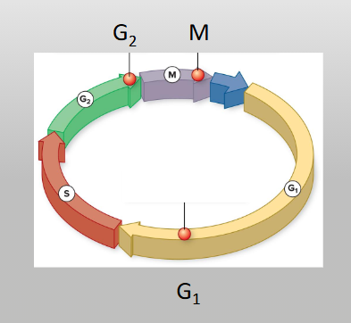

<p><span>At the end of metaphase</span></p><ul><li><p><span>Determines whether all sister chromatids are attached to the spindle</span></p></li><li><p><span>The cycle will not proceed until kinetochores of sister<br>chromatids are firmly attached to two spindle fibers</span></p></li><li><p><span>Failure to correct this, can lead to new cells getting wrong number of chromatids </span></p></li></ul><p></p>
97
New cards

The M checkpoint

At the end of metaphase

  • Determines whether all sister chromatids are attached to the spindle

  • The cycle will not proceed until kinetochores of sister
    chromatids are firmly attached to two spindle fibers

  • Failure to correct this, can lead to new cells getting wrong number of chromatids


<p>At the end of metaphase</p><ul><li><p>Determines whether all sister chromatids are attached to the spindle</p></li><li><p>The cycle will not proceed until kinetochores of sister<br>chromatids are firmly attached to two spindle fibers</p></li><li><p>Failure to correct this, can lead to new cells getting wrong number of chromatids</p></li></ul><p></p>
98
New cards

G1 Checkpoint (restriction point) Summary

  • G₁ Checkpoint (Restriction Point )

  • Late G₁ phase