Microscopic Anatomy, Organization, and Gross Anatomy of Skeletal Muscle
 Call Kai
 Learn
 Practice Test
 Spaced Repetition
 Match
 Flashcards
 Knowt Play
1/113

Earn XP
Description and Tags
Comprehensive vocabulary flashcards covering skeletal muscle microscopic anatomy, sarcomere architecture, connective tissue sheaths, the neuromuscular junction, muscle functional classifications, and the gross anatomy of head, neck, trunk, upper limb, and lower limb human skeletal muscles.
114 Terms
Skeletal Muscle Fiber
A relatively large, long cylindrical multinucleate skeletal muscle cell ranging from 10 to 100μm in diameter and up to 30cm in length.

Sarcolemma
The plasma membrane of a muscle fiber, beneath which multiple oval nuclei are located.

Sarcoplasm
The cytoplasm of a muscle fiber, nearly filled with longitudinally arranged myofibrils.

Myofibril
A long, rod-shaped organelle with alternating light (I) and dark (A) bands that nearly fills the sarcoplasm and pushes nuclei peripherally.

Myofilaments
Threadlike structures within myofibrils composed largely of two varieties of contractile proteins: actin and myosin.

Sarcomere
The actual contractile unit of muscle, extending from the middle of one I band (its Z disc) to the middle of the next along the length of a myofibril.

Thick Filament
A myofilament composed of myosin; in overlap zones, each is surrounded by six thin filaments.

Thin Filament
A myofilament composed largely of actin; in overlap zones, each is surrounded by three thick filaments.

A Band
The dark band along the length of a myofibril where thick and thin filaments overlap, bisected by the H zone.

I Band
The light band along a myofibril containing thin (actin) filaments only, bisected by a Z disc.

Z Disc
A dark protein disc in the middle of an I band that anchors thin filaments and serves as the boundary of a sarcomere.

H Zone
The lighter central region in a dark A band of a relaxed sarcomere that contains thick filaments only.
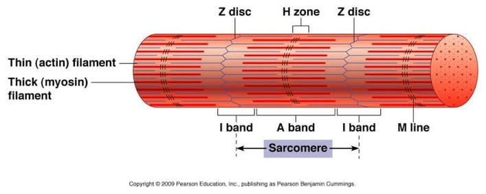
M Line
A dark line running down the middle of the H zone consisting of thick filaments linked by accessory proteins.

Transverse Tubule (T Tubule)
An invagination of the sarcolemma formed at each junction of the A and I bands that runs deep into the muscle fiber.

Sarcoplasmic Reticulum (SR)
An elaborate smooth endoplasmic reticulum of the muscle cell composed of tubules and cross channels that regulate calcium storage.

Terminal Cisterns
Cross channels of the sarcoplasmic reticulum that border a T tubule on each side.

Triad
A structural grouping in a muscle fiber formed by a central transverse (T) tubule bordered by two SR terminal cisterns.

Endomysium
A delicate sheath of areolar connective tissue enclosing each individual muscle fiber.

Perimysium
A collagenic membrane that wraps several sheathed muscle fibers to form a bundle called a fascicle.

Fascicle
A bundle of skeletal muscle fibers wrapped by perimysium.

Epimysium
A coarse overcoat sheath of dense irregular connective tissue binding numerous fascicles and enclosing the entire muscle.

Tendon
A tough, cordlike structure of dense regular connective tissue formed by converging muscle sheaths that attaches muscle to bone or another muscle.

Aponeurosis
A strong, sheetlike connective tissue structure attaching muscles to each other or indirectly to bones.

Origin
The fixed or immovable attachment of a muscle.

Insertion
The more movable attachment of a muscle.

Neuromuscular Junction
The junction formed between the axon terminal of a motor neuron and the sarcolemma of a muscle fiber.

Motor Unit
A functional structure consisting of a motor neuron and all the muscle fibers it stimulates.

Synaptic Cleft
A small fluid-filled gap separating the membranes of the axon terminal and the muscle fiber.

Acetylcholine (ACh)
The neurotransmitter stored in synaptic vesicles of the axon terminal and released by exocytosis to depolarize the sarcolemma.

Prime Mover (Agonist)
A muscle that is most responsible for producing a particular movement.

Antagonist
A muscle that opposes or reverses a particular movement; it is stretched and relaxed when the prime mover is active.

Synergist
A muscle that aids an agonist by assisting with the same movement or by reducing undesirable or unnecessary movements.

Fixator
A specialized synergist that immobilizes the origin of a prime mover so that all tension is exerted at the insertion.

Epicranius
A bipartite scalp muscle consisting of a frontal belly that raises eyebrows and wrinkles the forehead, and an occipital belly that pulls the scalp posteriorly.

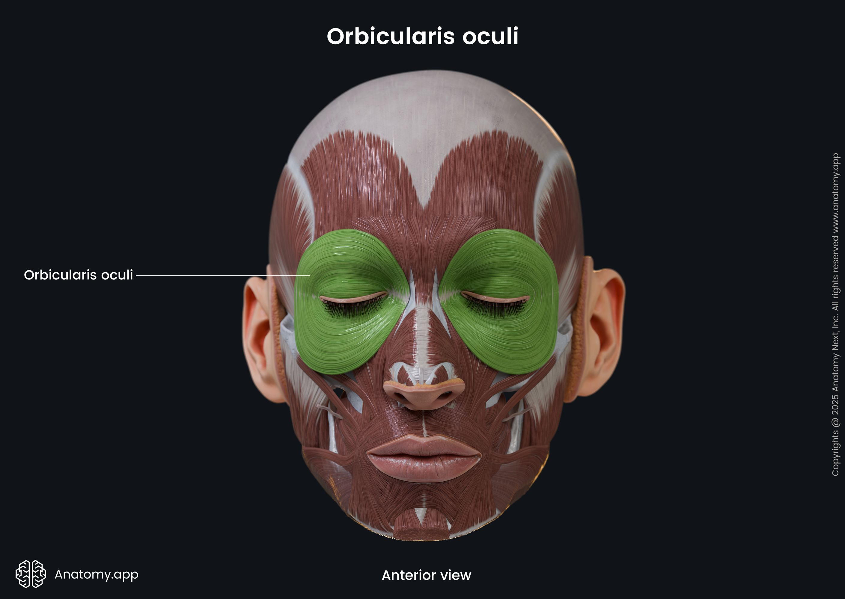
Orbicularis Oculi
A tripartite sphincter muscle of the eyelids that closes the eye and produces blinking, squinting, and draws eyebrows inferiorly.
Corrugator Supercilii
A small muscle acting with orbicularis oculi to draw eyebrows medially and inferiorly, wrinkling forehead skin vertically.

Levator Labii Superioris
A thin facial muscle between orbicularis oris and the inferior eye margin that raises and furrows the upper lip and opens lips.

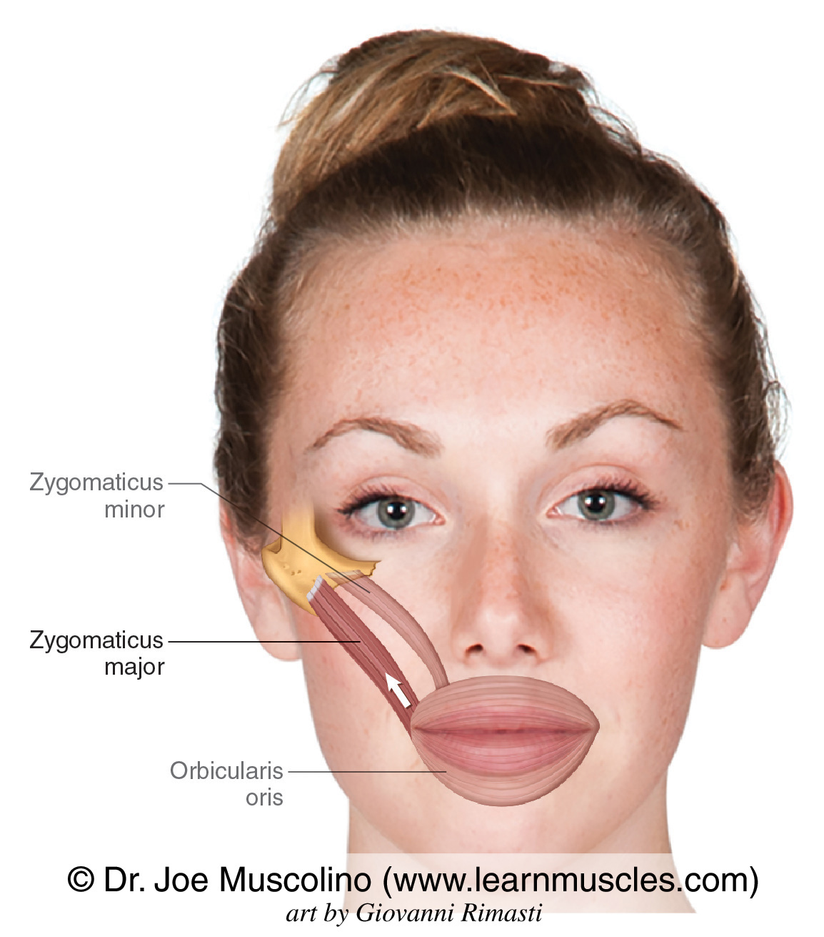
Zygomaticus
The smiling muscle extending diagonally from the corner of the mouth to the cheekbone, which raises lateral corners of the mouth upward.
Risorius
A slender muscle running inferior and lateral to zygomaticus that draws the corner of the lip laterally and tenses the lip.

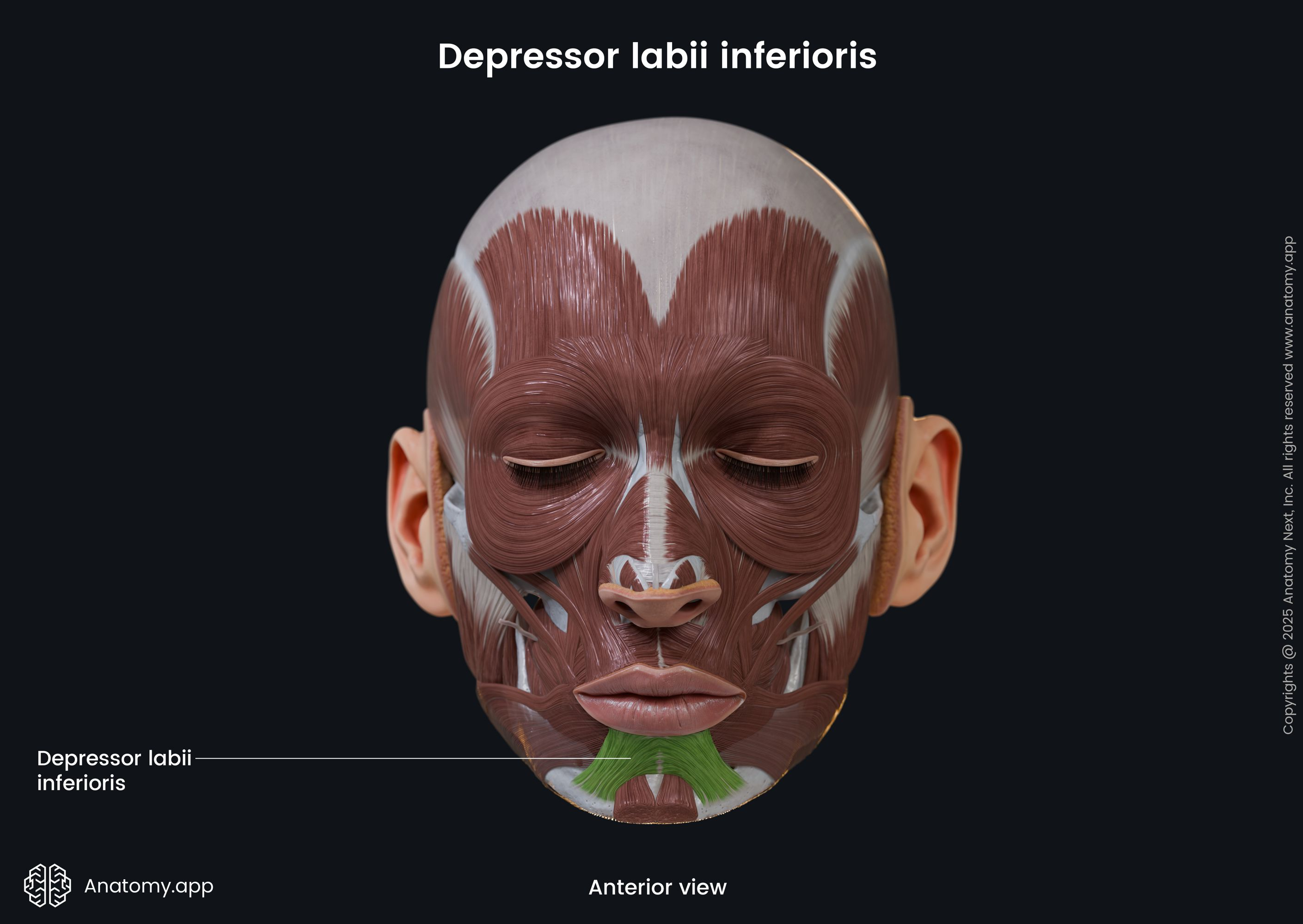
Depressor Labii Inferioris
A small muscle running from the lower lip to the mandible that draws the lower lip inferiorly.
Depressor Anguli Oris
A zygomaticus antagonist lateral to depressor labii inferioris that draws corners of the mouth downward and laterally.

Orbicularis Oris
The circular muscle of the lips (kissing and whistling muscle) that closes, purses, and protrudes the lips.

Mentalis
One of a muscle pair forming a V-shaped muscle mass on the chin that protrudes the lower lip and wrinkles the chin.

Buccinator
The principal muscle of the cheek running horizontally deep to the masseter that compresses the cheek and holds food between teeth during chewing.

Platysma
An unpaired, thin, sheetlike superficial neck muscle that tenses the skin of the neck, depresses the mandible, and pulls the lower lip back and down.

Masseter
A muscle covering the lateral aspect of the mandibular ramus that acts as the prime mover of jaw closure and elevates the mandible.

Temporalis
A fan-shaped muscle overlying parts of the frontal, parietal, and temporal bones that closes the jaw and elevates and retracts the mandible.

Medial Pterygoid
A deep mastication muscle running along the internal surface of the mandible that elevates the mandible and promotes grinding movements.

Lateral Pterygoid
A deep mastication muscle superior to the medial pterygoid that protracts the jaw and aids in grinding movements of the teeth.

Digastric
A suprahyoid muscle with two bellies united by an intermediate tendon that elevates the hyoid bone, opens the mouth, and depresses the mandible.
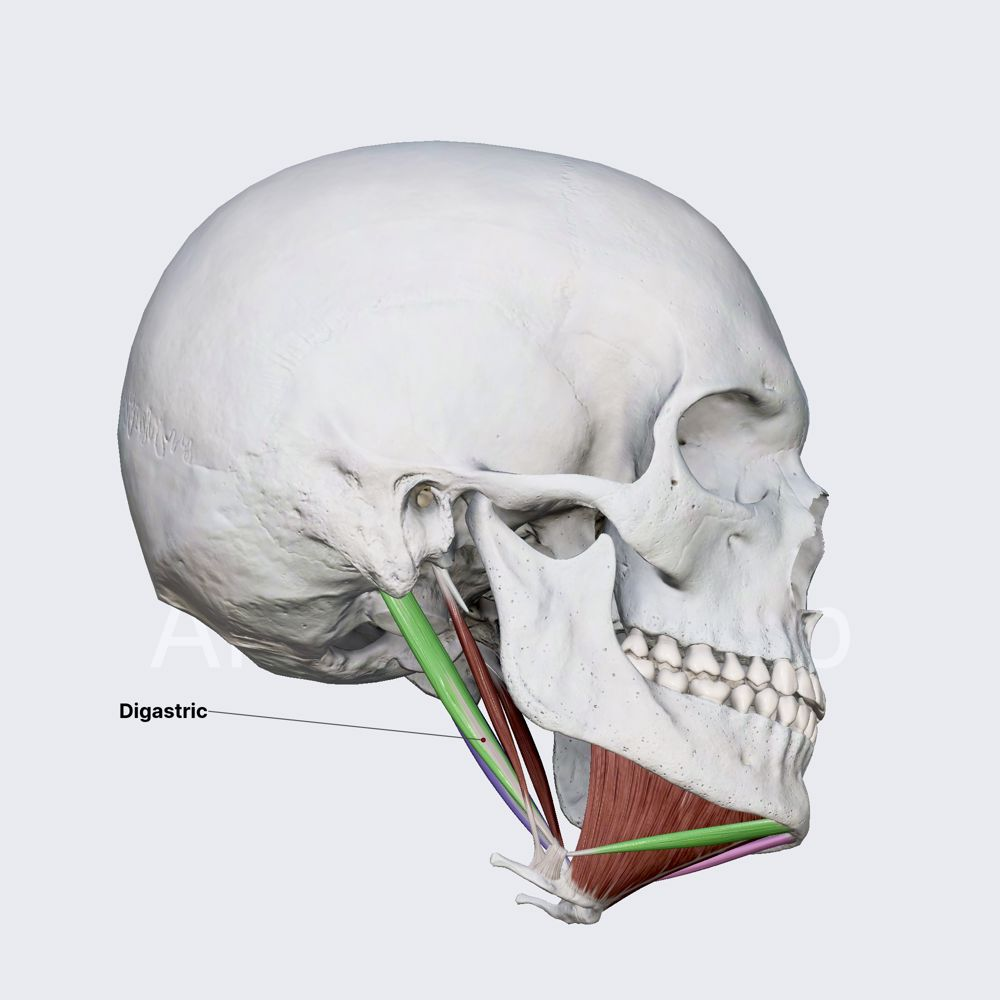
Mylohyoid
A suprahyoid muscle deep to the digastric forming the floor of the mouth that elevates the hyoid bone and base of the tongue during swallowing.

Sternocleidomastoid
A two-headed neck muscle that flexes the neck during simultaneous contraction, or rotates the head toward the opposite shoulder when acting independently.

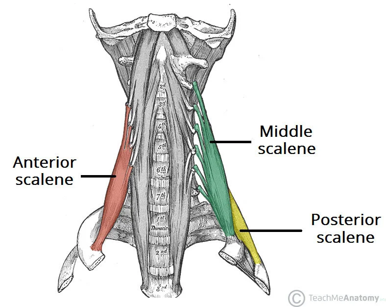
Scalenes
Lateral neck muscles (anterior, middle, posterior) that flex and slightly rotate the neck and elevate ribs 1-2 to aid in inspiration.
Splenius
A superficial posterior neck muscle (capitis and cervicis parts) that extends or hyperextends the head and rotates/bends the head toward the active side.

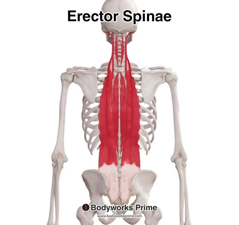
Erector Spinae
A long tripartite muscle column of the back (iliocostalis, longissimus, spinalis) that extends and bends the vertebral column laterally.
Quadratus Lumborum
A muscle forming the greater portion of the posterior abdominal wall that flexes the vertebral column laterally and extends the lumbar spine.

Pectoralis Major
A large fan-shaped muscle covering the upper chest that serves as the prime mover of arm flexion and adducts and medially rotates the arm.

Serratus Anterior
A fan-shaped muscle deep to the scapula that protracts and holds the scapula against the chest wall and rotates it upward.

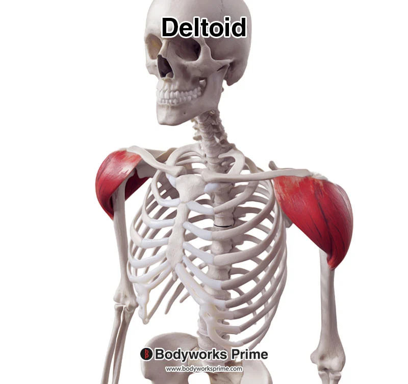
Deltoid
A fleshy triangular muscle forming the shoulder mass that acts as the prime mover of arm abduction and serves as an intramuscular injection site.
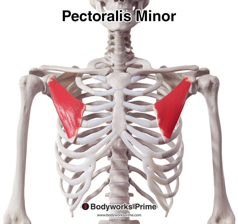
Pectoralis Minor
A flat, thin muscle deep to pectoralis major that draws the scapula forward and inferiorly with ribs fixed, or elevates the rib cage with scapula fixed.
External Intercostals
Eleven pairs of respiratory muscles between ribs with fibers running downward and forward that elevate the rib cage to aid in inspiration.
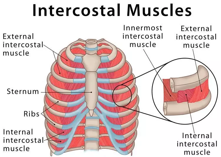
Internal Intercostals
Eleven pairs of respiratory muscles running deep and at right angles to external intercostals that depress the rib cage during forced expiration.

Diaphragm
A broad, dome-shaped muscle forming the thoracic floor that flattens upon contraction as the prime mover of inspiration.

Rectus Abdominis
A medial superficial segmented abdominal muscle extending from pubis to rib cage that flexes the vertebral column and stabilizes the pelvis during walking.

External Oblique
The most superficial lateral abdominal muscle with fibers running downward and medially that flexes the vertebral column and compresses the abdominal wall.

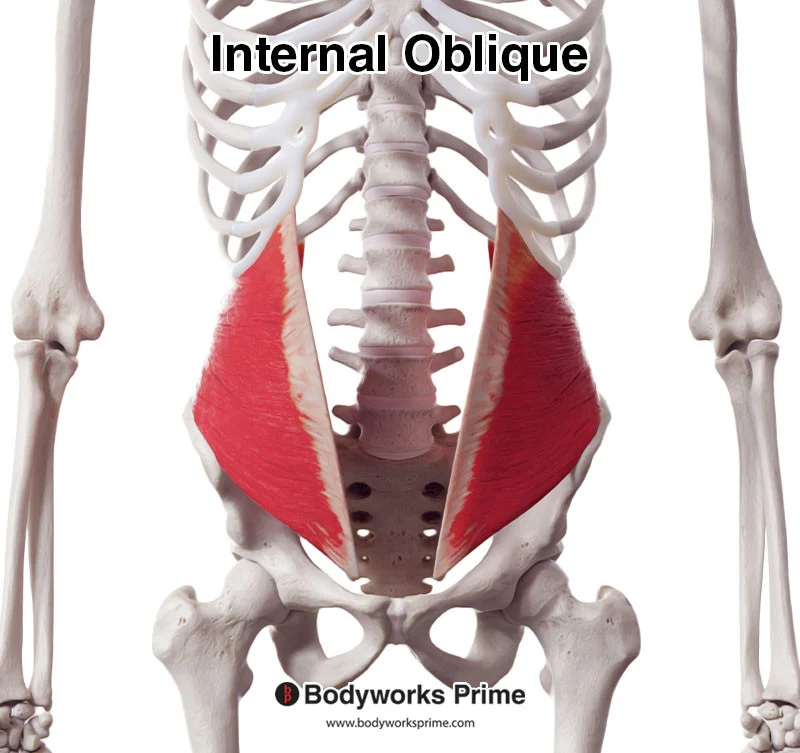
Internal Oblique
An abdominal muscle underlying the external oblique with fibers running at right angles to it that compresses the abdominal wall and assists in trunk rotation.
Transversus Abdominis
The deepest muscle of the abdominal wall with fibers running horizontally that compresses abdominal contents.

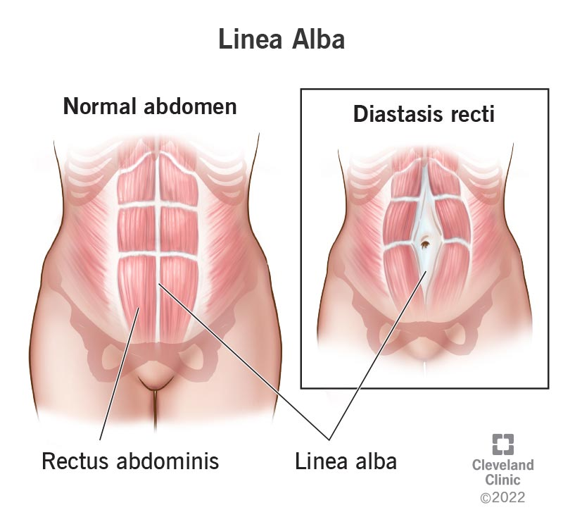
Linea Alba
A narrow tendinous sheath running down the midline of the abdomen from the sternum to the pubic symphysis, formed by the fusion of abdominal aponeuroses.
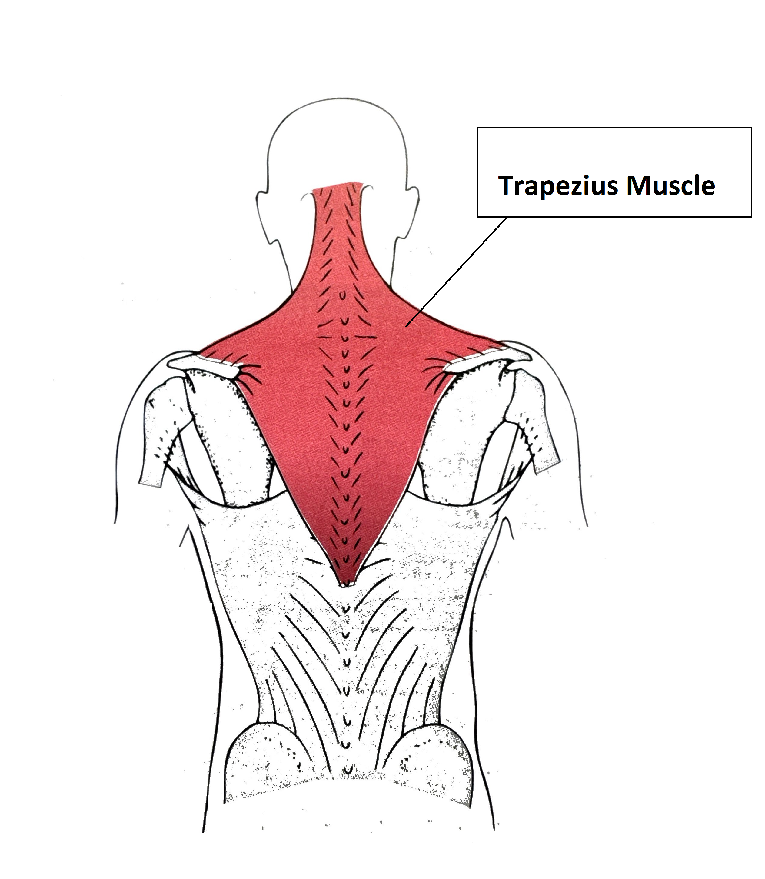
Trapezius
The most superficial muscle of the posterior thorax that extends the head and raises, rotates, retracts, elevates, or depresses the scapula.
Latissimus Dorsi
A broad, flat muscle of the lower back that serves as the prime mover of arm extension and adducts and medially rotates the arm.

Infraspinatus
A rotator cuff muscle located on the posterior scapula that laterally rotates the arm and stabilizes the shoulder joint.

Supraspinatus
A rotator cuff muscle obscured by the trapezius that initiates abduction of the arm and stabilizes the shoulder joint.

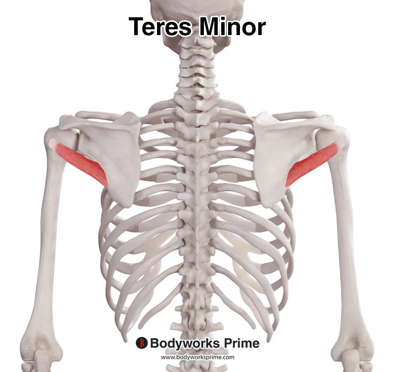
Teres Minor
A small rotator cuff muscle inferior to infraspinatus that laterally rotates the arm and stabilizes the shoulder joint.
Teres Major
A muscle located inferior to teres minor that extends, medially rotates, and adducts the arm as a synergist of latissimus dorsi.

Triceps Brachii
A large, three-headed muscle of the posterior humerus that serves as the powerful prime mover of forearm extension.

Biceps Brachii
A two-headed muscle of the anterior humerus that powerfully flexes and supinates the forearm and weakly flexes the arm.

Brachialis
A muscle located immediately deep to the biceps brachii that flexes the forearm.

Brachioradialis
A superficial muscle of the lateral forearm forming the lateral boundary of the cubital fossa that acts as a synergist in forearm flexion.

Pronator Teres
An anterior forearm muscle that acts synergistically with pronator quadratus to pronate the forearm and weakly flexes the forearm.

Flexor Carpi Radialis
A superficial muscle running diagonally across the anterior forearm that acts as a powerful flexor and abductor of the hand.

Palmaris Longus
A small fleshy forearm muscle with a long tendon that weakly flexes the hand and tenses the skin and fascia of the palm.

Flexor Carpi Ulnaris
A superficial muscle located on the medial anterior forearm that flexes and adducts the hand.

Flexor Digitorum Superficialis
A deeper anterior compartment muscle that flexes the hand and middle phalanges of fingers II-V.

Flexor Pollicis Longus
A deep muscle of the anterior forearm that flexes the thumb.

Flexor Digitorum Profundus
A deep muscle of the anterior forearm that is the sole muscle that flexes the distal phalanges of fingers II-V.

Pronator Quadratus
The deepest muscle of the distal anterior forearm that pronates the forearm.

Extensor Carpi Radialis Longus
A superficial muscle paralleling brachioradialis on the lateral forearm that extends and abducts the hand.

Extensor Digitorum
A superficial posterior forearm muscle that serves as the prime mover of finger extension, extends the hand, and flares the fingers.

Extensor Carpi Ulnaris
A superficial muscle on the medial posterior forearm that extends and adducts the hand.

Supinator
A deep muscle at the posterior aspect of the elbow that acts as a synergist of biceps brachii to supinate the forearm.

Iliopsoas
A composite muscle consisting of the iliacus and psoas major that flexes the trunk at the hip joint and flexes the thigh.

Sartorius
A straplike superficial muscle running obliquely across the anterior thigh (tailor's muscle) that flexes, abducts, and laterally rotates the thigh and flexes the leg.

Adductor Magnus
A large muscle of the medial thigh that adducts, medially rotates, and flexes the thigh; its posterior part acts as a synergist in thigh extension.

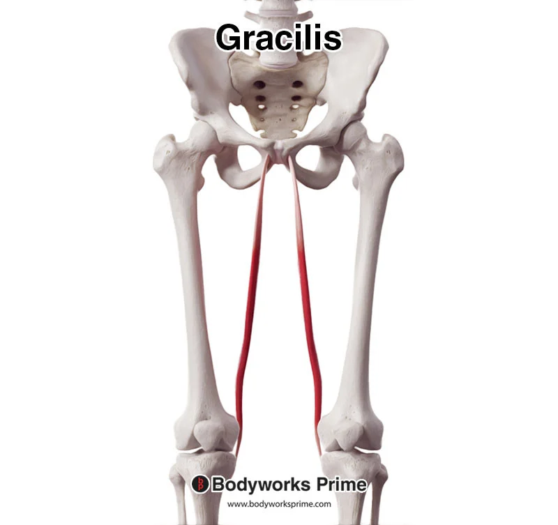
Gracilis
A straplike superficial muscle of the medial thigh that adducts the thigh and flexes and medially rotates the leg.
Rectus Femoris
A superficial anterior thigh muscle that extends the leg and is the only muscle of the quadriceps group to cross the hip joint and flex the thigh.

Vastus Lateralis
A large muscle forming the lateral aspect of the thigh that extends the leg, stabilizes the knee, and serves as an intramuscular injection site.

Vastus Medialis
A muscle forming the inferomedial aspect of the thigh that extends the leg and stabilizes the patella.

Vastus Intermedius
A muscle of the quadriceps group lying deep to rectus femoris between vastus lateralis and vastus medialis that extends the leg.

Tensor Fascia Lata
A muscle enclosed between fascia layers of the anterolateral thigh that flexes, abducts, and medially rotates the thigh and steadies the trunk.

Gluteus Maximus
The largest and most superficial gluteal muscle that acts as a powerful thigh extensor and intramuscular injection site.
