Body Logistics:
 Call Kai
 Learn
Learn Practice Test
 Spaced Repetition
 Match
Match Flashcards
 Knowt Play
1/34

There's no tags or description
Looks like no tags are added yet.
Name | Mastery | Learn | Test | Matching | Spaced | Call with Kai | Chat |
|---|
No analytics yet
Send a link to your students to track their progress
35 Terms
Define the term homeostasis with respect to water, temperature and pH
Homeostasis is the process by which the body maintains a stable internal environment despire external or internal changes.
Homeostasis means ‘steady state‘ while body is never static and continually adjusts to maintain dynamic equilibrium especially key variables like temperature, fluid levels and pH.
Explain the concepts behind positive and negative feedback loops:
Negative feedback loops, restore balance
Positive feedback: Amplify a response
Basic system:
Stimulus → receptor → sensory neuron → CNS → Motor neuron → Effector
State the ‘normal range‘ for core body temperature, and what happens when body is not in normal temperature range:
36.5 - 37.5
When body not in normal temperature range:
Hypothermia: Core body temperature below 35 degrees
Passive rewarming: removing patient from cold environment, remove wet clothes, cover with dry blankets (head to toe)
Active external rewarming: Heat packs and lamps, warm blankets, bair hugger system
Active core rewarming: warmed intravenous fluids, warm humidified oxygen
Hyperthermia: Core body temperature above 37.5 degrees
Body’s reaction to infection (more than 38 degrees = pyrexia), extra heat can be helpful for immune system to deal with pathogens
State the mechanisms in broad terms that regulate core body temperatures:
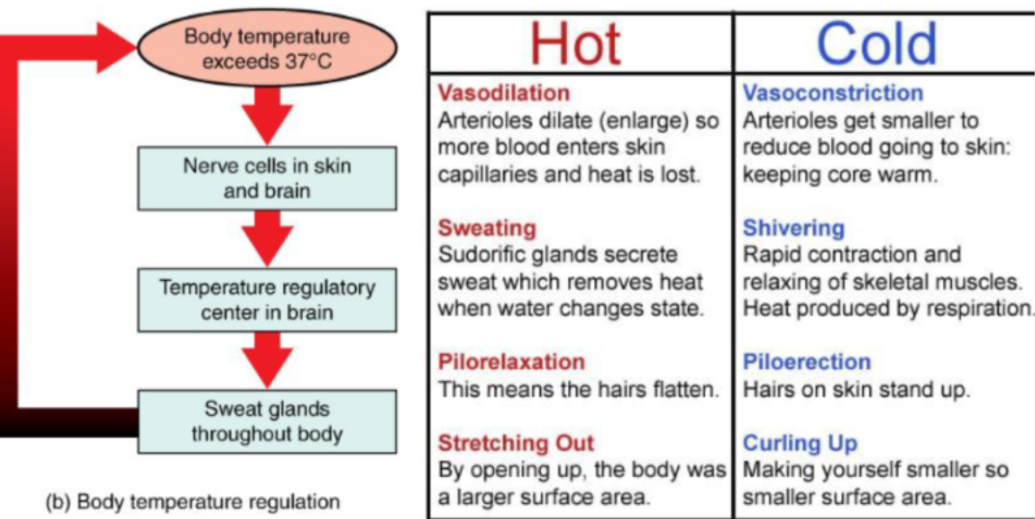
Sensors in the skin and hypothalamus detect temperature. The hypothalamus triggers responses such as:
When cold: Shivering, piloerection (hairs standing), vasoconstriction
When hot: Sweating, pilorelaxation (hairs lying flat), vasodilation
State the ‘normal range‘ for pH in tissues including blood:
Normal blood pH is 7.35 -7.45
Explain the importance of acid-base balance for normal human physiology:
Below pH 7.35 is acidosis
Above pH 7.45 is alkalosis
The kidney maintains pH in safe range:
Metabolic = the kidneys (remove the HCO3) => base
Respiratory = The lungs (remove CO2) => acidic
The conditions:
Metabolic acidosis = Build up of acidic metabolites such as lactic acid in severe infection or ketoacids in untreated diabetes
Metabolic alkalosis = Loss of H+ ions the acid through vomitting // Excess HCO3- lost through urine
Respiratory acidosis = Inability to breath out enough carbon dioxide // builds up as carbonic acid
Respiratory alkalosis = Breathing out too much carbon dioxide // hyperventillation during panic attack
Recall the percentage of water in a standard 70kg male:
60% of body weight
of which 40% is intracellular (2/3) fluid and 20% is extracellular (1/3)
State the body compartments where fluid accumulate:
Intra = inside // This is water inside the cell // contain 2/3 of total body water
Extra = outside // This is water outside cells (blood plasma, interstitial fluid - fluid between cells, small amounts of other body fluids such as lympth) // 1/3 of total body water
Interstitial fluid 75% of the extracellular fluid
Plasma fluid is 25% of the extracellular fluid => the plasma part of the blood
Compare and contrast the water balance in males, females, in early life and in older populations:

Factors effecting body water:
Age (main factor): water content decreases with age as body fat increases
Gender: Men have higher water percentage than women due to less body fat
Body composition: Higher body fat means, less water percentage (since fat is store anhydrously)
Fitness level: Athletes may have slightly higher water percentage due to increased lean muscle mass
Definitions:
Osmotic pressure
Osmole
Osmolarity
Osmolality
Definitions:
Osmotic pressure = the force of pulling where there is low solute concentration to high solute concentration
Osmole = Number of moles that a solute contributes to the osmotic pressure of a solution
Osmolarity = is the number of particles per L
Osmolality = is the number of particles per kg (double ll = heavyyy )
Overall, osmolarity and osmolality is a form of osmotic direction, high osmolarity/ osmolality to low osmolarity/osmolality
Outline the physiological differences between dehydration and water toxicity and their effect on osmolality:
Dehydration: Water pressure outside the cell is lower than inside the cell, so water moves out by osmosis, increased blood viscosity since same number of RBC in less water which increases chance of muscle clots, because thicker blood moves more slowly increases heart pressure to pump and this causes the clotting factors to take place, fast heart rate, muscle cramp. An increase in plasma osmolality triggering thirst.
Water toxicity: Excess water dilutes sodium → oedema, especially dangerous in brain (since high water intake can effect brain immediately) = causes seizures, coma and eventually death
Describe the process of odema:
When there is right hand heart failure, there is not effective pumping of the right heart to the lungs, so there is a build up of blood in the capillaries, this causes high hydrostatic pressure, so more fluid leaves capillaries
Large DVT = deep vein thrombosis, the pathway [Leg tissue → Leg vein → larger veins → heart], if there is a clot in the vein, the blood can’t go to the heart, and more blood travels to the vein via arteries. So, build up of blood, high hydrostatic pressure, so more fluid moves out of capillaries
Decreased levels of albumin in blood, less pull of water at venous end, so more fluid reabsorbed via lymph
Define histology:
‘histos‘ = tissue
Logy = study
The study of tissues under the microscope to understand how cells are organised
Describe common biopsy techniques, giving examples of tissues which can be sampled by each method
Tissue samples (biopsies) can be taken in several ways:
• Scraping: e.g., curettage for the endometrium.
• Needles: e.g., fine needle aspiration (thyroid), pipelle (uterus), trephine
(bone marrow), venepuncture (blood).
• Surgical excision: larger samples for solid organ tumours.
• Transvascular: access via blood vessels, e.g., heart or kidney.
• Patient collection: samples like sputum, semen, urine or stool. These are
not ‘true’ biopsies but are still samples which can be assessed and
visualised microscopically
Outline the steps involved in tissue processing from a biopsy sample to a slide which can be examined microscopically, and state why each step is necessary
Fixation: Preserves tissue structure (usually use formalin which contains
formaldehyde).
Dehydration: Removes water and prepares tissue for embedding (using alcohol
and solvents like xylene/toluene).
Embedding: Often in paraffin wax, infiltrates and surrounds tissue in a block.
Sectioning: Very thin slices cut using a microtome.
Mounting: Sections placed on glass slides.
Staining: Adds contrast for microscopic viewing.
These steps prevent decay, preserve fine detail, and allow light to pass through the
sample for microscope viewing.
Describe how tissue processing can lead to the formation of shrinkage and other artefacts
Processing may shrink tissue or create distortions (artefacts/artifacts). There are
many different types of artefacts in histology – the main artefact to be aware of is tissue folding, which can cause layers of cells to overlap or shift out of focus.
Outline the advantages and disadvantages of using frozen sections and formalin-fixed paraffin-embedded (FFPE) sections for microscopic examination:
Frozen vs FFPE Sections
FFPE (Formalin-Fixed, Paraffin-Embedded) sections are not always the best choice
for processing a biopsy sample. Frozen sectioning provides an alternative method.
Frozen: quicker process but lower quality; used during surgery while patient is
under anaesthetic. Will not remove fats or sugars from sample so related structures
can still be viewed. More prone to artefacts such as ice crystal damage.
FFPE: higher detail achieved but takes longer. Fats and sugars removed by
solvents. Stains better and can be stored indefinitely at room temperature.
Discuss the value of histological staining and state the components of tissue stained by routine stains and specalist methods
Haematoxylin: a basic dye that binds to acidic (basophilic, base-loving)
structures. Blue/purple colour. Highlights nuclei (due to DNA/RNA).Eosin: an acidic dye that binds to basic (eosinophilic, acid-loving)
structures. Red/pink colour. Highlights cytoplasm, muscle fibres, connective
tissue (due to mainly basic proteins).
Describe the main differences in preparing tissue for electron microscopy compared to light miscroscopy
D
Define the term ‘limit of resolution‘
Explain why electron microscopes are capable of finer resolution than light miscroscopes
Depending on the situation, light or electron microscopy can be used to view the
final sample/slide. Keep in mind, tissue preparation differs for electron microscopy.
• Fixed with glutaraldehyde instead of formaldehyde
• Embedded in epoxy resin instead of paraffin wax
• Stained with electron-dense chemicals e.g. osmium tetroxide
• TEM samples sliced on special microtome using diamond knife to 30-60nm
depth
Conversion between milli-, micro-, nanometres, pico

Define ‘tissue‘ and ‘organ‘ and ‘organ system‘:
Tissue: A collection of cells adapted to perform a specific function
Organ: A structural unit composing of two or more tissue types working together to perform a function
Organ system: When several organs work together to form a system which performs complex functions
State the four types of tissue:
Epithelia - lining cells, glands
Connective tissue - Bone, blood, fat, cartilage
Muscle tissue = Cardiac, skeletal, smooth
Nervous tissue - Peripheral nerves, spinal cord, brain
Describe how cells are held together at the molecular level: HORIZONTALLY
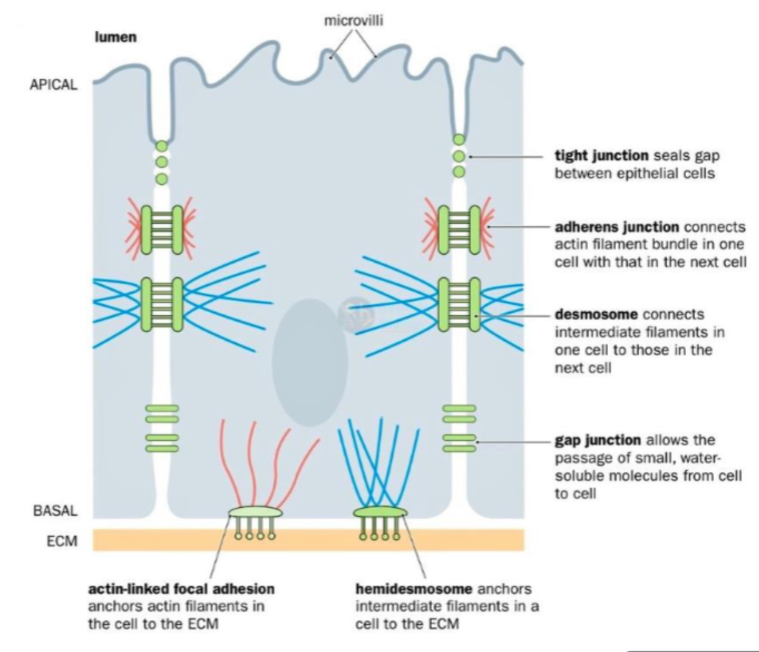
Tight junctions (Zonula occludens):
Structure: Belt-like seals between neighbouring cells, nearest to the apical surface
Function: Prevent paracellular leakage (leakage between cells), control passage of ions, water and solutes from => so act as selective barriers, restricting diffusion of water-soluble molecules.
Adherens Junctions (Zonula adherens):
Structure: Belt-like adhesion complexes just below tight junctions, about ½ way down from apical surface
Function: Provide mechanical stability and additional transport barrier
Desmosomes (macula adherens):
Structure: Spot-like junctions with cadherin molecules anchored to intermediate filaments, strongest cell-cell adhesion, found ½ way down lateral membranes
Function: Provide strong adhesion, especially in tissues under mechanical stress (skin and heart)
Gap Junctions:
Structure: Most widespread of cell-cell junction, made up of connexin proteins towards the base of the lateral membrane
Function: Allow direct cell-cell communication via small molecules and ions, can be opened/closed by changes in pH and calcium concentration (important in muscle contraction)
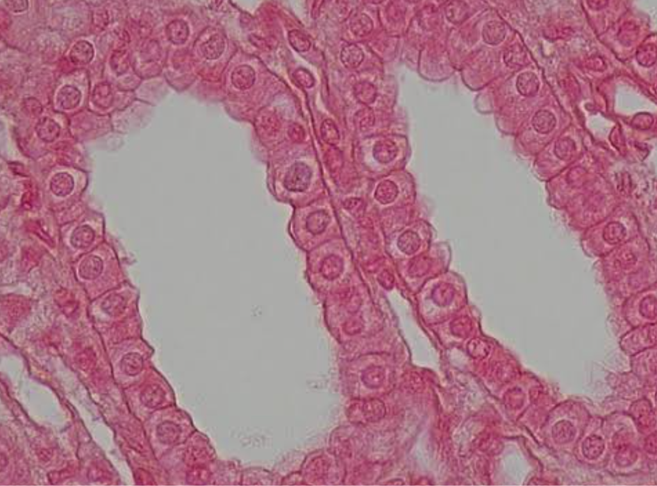
Define epithelia and explain classification terminology:
Epithelia are layers of closely packed cells that line the surfaces and cavities of the body. They serve as protective barriers and are involved in absorption, secretion, filteration and sensation.
Classification:
Simple: One layer of cells above the basement membrane
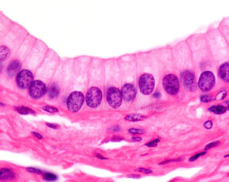
Stratified: Multiple layers of cells above the basement

Pseudostratified: May appear to have multiple layers of cells (with nuclei at different heights), but actually only have one layer.

Shapes:
Squamous (flat):

Cuboidal:
Columnar:

Explain the position, structure and function of basement membrane
The basement membrane is beneath the epithelium and above the lamina propria, anchoring epithelial cells to underlying connective tissue.
Function:
Anchors epithelial cells
Supports epithelium
Separates epithelium from connective tissue

Explain the reason for the different rates of renewal of each type of epithelium:
Epithelial Renewal Rates
• High renewal: oral mucosa, oesophagus, vagina, intestines
• Moderate: transitional (urothelium), pseudostratified respiratory, bile duct
lining (simple cuboidal/columnar), epidermis (skin)
• Low: renal tubules (simple cuboidal), blood vessel lining (simple squamous)
Why?
High renewal rate: Due to abrasion, friction and microbes
Low renewal rates: Relatively protected, renew slowly
Differentiate between simple and stratified (compound) epithelia from micrographs:


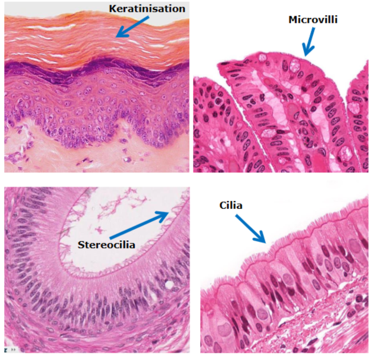
Recognise the different types of surface specialisation found on epithelial cells (keratin, microvilli, stereocilia and cilia) and descrive how their structure relates to function
Keratinisation (skin surface):
Location: Epidermis of the skin, especially thick on palms and soles
Structure: surface cells become filled with keratin (tough protein), where cells eventually die and form a protective outer layer
Function: Protection against abrasion, waterproofing, reduce water loss, barrier against pathogens
Microvilli (brush border in gut):
Location: Small intestines, kidney tubules
Structure: Tiny finger-like projections of cell membrane, contain actin filaments, form a brush border.
Function: greatly increase surface area, increase absorption of nutrients
Cilia (respiratory tract):
Location: Trachea, Bronchi, Larger airways
Structure: Hair-like motile projections, beat in coordinated fashion
Function: Move mucus and trapped particles towards the throat, so lungs are clean
Stereocilia (epididymis):
Location: Epididymis (attached to the back of the testis), part of inner ear
Structure: Very long microvilli, not motile
Function: Increase surface area for absorption
Describe the functions of goblet cells, club cells and microfold (M cells)
Goblet cells: Found in the GI and Respiratory that secrete mucus rich in mucins. These form a protective barrier that traps pathogens and particles

Microfold (M) cells: They are located in the epithelium of Peyer’s patches (GALT) in the gut => located around the small intestine, they transport microorganisms from intestinal lumen to immune cells

Club cells: Bronchiolar secretion and detoxification => Non-ciliated found in bronchioles of lungs. They produce components that detoxify harmful substances via enzymes and contribute to epithelial repair after injury

Explain briefly the genetic cause of cystic fibrosis, how epithelial cells are affected by smoking and define different examples of common respiratory diseases.
Cystic Fibrosis:
A mutation in deltaF508, leads to the production of faulty CFTR proteins, so this protein can’t regulate the chloride and sodium ion transport across epithelial cells.
So, leads to thick, sticky mucus to accumulate in lungs, pancreas and other organs leading to chronic respiratory infections and digestion problems.
Smoking:
Smoking damages the respiratory epithelium: Destroying cilia, increase muscus prodiction and goblet cell hyperplasia, promoting inflammation and oxidative stress which causes permanent damage to airway lining
Distinguish between mucous and serous membranes
Mucous membranes (mucosae)
Line body passages that communicate with the outside environment
Examples: gastrointestinal, respiratory and urinary tracts
Consist of:
Epithelium
Basement membrane
Lamina propria: underlying connective tissue
In some regions, a muscularis mucosae
Often produce or are covered by mucus
Functions include protection, secretion and absorption, depending on location.
Serous membranes (serosae)
Line closed internal body cavities and cover organs within them
Consist of:
Mesothelium: simple squamous epithelium
Thin layer of connective tissue
Produce serous fluid, which lubricates surfaces and reduces friction as organs move.
Examples:
Pleura: lungs
Pericardium: heart
Peritoneum: abdominal organs
Mucous membrane = lines passages connected to outside
Serous membrane = lines closed internal cavities and covers organs
For the gastrointestinal, urinary and respiratory mucosae:
Describe the structural relationships between the epithelia and
closely associated tissues (glands, underlying connective tissue,
lamina propria and muscularis mucosae)Discuss how the structure of their constituent tissues relates to
mucosal function
1. Epithelium
Forms the surface facing the lumen
Provides a barrier
Its type varies depending on function.
2. Basement membrane
Thin layer underneath the epithelium
Anchors/supports the epithelium and separates it from underlying connective tissue.
3. Lamina propria
Loose connective tissue underneath the epithelium
Supports the epithelium
Contains structures such as blood vessels and immune cells
Glands may also be present depending on the location.
4. Muscularis mucosae
Thin layer of smooth muscle
Allows local movement of the mucosa
Especially important as a distinct component of the GI mucosa

Describe how cells are joined together (2)
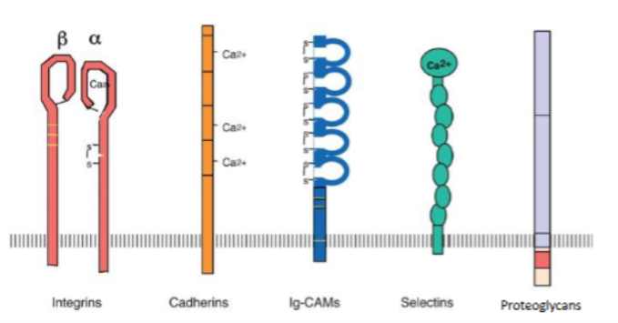
Integrins: Heterodimeric transmembrane receptors (alpha and beta subunits), function is to bind with ECM proteins (fibrinogen, collagen). Bidirectional signalling between inside and outside cells, regulating process like differentiation and cell death (Apoptosis).
Cadherins: Mediate cell-cell adhesion, through Adherens junctions and desmosomes, they link to cytoskeleton and maintain tissue architecture
Selections: Important for immune system
Proteoglycans: Core proteins with glycosaminoglycan (GAG) side chains. Function: Important in skin// hydration of extracellular matrix, mediating adhesion. Regulate growth factor availability which controls cell turnover and tissue repair.