microLAB 2: Bacterial Morphology and Staining
 Call Kai
 Learn
Learn Practice Test
 Spaced Repetition
 Match
Match Flashcards
 Knowt Play
1/14

There's no tags or description
Looks like no tags are added yet.
Name | Mastery | Learn | Test | Matching | Spaced | Call with Kai | Chat |
|---|
No analytics yet
Send a link to your students to track their progress
15 Terms
Bacteria are ___. what size? are their organelles membrane bound? what kind of chromosome?
prokaryotes.
smaler in size ~ 1mm
no membrane bound organelles
Single circular chromosome
Bacteria basic shapes
Bacilli (rod- shaped): streptobacilli (chains)
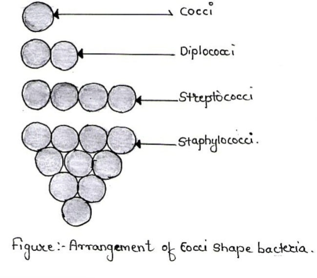
cocci (sphere-shaped): streptococci (chains): straphylococci (clusters)
spirilla (spiral-shaped)
spirochaete (corkscrew)
vibrio (comma-shaped)
Arrangement of cocci
• Diplococcus: cocci in pairs
• Streptococcus: cocci arranged in chains
• Streptococcus: cocci in chain
• Staphylococcus: grape like clusters
Why Do Staining?
• Most bacteria are colorless and provide minimal contrast
• Staining makes bacteria visibly observable
• To determine the morphology (form and structure),
arrangements and size of bacteria
types of staining:
• Negative staining: background is stained
• Simple staining: only 1 dye used
• Differential staining: to differentiate different
types of cells: eg. Gram stain, acid fast stain,
endospore Staining
• Special staining- more than one dye used-
special structures are seen. Eg. capsule
staining, flagella staining
NEGATIVE STAINING
• Negative, indirect or background staining is achieved by mixing bacteria
with an acidic stain or dye called a chromogen, such as Nigrosin, India
ink, or eosin.
• Spread of the mixture over a slide to form a film.
• No stain penetrates bacterial cell due to repulsion between the negative
charge of the stain and the negatively charged bacterial wall.
• Useful in observing bacterial morphology shape and size
negative staining procedure:
• No heat fixing required.
• Mix bacterial suspension bacteria in liquid media with acidic
stain (Nigrosin) & spread mixture on slide.
• Bacterial cells are not penetrated by stains.
• Air dry and focused with 4x, 10x, 40x & 100x
• Produces image of light bacteria against dark background.
what staining is this?
negative stain

Basic initial steps before staining:
- To observe more details about cell shape, size and arrangements.
- Bacterial smear preparation
place a drop of bacterial cells on the central portion of a slide
in a circular fashion
Air dry, Heat fix the slide –we use hot plates/not Bunsen
burners
Bacteria attaches/adhere to the slide – appears dull white
SIMPLE STAINING
• Simple to perform – only one basic stain used. Eg.
Crystal Violet, Methylene blue, Carbolfuchsin..
• Then add dye to create a contrast between
bacteria & background the bacteria cell wall is (-)
• the simple stain is (+), it attracts to the (-)
bacterial cell wall
simple staining procedure:
• A bacterial smear is prepared , air dried and heat fixed.
• A heat fixed smear is flooded w methylene blue for 45-60
seconds, then washed w water
• Blot dry w bibulous paper and examine under the
microscope with 4x, 10x, 40x & 100x
GRAM STAIN
• Hans Christian Joachim Gram, a Danish scientist & physician (1853-
1938)
• The most widely used staining procedure in microbiology is the Gram
stain.
• Used to differentiate between Gram(+) and Gram (-) bacteria
gram stain procedure:
1. Primary stain -basic dye Crystal violet
2. Treat with iodine solution (Mordant) increases interaction
between bacterial cell & dye
3. Decolorize smear with 95% ethanol
4. Counterstain with safranin- a basic dye that stains-
- Gram negative pink/red (E. coli)
- Gram positive purple/blue (S. Aureus)
ACID FAST / NON ACID FAST STAINING
(Ziehl-Neelsen and Kinyoun)
Acid Fast microorganisms: high lipid contents in cell wall
- To stain Mycobacterium species especially M. tuberculosis
• Primary stain-Carbolfuchsin, counterstain-Methylene blue.
• Acid Fast microorganisms require heat that the stain can penetrate.
• Not easily decolorized, after primary stain with carbolfuchsin (red).
Non Acid Fast microorganisms: low lipid content in cell wall
• Easily decolorized and appear blue/brown after counterstain with
Methylene blue.
ENDOSPORE STAINING (Schaffer-Fulton or Wirtz-Conklin)
New structure that is highly resistant & capable of surviving in
unfavorable conditions or environment.
• Once conditions are favorable a new bacterial cell arises. This
structure is called an endospore, since it develops within the bacterial
cell.
• Endospores do not stain easily, but once they are stained, they strongly resist decolorization