Pleuropneumonia in Horses Presentation and Diagnosis
 Call Kai
 Learn
 Practice Test
 Spaced Repetition
 Match
 Flashcards
 Knowt Play
1/24

There's no tags or description
Looks like no tags are added yet.
25 Terms
pneumonia
infection of lower resp tract
bronchopneumonia
bronchi and parenchima infection
pleuropneumonia
pneumonia into pleural space
stages of pleuropneumonia
exudative
fibrinopurulent
organization
exudative stage of pleuropneumonia
sterile transudate increased in pleural space
fibrinopurulent stage of pleuropneumonia
-bacterial invasion and fibrin deposition
organisation stage of pleuropneumonia
fibroblasts grow into exudate (pleural peel)
risk factorsfor pneumonia
-after viral infections
-strenuous exercise
-transportation and elevation of head
-GA
-overcrowding and inclement weather
-dysphagia (aspiration)
risk factors for pleuropneumonia
-long distance transport
-elevated head for prolonged time→ decreces mucocilliary ckearance of normal resp secretions
risk factor for aspiration pneumonia
-dysphagia (pharyngeal and postpharyngeal)
-oesophageal obstruction
-GA
-cleft palate
bacterial causes of pneumonia (aetiology)
-Streptococcus equi subsp zooepidemicus
-staph aureus and s.pneumoniae
-actinbacillus
-e.colli, pasteurellam, enterobacter, klebsiella, b.bronchiseptica
-bacteroides fragilis
-fusobacterium and peptostrep anaerobius
actinobacillus spp
gram negative nonenteric
clinical signs of pneumonia
-tachycardia/tachypnoea
-resp distress
-fever
-anorexia, depression
-and/or nasal discharge
-exercise intolerance
-auscultation shows crackles and dull area → rebreathing bag
clinical signs of pleuropneumonia
-pain intercostal spaces
-reluctance to walk, colic
-grunting during resp
-abduction of elbow
-ventral oedema (cehst)
-other clin signs of pneumonia
adventitious lung sounds in penumonia
-crackles and wheezes
-dull areas
-radiation of cardiac sounds
Dull area follows flat line
Pleural rubs
lung sounds of pleuropneumonia
-dull areas follow flat line (line along lung)
-pleural rub
diagnostic approach for pneumonia
-suspicion based on PE and rebreathing bag
-haem and biochem
-endoscopy
-TTW and BAL
-thoracic US
-radiography
-thoracocentesis
-thoracoscopy
clin path of pneumonia
cbc
neutrophilic leukocytosis
leukopenia
anaemia (chronic cases)
increased fibrinogen and SAA
decreased iron
TTW
-culture and sensitivity (aerobic and anaerobic)
-cytology of both lungs
-obtain even if pleural fluid available

what to remember if doing TTW
stop antibiotics 48 hours before
Anaerobic culture media (do not refrigerate!)
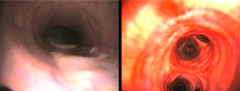
endoscopy
identify mucus→ bact pneumonia, asthma, others
identify affected site/ bronchi
abscesses
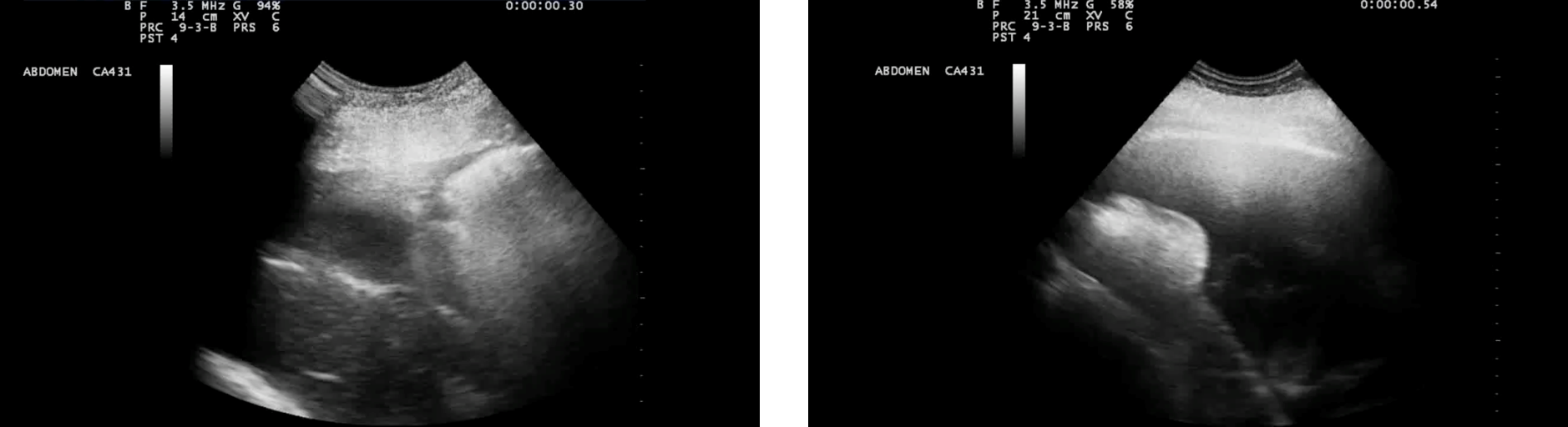
US show
-b lines (top images)
comet tails
consolidated lung with no air (middle images)
pleural fluid and fibrin (bottom 4 images)
on periphery of lung




will us always show pathology
if pathology further inside→ may not reach
radiography show
-alveolar pattern
-fluid lines

-multi nodular bronchoalveolar pattern

-abscesses

what should we do first if wanting to radiograph a horse with pleuropneumonia
remove fluid from thorax first to visualise structures better