NUC biliary and pancreas quiz (10/16)
 Call Kai
 Learn
 Practice Test
 Spaced Repetition
 Match
 Flashcards
 Knowt Play
1/94

There's no tags or description
Looks like no tags are added yet.
95 Terms
The gallbladder is a storage site for _____ which is secreted by the _____.
bile ; liver
The bile ducts are the vessels that carry the bile from the __________ of
the liver to the _____________ , and then to the intestine. The medical
term cholangio refers to the bile ducts (chol(e)-bile, angio=vessel).
lobules ; gallbladder
When we evaluate the Biliary System, we always include what?
the Gallbladder, bile ducts , pancreas, and liver
After a fatty meal, Food in the stomach and duodenum causes _________ ______ _______ to relax and gallbladder to _________, ejecting large amount of _____ into intestine where it aids digestion by emulsifying _____.
Sphincter of Oddi ; contract ; bile ; fats
Primary purpose of bile ducts and gallbladder is to transport bile
from the ______ to the __________ to regulate its flow
liver ; intestine
How much bile does the liver secrete each day?
1-2 liters
Bile drains into what and travels towards the intestine?
biliary ducts
When body is in the fasting state, the ______ ___ __________ keeps enough
tension of the _________ ____ _________ to prevent much bile getting into
duodenum
Sphincter of Oddi ; Ampulla of Vater
What lab value: Liver enzyme that is very sensitive to biliary obstruction.
Elevation indicates mechanical obstruction of the bile ducts
(like stone)
Alkaline Phosphatase (ALP)
What lab value: Elevation can indicate biliary tract obstruction
Alanine Aminotransferase (ALT)
What lab value: Elevation can indicate cholestasis
Aspartate Aminotransferase (AST)
What makes up the bile ducts?
Includes the Common Hepatic Duct, Cystic Duct and Common Bile Duct
What duct:
• Joins the main pancreatic duct at the ampulla of Vater to enter
the descending portion of the duodenum
• Located anterior to the main portal vein and lateral to the hepatic
artery
CBD
Bile backs up in the _____ and is forced into the ____________ which stores
and concentrates bile until the meal is eaten (particularly the fatty
meal)
CBD ; gallbladder
When ___________ empties and peristalsis in ___________ slows, Sphincter
of Oddi ________ and gallbladder __________, limiting flow of bile into
intestine once again.
stomach ; duodenum ; tightens ; relaxes
What causes the gb to contract & Sphincter of Oddi to relax?
Cholesystokinin
What causes liver to secrete more bile?
secretin
What hormones also stimulate the pancreas to secrete its digestive
enzymes?
secretin and cholecystokinin
what emulsify dietary lipids so they can be absorbed
much like detergent breaks down & surrounds dirt in the laundry?
Bile salts
What 4 things describe the cystic duct?
• Drains the gallbladder
• Contains spiral valves of Heister
• Joins the CHD to form the Common Bile Duct
• Not typically seen with ultrasound
The cystic duct is connected to the _____ of the gallbladder.
neck
What is located anterior to the main portal vein and lateral to the hepatic
artery?
common bile duct
What is the normal diameter of the CBD?
6mm or less
The gallbladder is an _____________ organ located in the __________ __________.
intraperitoneal ; gallbladder fossa
The gallbladder lies lateral to the______ and anterior and medial to the ________ __________.
IVC ; right kidney
What are the divisions of the GB?
• Fundus
• Most inferior and anterior portion
• Blind ending
• Body
• Mid portion between fundus and neck
• Neck
• Narrowed area that tapers to connect with the cystic duct
What is the normal wall measurement for the GB?
3mm or less
What is the normal A-P measurement for the GB?
5cm or less in the transverse plane
What is the normal length of the GB?
8-12cm
What is the Phrygian cap?*
• Fold of gallbladder fundus against body
• Common
What is the required patient prep for a GB exam?
- The patient MUST be NPO—NOTHING to eat or drink (some places will let them have sips of water) for at least 8 hours.
- Typically, the patient is instructed to have nothing to eat or drink after midnight the night before the sonogram
- if the patient is not NPO, the gallbladder will usually appear small and thick-walled due to its contracted state.
- The problem is that pathologic conditions of the gallbladder such as cholecystitis also have this appearance.
- Sonograms done on patients who are not NPO can result in a false positive diagnosis of pathology or the report stating that the condition could not be ruled out.
Why is the GB imaged in two different positions?
The purpose of these maneuvers is to
demonstrate the mobility of gallstones and to
make sure that no stones are lodged in the neck
or cystic duct
In order to make the confirmed diagnosis of ____________(as
opposed to polyp or tumor), you MUST document that their
position in the gallbladder changes when the patient changes
position
cholelithiasis (stones in GB)
The gallbladder is attached to the bile duct by the_______ _________ and
normally lies in the gallbladder fossa of the liver, but it is not
_________ ___________ to the liver, so its position is __________ dependent
cystic duct ; tightly fastened ; gravity
- When the patient rolls to the left, the gallbladder fundus moves to the left in response to gravity.
- When the patient sits upright, the fundus of the gallbladder moves inferiorly.
if the GB wall measures more than 3mm what could it indicate?
cholecystitis
For the CBD measurement it should be 6mm or less. Once the patient reaches age __, the normal measurement increases 1mm per ________.
60 ; decade
So, normal CBD size for someone in their 60's is 6-7mm, 70's=7-8mm, etc.
Post-cholecystectomy, the normal CBD size can reach _____ mm.
8-10
Often the GB folds at the junction of the neck and the body, creating a small echogenic linear structure within the GB, which often creates a shadow. This is called a __________ _________.
junctional fold
It is important to
recognize this as a normal
structure and to note that
small stones can be
disguised by its presence.
Rolling the patient onto
his or her side can often
eliminate the problem by
"unfolding" the fold.

In the long/sag view the GB appears more ______. In trans the GB appears more ___________.
stretched out ; circular
What connects the GB and the portal vein?
Main lobar fissure
Describe the Hartmann pouch
Posterior pouch between the neck and junctional fold
What are the rare congenital GB anatomic variants?
- Septations within the GB
- Rare
- Duplicated GB
- Extremely rare
- Confirmed with nuclear medicine hepatobiliary scan
- Agenisis of the GB
- Extremely rare
- Usually confirmed with exploratory laproscopy or laparotomy
The pancreas is located in the ________ _______ space and is a ____________ structure. However, there is a small portion of the __________ __________ that is in the ____________ cavity.
Anterior pararenal ; retroperitoneal ; pancreatic head ; peritoneal
The pancreas lies in a __________ ___________ plane and extends
from C-loop of __________ to hilum of _________.
horizontally oblique ; duodenum ; spleen
What are the posterior landmarks of the pancreas? Hint: 5
Aorta, IVC, SMA, Splenic Vein, and Portal Confluence
__________ __________ arises from aorta just superior to
pancreas body
Celiac Axis
__________, ____________, and ____________ __________ lie
________ and ___________ to pancreas, which can make
it difficult to image due to overlying bowel gas.
Stomach ; duodenum ; transverse colon
anterior ; lateral
What is the normal length of the pancreas?
12-18 cm
Dependent on age and body habitus
What is the pancreas divided into? Hint: 5
head, neck, body, tail, and uncinate
process
What is the primary secretory duct mainly seen in the body and what are the abnormal measurements? Pancreas
Duct of Wirsung ; greater than 2mm in body is considered and greater than 3mm is considered abnormal for head/neck
What is the secondary secretory duct for pancreas?
Duct of Santorini
What surrounds CBD and pancreatic ducts as they go into
duodenum?
Sphincter of Oddi
Arises anteriorly from aorta at superior border of
pancreas (Seagull in transverse)
celiac axis
Tortuous artery that runs along superior/anterior border
of body and tail of pancreas
splenic artery
(just ABOVE where the splenic vein is visualized)
Runs along superior border of neck/head of pancreas
Common hepatic artery
Runs along anterior/lateral portion of
pancreatic head before dividing into superior
pancreaticoduodenal branches
Gastroduodenal artery
Arises from the aorta posterior to the
pancreatic body
Superior mesenteric artery
Runs along posterior border of pancreatic
tail and body from splenic hilum to portal
confluence
splenic vein
- Runs posterior to the neck of the pancreas
and anterior to the uncinate process
- Long axis of this vessel is in the sagittal plane
Superior mesenteric vein
Splenic Vein and Superior Mesenteric Vein
join at portal confluence posterior to
pancreatic neck
This vessel proceeds posterior to pancreatic
head into porta hepatis
Portal vein
Runs anterior to portal vein and lateral
(right) of hepatic artery
Runs through groove on inferior/posterior
portion of pancreatic head
Joins pancreatic duct at Ampulla of Vater
CBD
Pancreas has both _________ and ____________
functions
endocrine ; exocrine
The primary function of pancreas is __________
exocrine
○ Involves secretion of digestive enzymes
(pancreatic juice) into duodenum via Ampulla of Vater
○ These secretions are necessary for normal digestion and absorption of food
What secrete hormones involved in endocrine function of pancreas?
Islets of Langehans
What are the 3 types of Islets of Langerhans cells and what are their basic functions?
- Beta Cells: secrete insulin
- Most numerous type
- Insulin stimulates liver to form glycogen from glucose
- Also stimulates certain cells to take up more glucose from bloodstream
- Net effect of insulin is to decrease levels of glucose in bloodstream
- Alpha Cells: secrete glucagon
- Stimulates cells to release glucose to meet energy needs of body
- Antagonistic to insulin
- Stimulates liver to break glycogen down to glucose for use
- Net effect of glucagon is to increase levels of
glucose in bloodstream
- Delta Cells: secrete somatostatin
- Inhibits production of both insulin and glucagon
- Helps to keep glucose levels in blood from making large swings
What is the congenital anomaly: annular pancreas?
Pancreas encircles the duodenum
Thought to occur during rotation of the
ventral bud to meet the dorsal bud
Ventral bud as split (bifid) formation that is
not usual
May cause duodenal stenosis

What is the congenital anomaly: Ectopic Pancreatic Tissue
Pancreatic tissue found within the wall of
the bowel
Can be found in the foregut or midgut
Functioning tissue which can develop
inflammation or tumors just like the
normal pancreas
What is the congenital anomaly: Pancreas Divisum
Occurs in about 10% of the population
* The Duct of Wirsung & the Duct of
Santorini do not fuse, and each drain
separately into the duodenum

What is a special thing a patient can do to better help visualize the pancreas?
Drink a lot of water
- If pancreas is extremely difficult to visualize,
have patient drink at least 12-16 oz. of water
○ Water will displace gas in stomach and
provide an acoustic window (like bladder
provides a window to visualize the uterus)
○ Water should ideally come from a bottle and
should be drank through a straw to minimize
air bubbles being swallowed
○ The patient should also be in upright or left
lateral decubitus position
_______, _________, _________, _________, and ______ of the
pancreas should be evaluated, comparing
it to the liver for echogenicity and texture
Size, shape, texture, contour, and lie
When the pancreas is measured in long plane that is the pancreas in the ________ section.
transverse
What landmark would you see: Long Pancreas Head
IVC
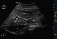
What landmark would you see: Long Pancreas Neck/UP
SMV
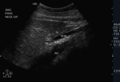
What landmark would you see: Long Pancreas Body
Aorta, Celiac axis, SMA

What landmark would you see: Long Pancreas Tail
just past posterior dive of splenic artery and vein

CBD
1

Cystic duct
2

Cystic artery
3

Right and left hepatic duct
4

Right and left hepatic arteries
5
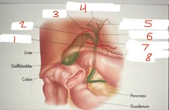
Common hepatic duct
6

Proper hepatic artery
7

Right gastric artery
8

Ampulla of vater
1

fundus of gallbladder
2

Body of gallbladder
3

Hartmanns pouch
4

Neck of gallbladder
5

Heister's valve
6

right hepatic duct
7

Left hepatic duct
8

common hepatic duct
9

Cystic duct
10

Common bile duct
11

Pancreatic duct
12
