MICI 3114- Virology Midterm 2
 Call Kai
 Learn
 Practice Test
 Spaced Repetition
 Match
 Flashcards
 Knowt Play
1/137

There's no tags or description
Looks like no tags are added yet.
138 Terms
What are the features of RNA virus macromolecular synthesis?
RNA viruses do everything that DNA viruses do of APUTTGAR but all in the cytoplasm (except retroviruses)
no nuclear activity
central dogma is that DNA viruses need to make mRNA → protein
RNA virus make mRNA→ protein + and - RNA viruses (all need to make mRNA and templates)
+RNA makes antisense genome
viruses are more autonomous than DNA
all reading 3’ to 5’
3’ end where RdRp binds and makes poly U tail and start decoding

What are virus classifications?
The Baltimore system is a method of classifying viruses into seven groups according to:
• Their type of genetic material: DNA or RNA
• Whether the genome is single- or double-stranded
• How they produce messenger RNA (mRNA) for protein synthesis
diff shapes and varieties
RNA viruses have diverse structures and genome arrangements

What is the process of + Strand RNA virus replication?
replication: makes a - strand to then be able to make sgmRNA from the -strand

What is the process of - strand RNA virus replication?
undergoes mRNA synthesis to make + strand
makes + full length complement

What is the process of ambisense RNA virus replication?
makes sgmRNA and antigenome to make partial components
or dsRNA can make a full strands for each sense

What are the general features of Virus Macromolecular Synthesis?
• cytoplasmic replication (except orthomyxovirus)
• no "classical" promoters or enhancers to regulate mRNA production
• must encode their own RdRp for mRNA production and genome replication
• no use of host transcription machinery which resides in the nucleus
• no use of host post-transcriptional machinery (except orthomyxovirus)
• must control RNA template utilization (Tc, TL, genome replication, packaging)
• transcription and genome replication
• genome replication independent of host cell cycle
Since their genomes are unlike the host genome, transcription and genome replication occur independent of most of the pathways host cells use for these processes.
They don't need to subvert this machinery; they do need to subvert the translation apparatus.
What are the features of RdRp?
• Viral RNA genomes must be copied within the infected cell to provide both genomes for assembly into progeny virus particles and messenger RNAs (mRNAs) for the synthesis of viral proteins.
• All RNA viruses except retroviruses (RT) and hepatitis delta virus encode an RNA-dependent RNA polymerase (RdRP).
• Negative strand RNA viruses must package an RdRP molecule in each assembled virion to be infectious.
• Positive strand RNA genomes encode an RdRP and are infectious when directly transfected into the host cell
• Viral RdRP’s are not inhibited by α-amanitin which inhibits cellular RNA pol II.
• Viral RdRP’s do not have proof reading ability and they make mistakes leading to mutations; rate varies with the viral RdRP species type
What are the physical features of RdRp?
resembles a right hand consisting of palm, fingers, and thumb domains with the active site located in the palm
cleft in where the template goes
nascent strand associate and hybridized to template
ds rough intermediate
The structures of RdRPs differ in detail from those of other polymerases, presumably to accommodate different templates and priming mechanisms. All nucleic acid polymerases have a similar core catalytic domain configuration and evolved from a common ancestor.

Do ribosomes and RNA polymerase collide during RNA virus replication?
At a certain point in infection, the RNA polymerase copies the RNA in a 3 → 5′ ′ direction while ribosomes traverse it in an opposite direction, raising the question of whether the viral polymerase avoids collisions with ribosomes.
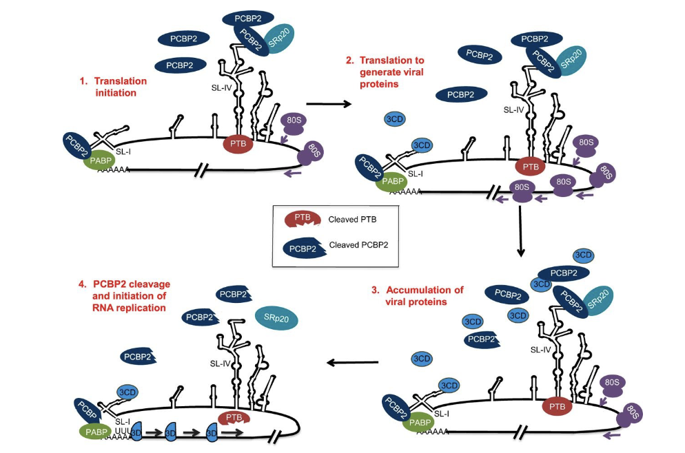
Binding of cellular poly(rC)-binding protein 2 (PCB2) within the 5 UTR initially stimulates translation. Once the viral protease has been synthesized, it cleaves poly(rC)-binding protein (PCB2), and binding of the cellular protein is reduced to favor replication.
Ambisense arenaviruses have a stem loop region IGR that blocks opposing ribosomal translation and RdRP movement until enough NP is made to melt the loop.
Alternatively replication occurs in membrane vesicle compartments and translation occurs in the cytosol
if keep ribosomes away from replication→ replication vesicles that have factories of replication
ambisense: genome can make mRNA in two directions
cleaved poly(rC)-binding protein can still bind to a different segment of the 5′ untranslated region (the cloverleaf) and promote viral genome synthesis.

What are the features of RNA capping in RNA virueses?
Most viral mRNA’s have a 5’ terminal cap structure and they use one of three ways of forming these caps:
1) Cap snatching 5’ends from existing cellular pre-mRNAs or mRNAs (e.g. influena or Bunyamwera viruses)
2) De novo synthesis using cellular enzymes to produce 7’meGuansine caps
3) Use of viral coded enzymes such as methyl, guanyl, uridyl tranferases associated with RdRP or other non-structural proteins
4) Addition of internal ribosome entry sites (IRES)
5) Addition of a protein (Vpg) or incorporation of an IRES elment at the 5’ end of the mRNA or genome
What are the functions of 5’ caps?
cap structure directs pre-mRNAs to processing and transport pathways, regulates mRNA turnover, and is required for efficient translation by the 5′-end-dependent mechanism. 5′ untranslated regions often contain secondary structures (e.g., hairpin loops) formed by base pairing of the RNA. These double-helical regions must be unwound to allow passage of 40S ribosomal subunits during translation.
What are the features of elongation in RNA viruses?
Nucleic acid synthesis begins with the formation of a complex of RdRP, template-primer, and initiating NTP. The NTP α-phosphate undergoes nucleophilic attack by the 3 -OH of the primer strand. The nucleotidyl transfer′ reaction then takes place, pyrophosphate is released, and the template-primer moves by one base. Viral helicases encoded asNS proteins can unwind secondary RNA structure
What are the features of IRES (internal ribosome entry sites)
• Different sets of translation initiation proteins are required for the function of various IRESs; the structures very much resemble tRNAs
• Internal ribosome binding on the hepatitis A virus IRES requires all the initiation proteins, including eIF4E. At the other extreme, the intergenic IRES of cricket paralysis virus requires none of them
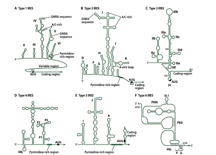
What are the features of 40S ribosomal subunit binding to IRES?
Forms initiation complex
can shut down host protein synthesis
elF4G (fragment) paralyses 5’ cap
poly C binding protein interacts with eIF4A
IRES in the top is interacting with initiation factor eIF3 and eIF4G
In the case of poliovirus, its protease cleaves a portion of eIF4G but a large portion is retained which binds to loop 4 of the IRES Type 1.

What are the diverse ways of maximizing number of proteins from a Monocistronic Genome?
• Polyprotein synthesis – picornaviruses, flaviviruses, HCV togaviruses
• Start-Stop-PA-transcription – paramyxoviruses, rhabdoviruses
• Intergenomic secondary RdRP binding site – togaviruses (sgmRNA)
• Ambisense transcription of genome and antigenome RNA’s (arenaviruses and some bunyaviruses)
• Reinitiation – Flu B RNA Segment 7
• Leaky Ribosomal Scanning – P/C genes of Sendai, P/V/C gens of Measles (overlapping reading frames)
• Use of alternative initiation codons (ACG instead of AUG due to tRNA like secondary RNA structure)
• Ribosome shunting across a hairpin loop – jumping RdRP
• IRES (internal ribosome internal entry site) (Bicistronic mRNA);
ribosome entry – Picornavirus, Flavivirus, HCV
What are the features of polyadenylation signals on RNA viruses?
• In (-)strand non-segmented RNA viruses the ribosome complex can proceed in a 5’ direction and pause at a stop codon to add a poly A tail; the ribosome complex then reinitiates at the next AUG and continues to the next stop codon, pauses, and repeats the stuttering process. Within the intergene sequence between the stop codon and the next initiation codon (AUG) is a short 5-7 uridine (U) stretch that signals the polyadenylation. Stop-Start model.
• In (+) strand RNA viruses the polyadenylation of the genome is specified by a long stretch of U residues and is more specific in length depending on the virus species.
• In some instances, de novo polyadenylation can occur to repair damaged RNA genomes but it is inefficient and less common.
• Poly A tails stabilize mRNA, helps move it from the nucleus, and aids the mRNA and 40S ribosome subunit recruit initiation factor in association with the PABP.
• The poly(A)-binding protein plays a crucial role in mRNA translation, bringing together the ends of the mRNA; poly(A)-binding protein is required for IRES-mediated translation initiation and helps recruit factors. Cleavage of this protein in enterovirus-infected cells inhibits host cap mediated translation
What are the general features of ambisense viruses (arenaviridae)?
• Although arenaviruses (eg Lassa, LCMV) are considered (−) strand RNA viruses, their genomic RNA is in fact ambisense: mRNAs are produced both from (−) strand genomic RNA and from
complementary full-length (+) antisense strands.
• The short S and the long L genome segments are transcribed first from the 3’ ends to produce N nucleocapsid and L polymerase mRNA’s first; the mRNAs are capped with 3-4 nucleotide fragments from host cellular mRNAs (capsnatching) using the endonuclease associated with L (RdRP).
• The hairpin loop (IGR) prevents the RdRP from moving down the genome and terminates transcription; as you get more N made it melts the loop and allows the formation of anti-genomes.
• From the 3’ ends of the anti-genome the mRNA’s for pre-GP and Z are transcribed for synthesis of GP1, GP2, SSP, and Z proteins.
• S and L genomes are copied full length from the 2 antigenomes again in the presence of high amounts of N which form helical nucleocapsids.
• The Z protein is a virulence factor which aids in the packaging and budding of assembled virions from the host plasma membrane

What are they types of picornavirus?
• Enterovirus: Causes polio, human hand-foot-and-mouth disease, myocarditis, and meningitis.
• Rhinovirus: Causes the common cold and upper respiratory infections.
• Hepatovirus: Includes Hepatitis A virus, causing acute liver inflammation.
• Aphthovirus: Causes foot-and-mouth disease in cloven-hoofed livestock.

What are the features of the poliovirus replication cycle?
virus grows in cytoplasm (no nucleus)
+ssRNA replicates in membrane vesicles (can become double membraned)
pirate ER and Golgi membrane where the virus grows
virus that impact the nervous system

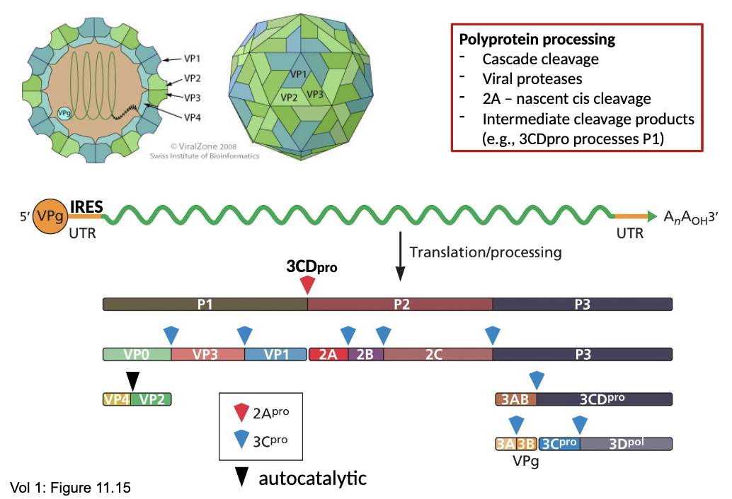
What are the features of the poliovirus genome organization (Poliovirus)?
Polyprotein processing
- Cascade cleavage
- Viral proteases
- 2A – nascent cis cleavage (cleaves P1 and P2)
- Intermediate cleavage products (e.g., 3CDpro processes P1)
3C protease cleaves the rest
How are polioviruses proteases involved with host shut off?
this virus makes the cap irrelevant causes host shut off and prevents cellular proteins from being made 2B and 3C cleave elF4G (important step leave it partially functioning for the IRES but leaves the eukaryotic feature out)
Type 1 IRES allow cap independent translation, it bypasses scanning and attracts host proteins and initiation factors.
dark green squiggle is the PCBP2 (critical host trans acting factor for type I IRES function)
elF4A with elF2 eIF4F
part that leaves is the 2Apro

What are the features of poliovirus attachment to cell receptor?
Attachment of poliovirus to the cell receptor and injection of viral genome RNA into host cell
Poliovirus binds to its receptor (CD155) on the cell surface via VP1, triggering a conformational change with externalization of VP4, pore formation, and extrusion of Vpg-RNA genome into the cytoplasm. On the other hand, Rhinoviruses and foot-and mouthdisease virus have been shown to enter the cell by receptor- mediated endocytosis. The capsid dissociates in the late endosomes and lysosomes in presence of their low pH, releasing the the Vpg-RNA genome into the cytoplasm
Pore formed by VP1 allows for RNA injection and collapsing of VP4 which enters the pore
VPg can be removed to expose the IRES

How do type I IRES’s function?
Allows translation to proceed in cap-independent manner
Critical for infection because viral proteases 2A, 3C cleave eIF4G and PABP to shut host cap-dependent translation.
Type I IRES bypasses scanning and attracts host proteins and initiation factors.
PCBP2 is the primary most critical host trans acting factor (TAF) required for type I IRES to function
Cap-binding factors eIF4G interacts directly with the IRES (often helped by PCBP2)
eIF4G recruits eIF3- alongside the 40S ribosomal subunit and initiator tRNA (Met) directly to initiation start site.
Vpg is cleaved from +ve genomes in the cytoplasm revealing IRES by TDB2 (phosphatase) prior to translation beginning
eIF-4E interacts with eIF-4G and it gets cleaved from the mRNA

What are the features of the poliovirus VPg protein?
The VPg (Viral Protein Genome-linked (protein 3B). It plays a role in safeguarding the viral genome (stability) and priming replication.
• Unlike cellular mRNAs that possess a 5' cap (7meGpp), the poliovirus (+) ssRNA genome is covalently linked at its 5' end to VPg.
• It shields the viral RNA from degradation by host cellular exonucleases and acts as a molecular switch, determining whether the genome enters translation (proteins) or replication (viral RNA genomes or antigenomes).
• VPg is permanently attached to the viral genomic RNA packaged inside mature virus particles (virions), but it must be cleaved off the incoming viral RNA before it can be translated by host ribosomes.
• A host cellular enzyme known as TDP2 (Tyrosyl-DNA Phosphodiesterase 2), acting as a "VPg unlinkase," specifically cleaves the phosphodiester bond linking VPg to the 5' terminal nucleotide.
• This exposes an Internal Ribosome Entry Site (IRES), allowing the host translation machinery to bind and initiate protein synthesis without a traditional 5' cap.
How does VPg work as a primer during replication of Poliovirus?
• The viral RdRp (protein 3D) catalyzes the transfer of two
uridine monophosphate (UMP) molecules onto VPg.
• This reaction yields VPg-pUpU, a uridylylated protein
intermediate with a free 3’ hydroxyl. This step occurs at a
specific internal structural signal within the viral genome
called the cis-acting replication element (cre) which is a highly
conserved loop containing AAACA.
• The 3’ hydroxyl group of the terminal UMP residues on the
VPg-pUpU complex serves as the primer to initiate synthesis.
• A phosphodiester bond forms between the hydroxyl group of Tyrosine residue 3 (Tyr-3) near the amino-terminus of the VPg peptide with the 5'-terminal phosphate of a uridine dinucleotide
What are the features of RNA Replication- Picornavirus Genome synthesis?
CRE (cis replication element) are stem loops with a consensus AAACA which can trigger the uridylation of VPg by 3CD and stimulate priming and replication
Replicative Intermediate (more double stranded circular because of poly A binding protein (PABP))
The number of + ve strands made exceeds number of negative strands
3’ antigenome starts to copy the A residues and forms a Poly U tail
travels from 3’ to 5’
RNA replicative intermediates have ds being formed
Poly U tail made and VPg on 5’ end where the original template
VPg attached to 3’ end of the negative antigenome RNA
can create multi stranded replication intermediates (makes them into + strands)

What are the features of the 3CD -PCBP2 translation replication switch in poliovirus?
• The viral protease 3CD acts as a molecular switch by cleaving
the host cell protein PCBP2 (poly(rC)-binding protein 2),
which stops viral translation and allows genome replication
to begin on the same RNA template. Dual Role of PCBP2:
• Translation Support: intact PCBP2 binds to stem-loop IV in
the 5' noncoding region of the poliovirus genome and
recruits components required for the Type I internal
ribosome entry site (IRES) to drive viral translation.
• Replication Interference: Because translation and RNA
synthesis cannot happen at the same time on a single
positive-sense RNA strand, active ribosomes block the RNA-
dependent RNA polymerase. Prevents Collision
What are the features of the 3CD-Mediated Cleavage Mechanism? (poliovirus/ picornaviridae)
• Targeted Cleavage: During the mid-to-late phase of infection, the
viral proteinase 3CD (and/or 3C) cleaves PCBP2 at a specific site
• Shutoff of Translation: The resulting cleaved fragment loses its ability to bind stem-loop IV of the IRES, destroying the functional IRES complex and causing ribosomes to fall off the RNA template.
• Activation of Replication: The cleaved PCBP2 fragment retains its capacity to bind to stem-loop I at the 5' end of the genome (clover leaf) and form a functional ternary complex with the viral 3CD protein. This complex shifts the template from a translation hub into a dedicated site for negative-strand RNA synthesis and genome replication

What are the features of the PABP-3CD Coordination (Genome Circularization) (in poliovirus/ picornaviridae)?
• Poliovirus replicates its positive-sense RNA genome into a negative sense anti-genome template forming a double-stranded replicative intermediate (RI), which requires 3Dpol.
• Bridge Complex: Poly(A)-binding protein (PABP) binds to the 3' poly(A) tail of the viral RNA. Simultaneously, 3CD binds to the cloverleaf structure (stem-loop I) at the 5' end of the same RNA molecule (assisted by the cleaved PCBP2 fragment).
• Circularization: PABP interacts directly with the 3CD-cloverleaf complex. This protein-protein bridge bends the single-stranded viral RNA into a closed circle loop.
• Polymerase Delivery: Circularization brings the 3' poly(A) tail into proximity with the 5' end. This positions the viral replication machinery—specifically 3Dpol (the active polymerase cleaved from 3CD) and the VPg priming protein (3B)— on the 3' end of the antigenome to begin negative-strand RNA synthesis.
• Initial synthesis at the 3' poly(A) tail
What are the features of membrane rearrangements (replication factory in poliovirus)?
• Poliovirus cannot replicate in the open cytoplasm; it must shield its double-stranded RNA intermediates from host immune sensors
(like RIG-I/MDA5). It achieves this by remodeling host intracellular
membranes into replication complexes/factories.
• Lipid Recruitment via 3CD/3CD-interactors: Viral proteins 2BC and 3A alter host lipid homeostasis. They recruit GBF1 (Golgi-specific GEF) and PI4KB (phosphatidylinositol 4-kinase beta) to the
replication site.
• PI4P and Cholesterol Enrichment: PI4KB synthesizes a unique lipid called phosphatidylinositol 4-phosphate (PI4P) on the target
membranes. High levels of PI4P then attract host OSBP (Oxysterol-
binding protein), which pumps cholesterol into these viral
membranes.
• 3CD Binding to Remodeled Membranes: The localized
concentration of PI4P and cholesterol creating vesicle clusters.
• 3CD binds directly to these PI4P-rich lipid platforms. This
anchoring concentrates 3CD, its cleaved products (3C and 3D), and
the circularized viral RNA template into localized "factories,"
multiplying replication efficiency.
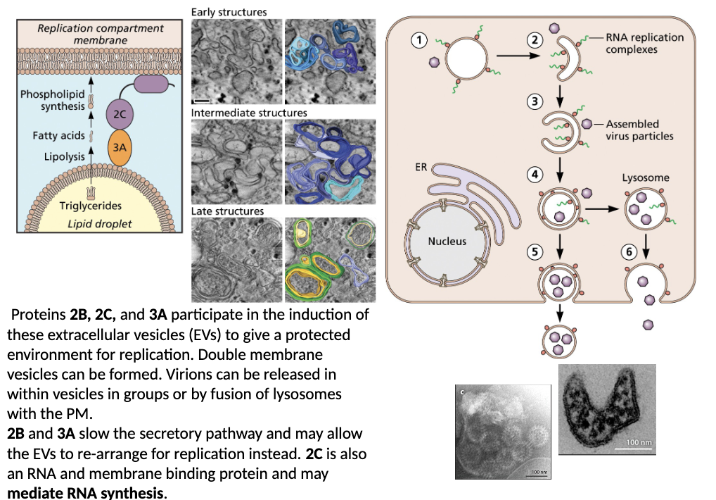
What are the features of poliovirus vesicle biogenesis
Proteins 2B, 2C, and 3A participate in the induction of these extracellular vesicles (EVs) to give a protected environment for replication. Double membrane vesicles can be formed. Virions can be released in within vesicles in groups or by fusion of lysosomes
with the PM.
2B and 3A slow the secretory pathway and may allow the EVs to re-arrange for replication instead. 2C is also an RNA and membrane binding protein and may mediate RNA synthesis
How does poliovirus overcome the features of monocistronic constraint, control, genetic economy and competition?
Monocistronic Constraint
- polyprotein production
Control
- standard cis/trans control (e.g., RdRp binding sites)
- IRES element controls cap-independent TL
- VPg regulates TL versus RNA replication
- 2BC and 3A alter vesicle biogenesis to generate replication complexes
- cascade cleavage generates intermediates with different functions
Genetic Economy
- cascade cleavage generates intermediates with different functions
- multi-functional viral proteases
- sacrifices economy with large IRES in exchange for host-cell competition (disrupts eIF4F complex)
Competition
- 2A cleaves eIF4G (inhibit cap-dependent host TL)
- 3C cleaves polyA binding protein (PABP) (inhibit cap-dependent host TL)
- 2BC and 3A alter cellular membrane biogenesis (pleiotropic effects on cell)
What are the features of Hepatitis A?
Liver infection
It commonly spreads through contaminated food or water or close contact with an infected person. Symptoms can include fatigue, nausea, loss of appetite, abdominal discomfort, dark urine, and jaundice. Unlike hepatitis B or C, hepatitis A does not usually cause chronic infection.
What are the features of coxsackie virus?
Coxsackievirus infections can cause hand-foot-and-mouth disease, herpangina, and nonspecific febrile/respiratory, gastrointestinal illness, neurological symptoms. Most infections are mild or asymptomatic
What are the features of rhinovirus?
common cold
Disease symptoms include, runny or stuffy nose, sneezing,
sore or scratchy throat, cough, mild headache or body
aches, low-grade fever, symptoms usually improve within 7–
10 days
What are the features of foot and mouth disease (FMD)?
FMD is a highly contagious viral disease of cloven-hoofed
animals including cattle, pigs, sheep, goats, and buffalo; not the same disease as hand, foot and mouth disease in humans.
What are the features of the genome of Calicivirus (norovirus)?
Most caliciviruses use VPg both as a primer for replication and also as a cap for translation. For the most part they do not use IRES elements. Contains a subgenomic RNA binding site to make a sgmRNA that codes for the structural proteins VP1 and VP2. VP2 is synthesized following a ribosomal termination-reinitiation site. VP1 has a protruding region (P) and an inner shell region (S). VP2 is a small organizing protein.
3 open reading frames
ribosomal termination-reinitiation (small less frequent protein)
VPg is not removed prior to translation (polio it is )
VPg both replication primer and can work like and IRES
to get the subgenomic RNA need viral antigenome (binding site which is for the RNA pol →sgRNA and makes mRNA (+ sense subgenomic))

What are the features of the open reading frames of Calicivirus (norovirus)?
ORF1: Non-structural proteins (proteases, VPg like protein, RdRp, elements for replication vesicles)
ORF2: Major structural proteins (Capsid, shell, protrusion)
ORF3: Minor structural proteins: low copy numbers helps stabilize structure

What are the features of Norovirus in relation to its primer and cap activity?
The VPg protein of noroviruses is a small covalently linked to the 5' end of all viral genomic and subgenomic mRNA. Norovirus relies on VPg to drive both viral genome replication and translation initiation.
What is the Mechanism of Genome RNA Synthesis Priming By Vpg in Norovirus?
During replication, the viral RdRP cannot initiate RNA synthesis de novo. Instead, VPg acts as a protein primer through the following steps:
• Uridylylation: The viral RdRp catalyzes the transfer of a uridine monophosphate onto a conserved tyrosine residue (Tyr-24) within VPg, forming a covalent VPg-pU intermediate.
• Primer Recognition: This uridylylated VPg presents a free ribose hydroxyl group that the RdRp recognizes and positions at the initiation site of the template RNA.
• Strand Extension: The RdRp extends the nascent RNA chain from the VPg primer, leaving VPg permanently attached to the 5' terminus of the newly synthesized strand (genome or antigenome).
What is the role of VPg in relation to translation initiation in Norovirus?
VPg also plays a critical role in hijacking the host cell's protein synthesis machinery:
• Initiation Factor Binding: VPg binds host translation initiation factors, specifically binding to eIF4E and eIF4G.
• Ribosome Recruitment: serves the function of a standard cap, recruiting the 40S ribosomal subunit to the viral genome RNA and sgmRNA, 60S ribosomal subunit, followed by elongation.
What are some antiviral drugs directed against norovirus?
RdRp active site allosteric inhibitors
Targeting intrinsically disordered regions (IDPs) tries to block interactions of residues before VPg can dock
Protease inhibitor (NS6)
What are the features of norovirus?
The physical symptoms are a consequence of the virus infecting the
small intestinal enterocytes. The acute symptoms of norovirus
infection—such as severe vomiting, watery diarrhea, nausea, and
abdominal cramps—are not caused by a single designated "toxin"
protein. Infections can be initiated by as few as 1-10 virus particles.
How is Norovirus hypervariable?
• High Genetic Diversity and Strain Variation: Norovirus is divided into multiple genogroups and dozens of genotypes. Infection with one strain often provides little to no cross-protection against other strains. Because so many different strains circulate in the human population, catching one type doesn't mean you are immune to the others. (no cross protection for other strands)
• VP1 and VP2 undergoes genetic drift.
• Immunity to a specific strain is short-lived 6-8 months.
• Perfect Pathogen - combination of rapid evolution, high contagiousness, low infectious dose, and short-lived immunity
What are the features of Matonaviridae (Rubiviridae)?
Rubella virus (humans); ruhubu virus (bats), rustrela virus (rodents); transmitted by aerosols; rubella virus is transmitted by aerosols
Rubiviruses: The Rubella virus causes rubella (German measles), characterized by a maculopapular rash and mild fever,
which poses severe risks of congenital defects if contracted during early pregnanc
What are the features of togaviruses?
A specific group of viruses that possess a membrane envelope or toga (Roman clothing)
Alphaviruses are transmitted by arthropods (mosquitoes)
Genome: (+) ssRNA
Capsid: icosahedral nucleocapsid core enclosing the viral genome
Envelope: A host derived lipid bilayer membrane studded with transmembrane heterodimers of viral glycoproteins (E1 and E2), which form heterotrimeric surface spikes responsible for host cell attachment and membrane fusion

How do togaviruses enter host cells?
Togaviruses enter host cells in clathrin coated pits during receptor
mediated endocytosis.
Virions enter the early endosome and the envelope fuses with the late endosome membrane at low pH and releases the nucleocapsid into the cytosol. Fusion of the viral envelope with the late endosomal membrane at low pH.
pore formed and fusion peptide that comes out membrane protein E1 and it goes into target membrane bringing the membranes close together creating hemifusion and that creates a pore
SFV and SINV also enter host cells in calveolin endosomes and by macropinocytosis depending on tissue and cell type

What are the features of Togavirus RNA (+) Genome and Viral Protein Products?
copied into subgenomic RNA and theres transcriptional control region between the polyproteins
non-structural proteins precursor 123 and nsP4 which is the polymerase (partial cleavage and the full cleavage is achieved to get full activity)
late replication nsP1,2,3,4
CP= capsid protein
E3= envelope protein
6K can be modified by frameshift and creates TF which contains part of 6K E1 has stop codon bc of this frameshift and this make small amounts of E1

What are the differences between alphavirus and rubivirus genomes?
Rubivirus has not 6K protein being made only 3 structural proteins and the non-structural only 4 proteins

What is the conserved sequence element (CSE) in alphavirus genomes?
Don’t need to know this in too much detail
packaging signals in RNA that help it package into nuclear capsid (diff signals for diff viruses)
Features of the alphavirus genome:
protein that caps the genome
RdRp
protein that help with fusion,
spike
TF: trans frame

What are the features of togavirus spike proteins?
E1 E2 that has been activated by cleavage and low pH environment has fusion peptide that goes into the target membrane (different than type 1 fusion protein) this type 2 and has a lot beta sheets activated by low pH
E2 has two functions: its cytoplasmic domain interacts with the nucleocapsid for assembly and its ectodomain is responsible for binding a cellular receptor

What are the features of togavirus spike proteins and virus entry?
p62 (aka pE2 aka E2/E3)- chaperone for E1
E2 - binding and assembly
E1 - fusion protein
6K - budding
Fusion in alphaviruses requires dissociation of the E1-E2 heterodimer and formation of E1 homotrimers Dimer dissociation results in the exposure of the E1 fusion loop E2/E1 dimer dissociation, insertion of E1’s fusion loop into the endosomal membrane, and E1 homotrimer formation

What are the features of Togavirus genome organization and replication?
antigenome being made:
P123+NS4 = lower grade polymerase and makes the antigenome (-) strand when fully cleaved have full activity and makes a lot of the genome and subgenome
binding region for RNA polmerase and bind at the red arrow
formation of subgenomic + strand mRNA and then translated to make the capsid
precursor E2 not fully cleaved
capsid can be cleaved during replication because it is autolytic (spans through the membrane)
further cleavage of structural proteins (cascade of cleavage)

What are the features of the togavirus downstream hairpin loop?
DLP RNA activates PKR which phosphorylates eIF2α and inhibits canonical translation
control element that acts like tRNA
normal cell has EIF2 function→ important initiation function
because it is a dsRNA it phosphorylated eIF2alpha and it gets removed
no eIF2
causes initiation and make more structural proteins because of this modification
DLP structure can stall 40S subunit scanning

What are the features of Togavirus Ribosomal Frameshifting (NS3/NS4 and 6k/TF) Junctions?
Frameshift signals include a followed by an heptanucleotide “slippery sequence” RNA pseudoknot
Secondary structure of pseudoknot stalls the ribosome and slippery sequence allows ribosome to slip back one nucleotide while maintaining the tRNA in the peptidyl tRNA binding or P site and then resume translating but now in a new -1 reading frame
6K region frameshift goes and takes part of the ORF out and shifts -1 don’t get the AUG that is normally there and don’t get an E1
slippery sequence → XXXX(any nucleotide)Y (A or U) and Z is (A,C or U)
ribosome shifts back
envelope protein in replication vesicle
E3 activation
C protein has signal leader activity signal peptides are cleaved

What are the features of Togavirus Transcription and Genome Replication?
cascade cleavage by nsp2 protease (like poliovirus) to generate three different RdRp complexes
RdRp is big and can only encode 1 (nsp4) so uses cleavage to modify RdRp function (control, genetic economy)
Nsp1/23/4 complex could ping pong back and forth on RI structure making more FL + and – until 23 gets cleaved

What are the features of Alphavirus 5’-end Capping?
NSP2 triphosphatase activity generates a diphosphate at the 5′ end of the newly synthesized genomic RNA. N
SP1 guanine-7-methyl transferase activity methylates GTP to N7-methyl guanosine triphosphate which is transferred to the 5’ end of the genome RNA by NSP1 m7-guanylyl transferase to generate a ‘type 0’ capped RNA. Pi, inorganic phosphate; PPi, inorganic pyrophosphate.

What are the features of Structural Protein Processing, Assembly, and Budding (Egress) in rubella virus?
The carboxy terminal region of‐ the capsid protein contains a
serine protease domain that autocatalytically self-cleaves
the capsid protein from the nascent polypeptide chain.
Signal peptidase (host)-white square- cleaves 6k away from
E1 and pE3-E2.
Furin (host late Golgi)- white circle activates fusion potential
by cleaving E3
Unlike the alphaviruses, rubella virus assembles and buds into
the Golgi factory membranes is released via secretory vesicles
(not shown).

What are the functions of the 6k protein in Togaviruses?
6K protein and its overlapping translation product, the TF (transframe) protein, are small
hydrophobic membrane proteins situated between pE2/E2 and E1.
1. The 6K Protein
The 6K protein is a classic viroporin (ion channel) that optimizes internal environment for viral replication and transport.
Glycoprotein Transport and Maturation: acts as a signal sequence and assists in the proper folding, trimerization, and trafficking of the E1 and E2
Budding and Virion Release: 6K is critical for efficient virus budding. Retained inside infected cells but mechanism is vague.
What are the functions of the TF protein in togaviruses?
2. The TF (Transframe) Protein
TF protein is produced when ribosomes undergo a -1 frameshift within the 6K open reading frame. Shares N-terminus of 6K but has a unique, palmitoylated terminal tail. Reduces amount of E1.
Incorporated into Virions: Unlike 6K TF is efficiently incorporated into budding and mature virions; is detected on the viral envelope surface.
Pathogenesis: TF is a major virulence factor. Mutations or deletions severely attenuate alphavirus replication and reduce virus production disease severity.
Host Subversion and Immune Evasion: TF has been implicated in suppressing host innate immunity by inhibiting type I interferon and host cell polarity proteins (Scribble-PDZ motifs); facilitates virus egress
How does togavirus produce host shut off?
Interferon stimulated genes (ISG’s) can trigger cell death
NS2 in Old World viruses can degrade RNA pol II subunit RPB1 and blocks cellular transcription
Capsid (NP) New World viruses can bind nuclear importin and exportin proteins and block themiagration and production of host cell mRNA (CRM1 and Nup98)
dsRNA and stress can activate PKR and PERK to phosphorylate eIF2 and inhibit host protein translation; DLP on sgmRNA overcomes this; allows viral proteins to be synthesized
NSP3 of many Old World alphaviruses interacts with‐ G3BP and its mosquito homolog Rasputin to form cytoplasmic complexes that block assembly of stress granules, preventing their associated antiviral response
NSP2 protease can block kinase and phosphorylation of proteins in the interferon Jak-STAT pathway
Viral sgmRNA out-competes host mRNA
What are the features of alphaviruses?
Arthritogenic Alphaviruses: Cause acute febrile illness accompanied by severe, debilitating, and long-lasting joint pain
(arthralgia).
Encephalitic Alphaviruses: Cause inflammation of the central nervous system, often leading to neurological damage or death.
How do togaviruses overcome the features of monocistronic constraint, control, genetic economy and competition?
Monocistronic Constraint
- polyprotein production and processing (stop codon readthrough)
- ribosomal frameshifting
- sgmRNA production
Control
- standard cis/trans control (e.g., RdRp binding sites)
- internal RdRp binding site (RBS, quantitative control)
- downstream hairpin loop (DLP, late virus TL)
- cascade cleavage generates multiple RdRp complexes
Genetic Economy
- cascade cleavage generates multiple RdRp complexes
- viral and cellular proteases
Competition
- downstream hairpin loop (TL with no eIF2)
Where does togavirus genome replication occur?
Genome replication occurs in association with spherules on cellular
membranes
Alphavirus nonstructural proteins, together with viral genome RNA, induce formation of membranous invaginations termed "spherules" on cellular membrane structures. The NS proteins along with nascent genome RNA assemble with the plasma membrane to form type I cytopathic vacuoles (CPV-1), where RNA synthesis occurs.
Type I has spherules invagination of ER membranes, rearrangement of membranes
Type II vacuoles later on → and bud into ECF and bud from the plasma membranes
What are some of the diseases of flaviviruses?
• Yellow Fever
• Dengue
• Japanese Encephalitis
• Tick-borne Encephalitis
• Powassan
• West Nile
• Zika
• Over 50 different flaviviruses around the world
What is the general structure of flavivurses?
precursor M organizing protein on the inside of the envelope
shell on the outside E protein make dimers

What are the features of Flavivirus Polyprotein Production and Processing?
+sRNA virus being cleaved by proteases
signal peptidase and Golgi protease and NS3 protease (viral proteases)
=core proteins the C M E NS1, NS3,NS4A, NS4B, NS5
cleavage needs to happen
polyprotein strings through ER membranes NS3 NS5 on cytoplasmic side
NS1 and E are in the ER
Proteolytically processed by both viral and cellular proteases
Golgi protease cleaves prM during assembly pathway

What are the features of Dengue virus genome?
all the flavivirus genomes don’t have poly A or IRES sequence
does have secondary structures at 5’ end and 3’ end lots of hairpins (ribosomes will recognize and bind these tRNA like structures) the cap is a 7 methyl guanosine cap
1. Capsid Protein (C): Binds and encapsidates ssRNA
2. Premembrane / Membrane Protein (prM / M): (prM) that acts as a chaperone for the E protein during assembly, later cleaved by host furin protease in the trans-Golgi network into the small mature membrane (M) protein. Prevents premature fusion of the viral Envelope protein
3. Envelope Protein (E): A major surface-anchored glycoprotein. Mediates attachment to host cell surface receptors and fusion with the host cell membranes.

What are the features of Flavivirus replication cycle?
invaginations open to cytoplasm Replication compartments formed by NS proteins and are ER-derived membrane
Sites of viral genome replication, protein synthesis and virion assembly in close proximity
Buds into the ER
flavivirus are not budding viruses they secrete the virus in secretory vesicles or varioles
attach to receptor
not budding exit of the cell
forms proteins in their factories
vesicles fuse with plasma membrane and then leaves the vesicles
What are the features of flavivirus entry pathway ?
Clathrin dependent endocytosis entry pathway
Need for triggered conformational changes in attachment/fusion protein complex (receptor binding, low pH)
Molecular gymnastics to ensure uncoating in correct location (metastable structures)
are disrupted and fusion loop is exposed to insert in target membrane E dimers
fusion, horizontal protein E2 dimer, at low pH the virus extrudes and fusion peptide enters the target membrane
low affinity receptors

How do flaviruses replicate?
replicate in the cytoplasm of host cells in a process intimately
linked to altered endoplasmic reticulum (ER) membranes.
enter the cell via receptor-mediated endocytosis, release their
positive-sense single-stranded RNA (+ssRNA) genome upon endosomal acidification, translate a single polyprotein, replicate their RNA within virus-induced membrane invaginations, and assemble immature particles that mature through the secretory pathway before release.
Do Flaviruses use IRES to drive translation? What are their 5’ caps? Is the genome polyadenylated?
Flaviviruses do not possess an IRES. Instead, they utilize a 5′ type 1 cap structure (m7GpppAm) to initiate translation in a standard, cap-dependent ribosomal scanning manner, mimicking host cell mRNAs.
(Note: This distinguishes them from other Flaviviridae genera like Hepacivirus [HCV] and Pestivirus, which do rely on an IRES)
Why does flavivirus not have 3’ poly A tails?
In place of a poly(A) tail, the 3 untranslated region′ (UTR) terminates in a highly conserved, stable 3 stem-loop (3 SL) structure′ ′ .
This structure performs the following functions:
Protects against degradation: It prevents 3 -to-5 exonucleolytic decay by host enzymes.′ ′
Facilitates translation & replication: It binds viral and cellular proteins to loop the genome (circularization), bringing the 5 and 3 ends together. This′ ′ circularization is required for the viral RNA-dependent RNA polymerase to initiate negative-strand RNA synthesis.
What do the non-structural proteins of flavivirus do in replication?
The flavivirus genome encodes seven non-structural proteins. They are responsible for altering host membranes, replicating the RNA genome, cleaving the viral polyprotein, and evading the host immune response.
RdRp, proteases
NS1 forms a pore, inner hydrophobic pore that can cause vascular leakage → endothelial dysfunction (good for diagnostic released in blood and can be identified via tests)
What are the features of Flavivirus RNA genome replication circularization?
Stem loop A (SLA) and 3’ stem loop (SL) most conserved regions of flavivirus genomes
Inverted 5’ and 3’ complementary sequences promote genome circularization
Circularization is a switch between translation and replication (RNA synthesis)
5’ stem loop A (SLA) and 3’ stem loop (SL) most conserved regions of flavivirus genomes
NS5 RdRp binds to 5’-SLA
complementary sequences at 5’ and 3’ ends mediate genome cyclization to allow transfer of RdRp from 5’-SLA to 3’-end of RNA

What are the types of flavivirus replication organelles?
Vesicle packets (VP) and Convoluted Membranes (CM)
What are the features of flavivirus vesicle packets?
invagination and get the replication complex started
→vesicles packets this is the location where RNA is being replicated, protected from RNA sensors and host defenses
VPs are the site of RNA replication
Physically segregate steps of viral life cycle in discrete juxtaposed membrane compartments
Increase the local concentration of components making viral genome replication and assembly more efficient

What are the features of flaviviruses of convoluted membranes?
bundles of virus that aren’t yet fully assembled vesicle packets
CMs are enriched in suggesting they are sites for polyprotein production and processing NS2B-NS3 protease
Final budding and assembly of DENV through ER membranes and maturation in Golgi
Polyprotein processing

What are the features of the flavivirus exit pathway?
lower pH then contracts and the low pH releases the precursor
Similar issues on when to activate fusion during entry apply to stopping fusion during assembly (e.g.,low pH in Golgi would activate E protein inside cell )
in Dengue serves as a to block fusion activity of E (cf togavirus pE2-E3/E1 relationship) prM protein clamp protein
Cleavage of prM by furin protease in Golgi allows folding of E into final metastable prefusion dimer conformation ready for fusion activation (M remains in viral envelope)

What are the features of yellow fever?
The main functional receptors for the Yellow Fever virus (YFV) belong to the low-density lipoprotein receptor (LDLR) family. Found in the liver.
What are the features of Dengue fever?
•Capsid (C): Packages the positive-sense single-stranded RNA genome to form the viral nucleocapsid.
•Precursor Membrane / Membrane (prM / M): Acts as a chaperone for the E protein during assembly. The precursor (prM) is cleaved by host furin protease in the Golgi apparatus to mature into the M protein, preventing premature fusion.
•Envelope (E): A major surface glycoprotein responsible for host cell binding (via receptor and fusion
the reason Dengue is such a threat is NS1 protein hexamer can bind to epithelial cells and puncture blood vessels→ hemarhoage
Onset of fever after 3-14 days following exposure to mosquito
What is the Importance of NS1 in Dengue Fever and Dengue Pathology?
NS1 acts as a primary toxin driving vascular leakage, immune evasion, and severe disease.
• Secreted soluble NS1 (sNS1) damages the endothelial barrier lining blood vessels. It disrupts intercellular tight junctions,
cytokine storm and inflammation
interferes with the host complement system
Platelet Disruption and Hemostasis
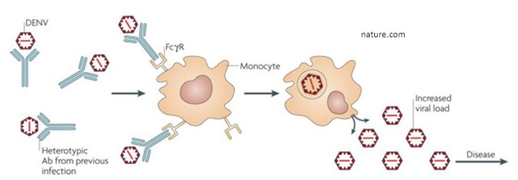
What is antibody-dependent enhancement (ADE)?
Antibodies from the first infection are cross-reactive with other DENV serotypes
Abs are subneutralizing
FcyR recognizes Abs and internalizes immunocomplexes
antibodies that were mild and recognized first strain can bind to new virus and the Fc of antibody can bind to monocytes and infects the monocytes
How does West Nile Virus and Dengue inhibit innate immunity?
can inhibit production of interferon and signal of interferon through JAK STAT pathway
West Nile virus (WNV) uses its non-structural (NS) proteins to shut down host innate immunity by targeting two key stages: the induction of type I interferons (IFNs) and the downstream JAK-STAT signaling cascade. By blocking these pathways, the virus prevents cells from expressing interferon-stimulated genes (ISGs) that would otherwise halt viral replication.

What are the features of the Zika virus?
Historically reported as a mild influenza-like illness that resolved within days and occurred in about 20% individuals
Guillain-Barre syndrome in a small percentage of adults –
inflammatory polyneuropathy presenting with paresthesis, weakness, pain; can progress to paralysis and death (autoimmune Ag mimicry, direct injury of peripheral nervous system)
Infects: Skin, Testis, OBGYN, embryonic brain tissue

What are the features of monocistronic constraint, control, genetic economy and competition in Flavivirus?
Monocistronic Constraint
- polyprotein production
Control
- standard cis/trans control (e.g., RdRp binding sites)
- 5’- and 3- stem-loops (SLA and SL) regulate RNA replication
- replication compartments: spatiotemporal control of TTG
Genetic Economy
- Simple virion structure
- Polyprotein production
- Viral and cellular proteases
- multi-protein complexes with different functions
Competition
- replication compartments (sequester PAMPS, optimize TTGAR)
What are the features of HCV
The virus is extremely hypervariable and produces chronic liver
disease, transmitted via blood; made the development of a
vaccine problematic
Lack of an animal model other than chimpanzee slowed drug
and vaccine development
What are the routes of infection for HCV?
Intravenous drug use
Contaminated blood supply and products
Improper therapeutic injections
Neonatal transmission
Occupational exposure, needle stick
Sexual intercourse
Tattooing, circumcision
What are the general structural features of HCV?
can grow virus in culture only infects humans and primates (hard to work with)

What are the features of the HCV genome and polyprotein cleavage?
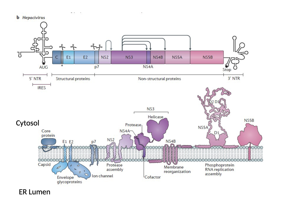
HCV polyprotein is cleaved by host cell proteases (), a virus specified cysteine protease (NS2), a virus specified serine protease (NS3); cleavage sites by viral proteases are indicated by arrows. IRES internal ribosome entry site. P7 ion channel
structural proteins on 5’ end
pore protein p7
signal peptidases that cleave: NS3 protease NS2 only cleaves itself

What are the features of structural proteins in HCV?
1. Core Protein (C) viral nucleocapsid; localizes to lipid droplets, which is essential for viral assembly; contributes to hepatic steatosis and oncogenesis.
2. Envelope Glycoprotein 1 (E1)membrane fusion protein during endosomal entry; forms a heterodimer with E2
3. Envelope Glycoprotein 2 (E2)receptor-binding protein for host
attachment to host cell surface receptors; contains hypervariable regions for immune evasion (evades antibodies)
lipid droplets and nucleocapsid help with assembly
What are the features of N-S proteins of HCV?
proteases
cofactors for proteases
ER membrane modifiers
phosphoprotein: Regulates RNA replication, virion assembly, and immune evasion;
hyperphosphorylation favors binding to lipid droplets to facilitate virion assembly;
RdRp
What are the features of HCV Receptors and Virus Life Cycle?
HCV enters host cell using a complex cascade of co-receptors and entry factors
uses multiple low affinity receptors first and makes it way to tight junction
lipoproteins associated with it and lipids are stripped for E1 E2 to bind
go to late endosome and E1 activity unfolds, activated at low pH and fusion with late endosome and release nucleocapsid and RNA genome associates with rough ER and makes capsid protein and structural proteins
get factories for RNA synthesis and some for lipids (separate) delivers nucleocapsid buds into the lumen
secretion through secretory vesicles and released by exocytosis

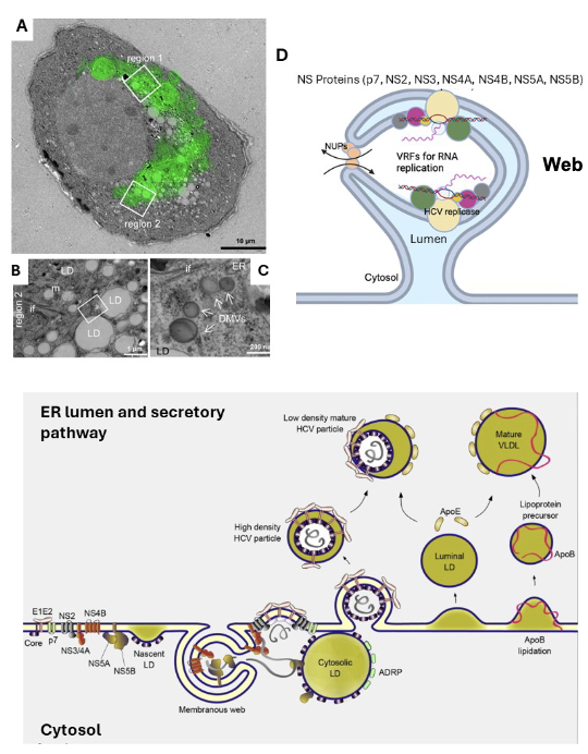
What are the features of the Membranous Web and HCV Replication?
viral factory, replication vesicles are larger (compared to flaviviruses) not invaginations they stick out from the ER into the cytosol
Virus assembly occurs in the membranous web composed of double membrane virion replication factories (VRFs) derived from the ER.
Virions bud into the ER lumen obtaining their lipid membrane and
ApoE-lipid components (where RNA synthesis and antigenome is made)
Lie in close proximity to lipid droplets which present core protein to
the new RNA
Apolipoproteins , lipoproteins, and lipids shield the virions from antibodies and cytotoxic T cells and macrophages.
nucleocapsids accumulating around the cytosolic lipid droplet (LD) and the nucleocapsids combine with the genome RNA being made and bud into the lumen of ER
LD and mature LDLs being formed in lumen and apolipoproteins (protein that accumulates at the surface of the lipoproteins)
What are the features of apolipoproteins in HCV?
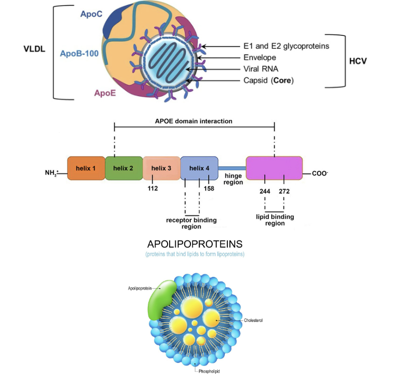
VLDL lipid viro particle
lipoproteins on the outside
has alpha helical regions which are amphipathic lipid binding region
micelle and inside there is cholesterol,
remember apolipoproteins and lipids are important for maturation and transport through secretory pathway P7 is cleaved and ion pore is made and virus is exuded via exocytosis
What is the importance of miRNA 122?
Important Liver Specific Factor Which Enhances HCV RNA Translation
miRNA 122 is involved with cholesterol metabolism
causes increased replication of Hep C RNA
What are the features of drugs that have been developed against 3 specific targets in HCV?
NS3/NS4A protease inhibitor
NS5A inhibitor
NS5B polymerase inhibitor

How can HCV inhibit innate immunity?
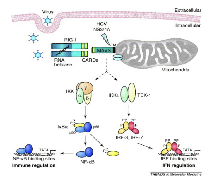
NS3/4A Protease Inhibits Activation of IRF-3 By Cleaving TRIF, A Component(Adaptor) of the TLR3 Pathway
NS3/4A can cleave TRIF adaptor (predicted by genetic search) and blocks the TLR3 pathway which senses viral ssRNA and normally stimulates IFN type I production. The viral protease blocks interferon and allows the virus to grow
NS3/NS4A cleaves MAVS to disrupt RIG-I function
and subsequent IRF-3 and NF-kB signaling
What are the features of HCV in relation to monocistronic constraint, control, genetic economy, competition?
Monocistronic Constraint
- polyprotein production, proteins cleaved by host and viral proteases
Control
- use of 5’ terminal IRES
- terminal 3’ RNA secondary structure required for replication
- no poly A sequence on genome, secondary structure and circularization of genome during replication
- replication compartments secure RNA during replication and synthesis
- NS5A can serve as a switch between translation and replication (RNA synthesis)
- antiviral drugs have been directed against protease, NS5A, and NS5B RdRP
Genetic Economy
- Simple virion structure with IRES and
- Polyprotein production
- Viral and cellular proteases
- multi-protein complexes with different functions
- associated with apolipoprotein E and VLDL for shielding viron
Competition
- NS3/4A protease can block host innate immunity and interferon I
production
- ApoE and lipoproteins associate with mature virion and shield it from antibodies and immune cells
-miRNA-122 can stabilize RNA at 5’ end of genome from host
What are the features of Coronavirus in relation to monocistronic constraint, control of template utilization, genetic economy, competition?
Control of template utilization
- Spatially segregate RNA replication and transcription in membrane
replication compartments
- sgmRNA production via discontinuous minus RNA synthesis
- RdRp holoenzyme complexes
Monocistronic constraint
- Polyprotein production and processing
- Ribosomal frameshifting makes two different polyproteins
- Nested set of monocistronic sgmRNAs
Genetic economy
- Simple virion structure
- One RdRp plus multiple cofactors makes holoenzyme complexes
Competition
- Nsp1 induces host shut-off
- Accessory proteins inhibit apoptosis and interferon responses
- Membrane replication compartments