Dermatology
 Call Kai
 Learn
 Practice Test
 Spaced Repetition
 Match
 Flashcards
 Knowt Play
1/79

There's no tags or description
Looks like no tags are added yet.
80 Terms
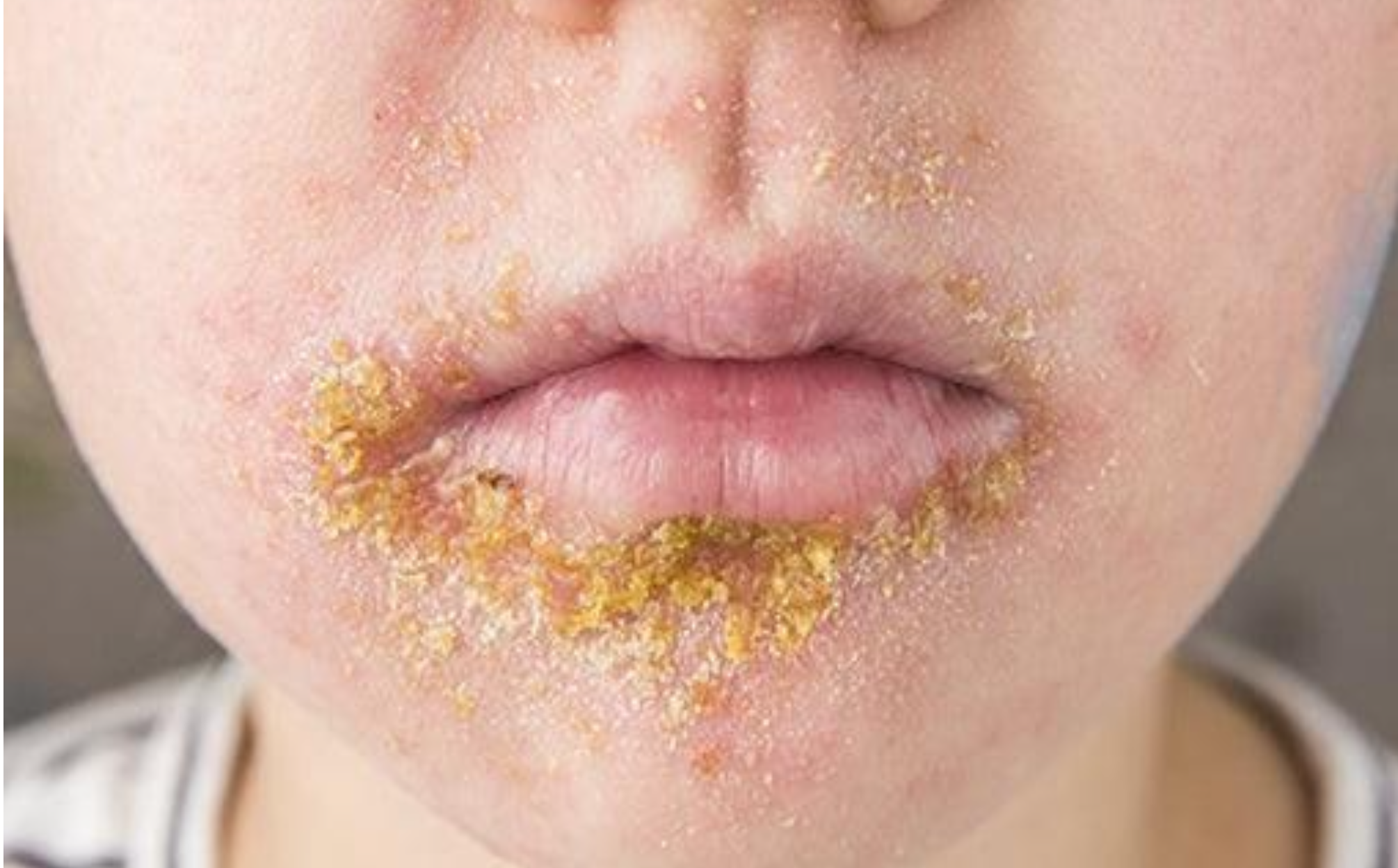
thin-walled vesicle on an erythematous base, then,
the vesicles rupture so rapidly that it is rarely seen as such.
• The exuding serum dries to form yellowish-brown, honey-colored, stuck on
crusts

initial lesion is a vesicle with an erythematous base and surrounding halo that
enlarges to 0.5-3 cm in size and then ruptures leaving a dark adherent crust
surrounded by a rim of erythematous indurated area
• Removal of the crust shows a saucer (dish) shaped ulcer with raised, indurated,
violaceous margin. The base extends deeply in the dermis.

initial lesion is thin walled yellowish-white pustule at the follicular orifices,
pierced at its center by a hair. It heals without scarring

initial lesions are single or multiple painful, erythematous, often
fluctuant, nodules and may have suppurative drainage

deep, painful cluster of interconnected boils (furuncles) that extends into the
subcutaneous tissue and discharges pus through multiple follicular openings

Initial lesion is in the form of recurrent discrete inflammatory papules or
pustules in the beard area pierced by hairs

Papules and pustules commonly occur on the
anterolateral aspect of the neck or on the angle of the jaw

The lesion is in the form of a well-demarcated red area. The lesion has an advancing
edge, and the surface may show vesicles, pustules or bullae

Ill-defined, indurated, red, tender hot area of skin appears at the affected site
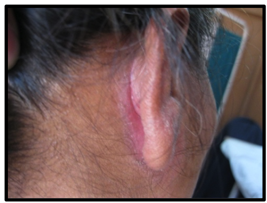
Streptococcal intertrigo: Longitudinal painful fissure at the angle of the skin
folds. The skin around is red, moist & may be crusted

Well-defined reddish- b r o w n patches with no active edge,
covered with fine scales that affect the intertriginous areas. It may be asymptomatic
or associated with mild itching

The affected digit is red, swollen and painful. Compression of the nail fold may drain pus.

One or several fingernails are affected. There is inflammation of the proximal nail
fold with erythema, edema and absence of the cuticle. Damage to the nail matrix results
in nail plate changes
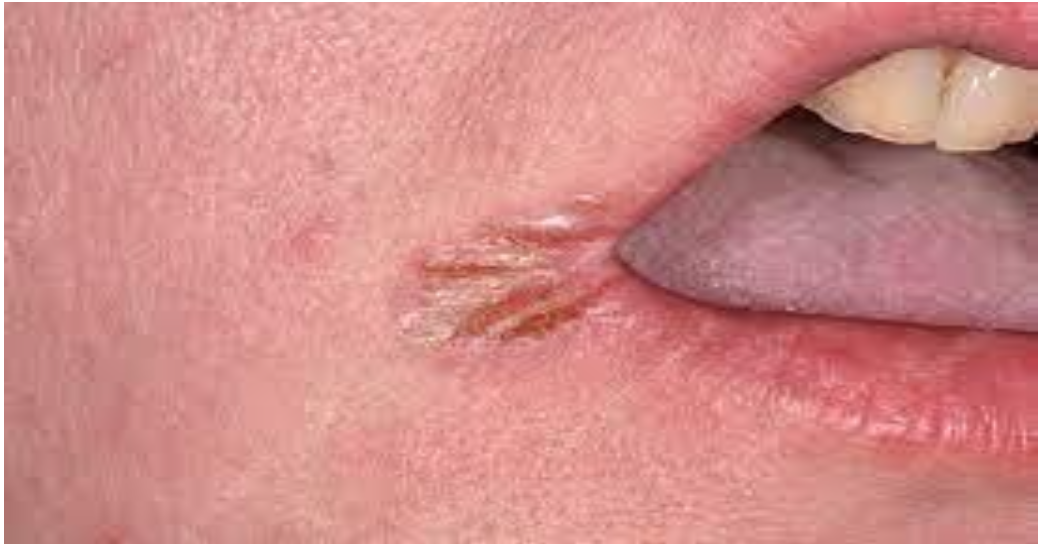
Characterized by
maceration, erythema and fissuring of the oral commissures

asymptomatic grayish papules which are few mm in diameter with
verrucous (irregular) rough surface

multiple skin-colored small
papules with flat smooth surface that affect mostly the face or
extensor surface of hands.

Skin-colored or grayish small papules or
pedunculated grayish cauliflower mass.

Skin colored lesion solid border with black dots &
distracted creases
*Creases is an important point to differentiate planter
wart (distracted) from callus (normal crossed creases)

a group of vesicular eruption on an erythematous base usually around
the mouth . Burning sensation may precede or accompany the eruption.

The eruption is itchy macules, papules, vesicles, pustules and crusts. The eruption
is polymorphic and centripetal in distribution together with mucous membrane
affection (eye, mouth, gingiva). The extremities are less involved.
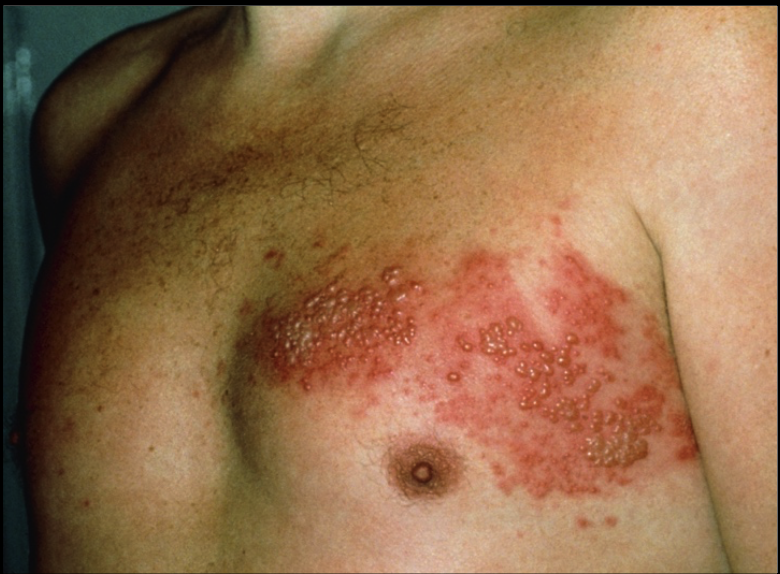
A group of vesicular eruption on an erythematous base appearing along the pathway
of cutaneous nerve (dermatome) . The vesicles contents dry up in 1-2
weeks forming crusts. Most uncomplicated lesions heal without scarring.
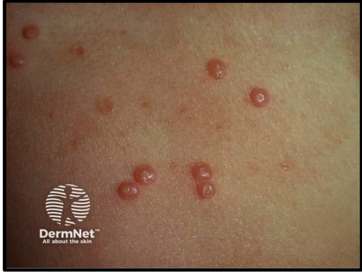
Highly contagious lesions.
- The primary lesion is painless, shiny, pearly white papule, usually umbilicated
in the center

Pleomorphic lesion including pustules & crusts with characteristic barrow
+/- scratch mark

Nets & lice presentation stuck on the hair
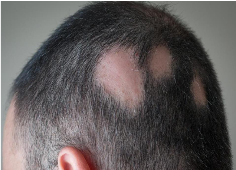
symptomatic single or multiple areas of sudden onset of hair loss.
• Characteristically, the scalp skin is completely normal with no crusts or scales.

Compulsive habit that induces the individual to pluck his or her own hairs.
• Occurs in neurotic individuals.

Skin lesions are in the form of "milky or chalk white", well-defined macules or
patches of variable sizes

Asymptomatic, light-to-dark brown macules or patches with irregular borders.

erythematous (salmon red) papule covered with laminated silvery
scales, which may coalesce to form sharply defined plaques. asymptomatic, bilateral, and symmetrical.

Small rounded drop-like lesion, red salmon
erythematous papule

Partial or complete red, dry and
thickened palms and/or soles, often with deep painful fissures. The skin
changes tend to have a sharp border and are often symmetrical

Diffuse scale or solitary plaques with no hair loss

Nail pitting,
subungual hyperkeratosis, onycholysis, oil drop sign, splinter hemorrhages or
leukonychia.

characterized by absent or decreased scales due to moisture and continuous
friction. There is usually considerable itching.

Napkin psoriasis (Figure 9.8): affection of napkin area in infants as a
presentation of Köebner phenomenon.
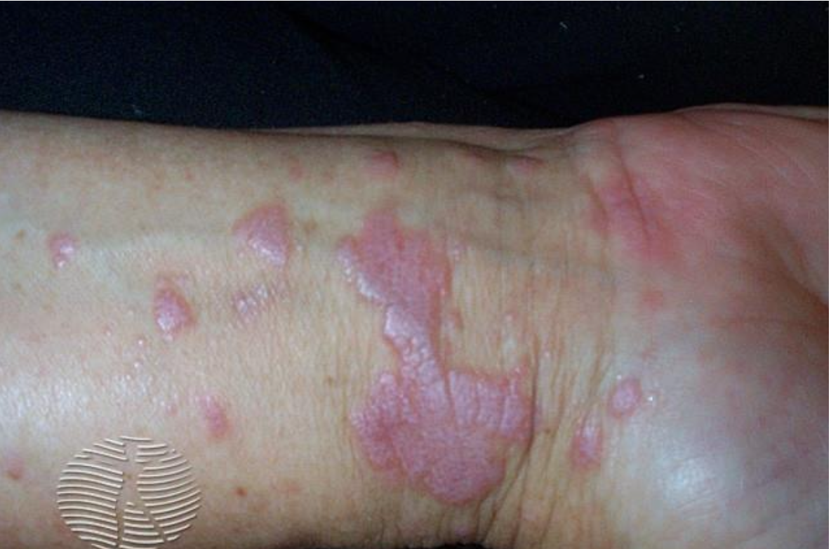
primary lesion is an itchy flat-topped polygonal papule. It is violet or
purple in color and shows whitish striations called Wickham’s striae, which
can be seen by a magnifying lens.
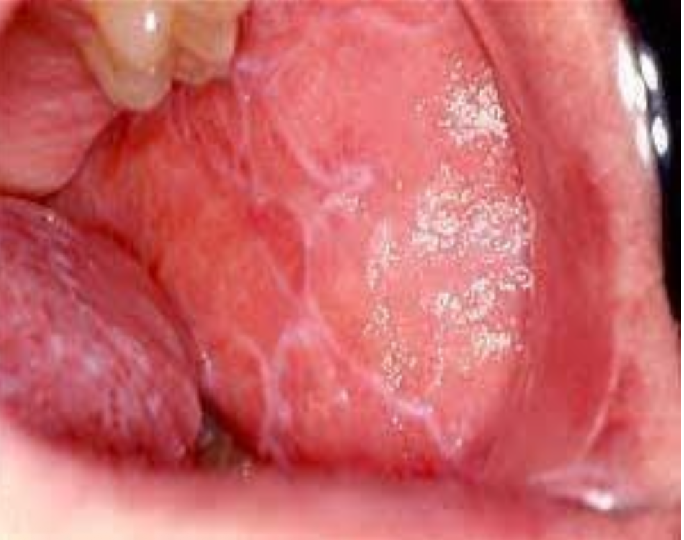
affects
buccal mucosa and lips in the form of white lines, streaks, or patches. Ulcers
may occur and can turn into squamous cell carcinoma in 1-2% of the cases.

Longitudinal ridging, pitting. Partial
or complete destruction of the nail may occur.
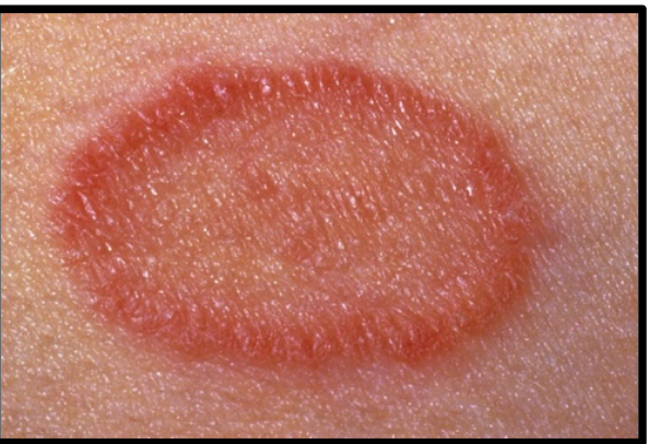
begins with a solitary macule or patch, that heralds the
eruption and thus is commonly referred to as the herald patch.
§ This initial lesion enlarges over a few days to become a patch with a collarette of
fine scale just inside the well-demarcated border.

secondary eruption is more generalized and symmetrical. as macules or maculo-papules,
erythematous and covered by fine scales. They enlarge gradually, become oval and
acquire the same appearance of the herald patch.

Lesions are pleomorphic: comedones (the characteristic primary lesion of acne),
papules, pustules, nodules and cysts. Post-acne erythema and scars may also
occur.
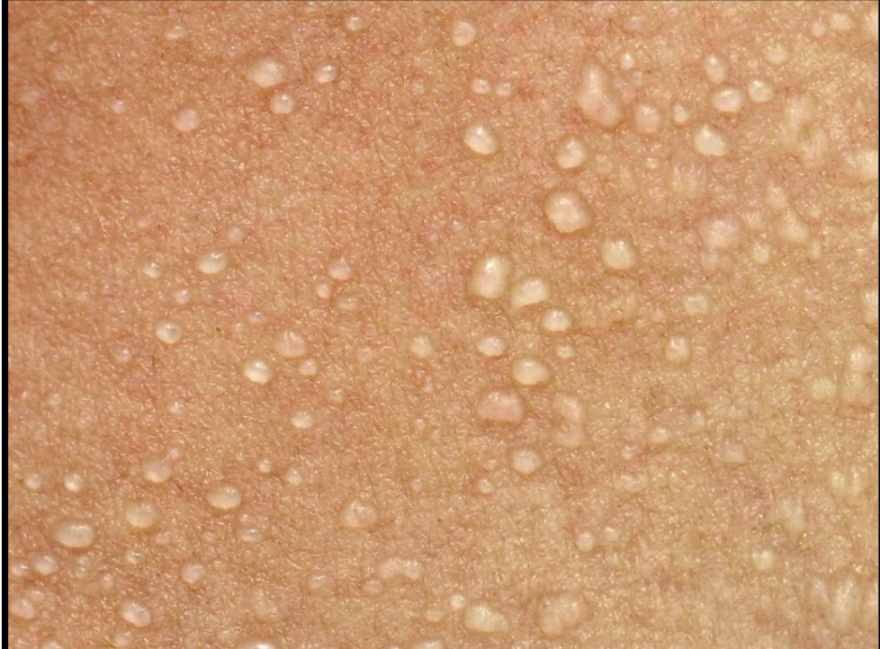
occlusion of the eccrine duct at the skin surface
resulting in accumulation of sweat under the stratum corneum. The lesion appears as
a clear drop or vesicles with little or no erythema.
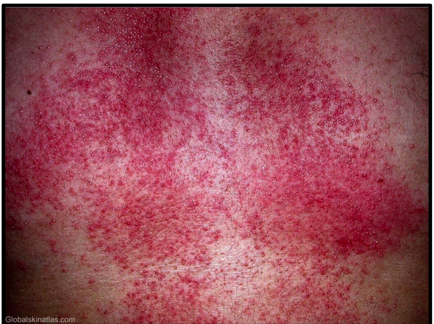
rom occlusion of the intraepidermal part of the
eccrine sweat duct, resulting in extremely pruritic erythematous papules.

cclusion of the eccrine sweat duct at
the dermoepidermal junction. produces
asymptomatic flesh-colored papules

-Ophiasis: alopecia areata involving the hair line

compulsive habit that induces the individual to pluck his or her own
hairs and occurs in neurotic individuals

radual hair loss caused by repeated or prolonged pulling tension on hair
roots.
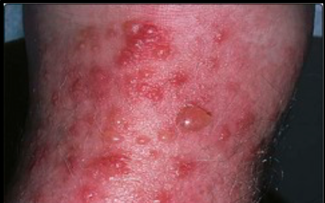
Acute stage: Ill-defined erythematous patches, papulovesicular eruption with
exudation of serum

Subacute stage: Erythema and scaling

Chronic stage: Dryness and lichenification

Allergic contact dermatitis is an example of type IV hypersensitivity
reaction and is diagnosed by patch test.

Lesions most frequently start on the cheeks, sparing perioral and para-nasal areas.
§ Lesions consist of intensely itchy and dry erythematous and edematous lesions. It
may become vesicular with oozing and crustations . Secondary
infection and lymphadenopathy are common.

Lesions consist of itchy erythematous, edematous papules which tend to be
replaced by lichenification

characterized by circular or oval coin shaped lesions with a well
demarcated edge

Lesions are present on the lower part of legs as an erythematous scaly o r
oozing area surrounded by small slate blue macules resulting from hemosiderin
deposits

Diffuse or localized scaly erythematous macules and patches, covered with
yellowish greasy scales

The lesions appear as rounded or oval erythematous, ill-defined patches with
fine white scales . Later, it subsides to leave areas of hypopigmentation.

characterized by itchy, transient (evanescent),
erythematous swellings of the dermis or subcutaneous tissues (wheals). Individual
lesions usually last less than 24 hours

special type of urticaria of sudden onset involving the
subcutaneous tissue leading to edema of the eyelids, lips, hands and genitalia. It
might/might not be associated with urticarial wheals elsewhere

Initially, an extremely itchy edematous papule develops at the site of the
insect bite, followed by a firm pruritic papule which usually persists for several
days. Lesions are often grouped in clusters and develop in crops at irregular
intervals and may vesiculate. Lesions are accompanied by scratch marks and may
be complicated by secondary infection.

characterized by sudden
onset of symmetrical multiforme lesions including erythematous macules, papules
vesicles and sometimes bullae. Some of the lesions evolve into target or iris lesion

The onset is sudden with extensive typical maculopapular rash
• generalized bullous eruption with affection of oral mucus membrane.

Exanthematous (morbilliform) eruption
is the most frequent type; causes a widespread, measles-like red rash mainly on the trunk
and arms

Sharply demarcated, round or oval red-to-violaceous macules, patches, or plaques
that consistently recur in the exact same location(s) upon re-exposure to a culprit
medication. Sometimes bullae on top.

Scaly patches with hair broken off appears as black dots

It presents as single or multiple scaly patches with the hairs cut into short stumps
1-2 mm above scalp level.

a single or multiple painless soft swellings of the scalp.
The hair is easily detached, and the surface of the swelling is studded with few
pustules.
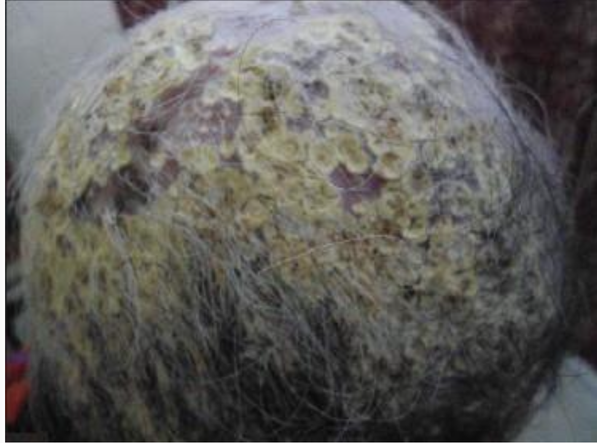
Affected hair is surrounded by thick yellow adherent crust that is
characteristically cup shaped (sulfur cups or scutula).
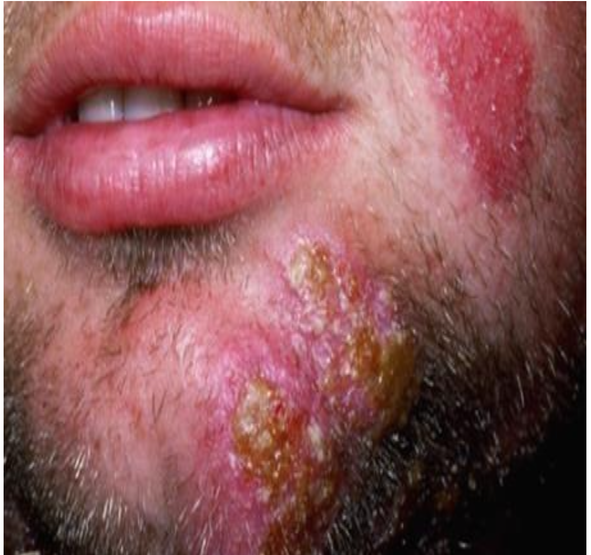
Soft swelling in the beard area hair easily detached with presented pustule
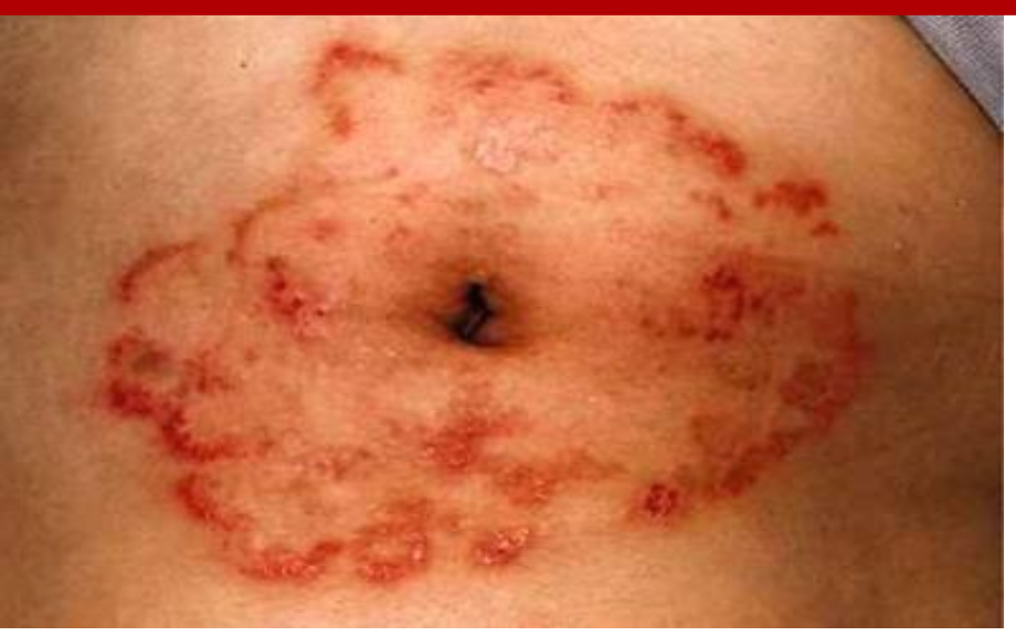
Well defined erythematous patch with raised active edges which
is severely itchy circinate lesion

Well defined erythematous patch with raised active edges with
healed center

Itchy interdigital maceration
of the 4th /5th toe-web space

Squamous/Chronic Hyperkeratotic: chronic plantar erythema with slight scaling to
diffuse hyperkeratosis. It can be asymptomatic or pruritic. Both feet are usually affected.
Typically, the dorsal surface of the foot is clear, but in severe cases, the condition may extend
onto the sides of the foot

intensely itchy or painful vesicles and bullae filled with clear
or purulent fluid, typically overlying areas of erythema on the instep or soles of the feet
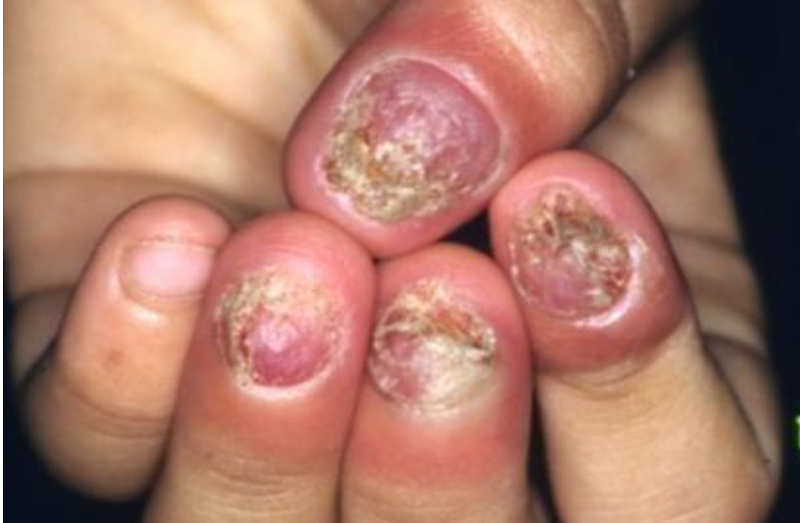
Brittle, friable, nails with subungual hyperkeratosis with distracted nail plate
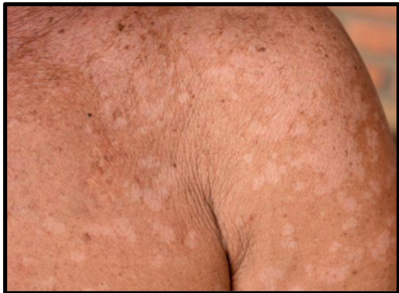
Well-defined hypo/hyper macules & patches
covered by fine scales

hitish curd-like patches (pseudo-
membrane) on tongue and buccal mucosa characterize it . The
patches can be scrubbed and leave erythematous base.

The
lesion is well-defined, moist and erythematous. The edge is festooned and small
satellite lesions (papules & pustules) are seen outside the edge

olds are involved. The edge is festooned and small
satellite lesions (papules & pustules) are seen outside the edge