NEUR-201 Exam 1
 Call Kai
 Learn
Learn Practice Test
 Spaced Repetition
 Match
Match Flashcards
 Knowt Play
1/97

There's no tags or description
Looks like no tags are added yet.
Name | Mastery | Learn | Test | Matching | Spaced | Call with Kai | Chat |
|---|
No analytics yet
Send a link to your students to track their progress
98 Terms
Population
entire group researcher wants to understand
Sample
subset of which measurements are collected
Descriptive Statistics
summarizes observable sample data
EX: mean firing rate of 50 recorded neurons was 22 Hz

Inferential statistics
draws conclusions about larger population
EX: treatment appears to increase firing rates in comparable neurons

Random Sampling
members have equal chance of being selected
EX: randomly selected individual from a population registry
Representative Sampling
deliberately matching important population characteristics
EX: recruiting participants in proportions matching population
Convenience Sampling
selecting easily accessible subjects (can skew data)
EX: studying volunteers from one class
Independent variables
what researcher manipulates
(EX: drug treatment)
Dependent variable
measured outcome
(EX: number of apoptotic cells)
Categorical variables
variables that can be put into categories
(EX: drug/control, cell type, etc)
Continuous variables
numerical quantities
(EX: membrane voltage, reaction time, neuronal firing rate, etc)
Experimental study
manipulates variable to test its effect
(EX: administering different drug dosages and measuring neuronal activity )
Correlational study
measures existing variables to determine whether they are associated
(no manipulation involved)
(EX: recording naturally occurring protein expression & neuronal activity to examine their association)
Somatic intervention
manipulates brain/NS to measure behavior
Behavioral intervention
manipulates behavior/experience to measure brain activity
Frontal lobe
involved in planning, decision-making, movement, impulse control, speech production
(impaired frontal lobe = impaired planning & behavioral regulation)

Parietal lobe
involved in somatosensory processing & spatial awareness
(impaired parietal lobe = impaired spatial processing)
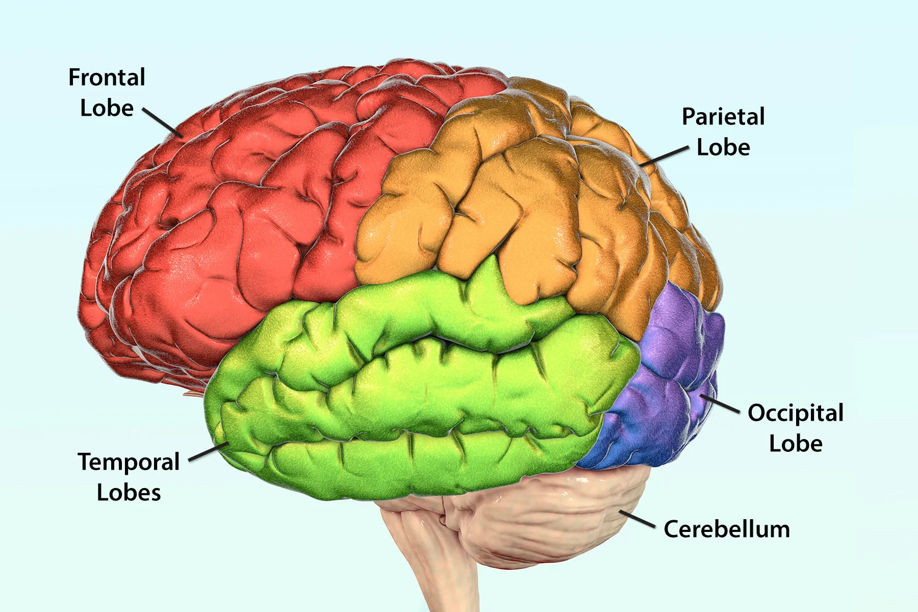
Temporal lobe
involved in hearing, language comprehension, memory, recognition, etc
(impaired temporal lobe = difficulty in memory/or language comprehension

Occipital lobe
visual processing
(impaired occipital lobe = visual processing difficulties)

Hippocampus
aids in formation of new memories & learning
(impaired hippocampus = memory impairment)

Amygdala
aids in emotional processing (especially threat)
(impaired amygdala = altered responses to emotionally significant stimuli)

Hypothalamus
area of temperature, thirst, hormone, sleep regulation
(impaired hypothalamus = disrupted homeostasis)

Thalamus
area of sensory & motor relay/regulation (except smell)
(impaired thalamus = altered sensory processing)

Basal ganglia
aids in movement control & reward-related processing
(impaired basal ganglia = movement regulation difficulties)

Neuron
receives & transmits electrical and chemical information
Astrocyte
supports neurons, supplies nutrients, and regulates NTs
Microglia
aids in immune surveillance, inflammatory responses, and synaptic pruning
Oligodendrocytes
produces myelin around CNS axons
Schwann Cells
produces myelin around PNS axons
OPCs
progenitor cell that aid toward producing oligodendrocytes
Histology
studying microscopic structure of tissues
(staining techniques allows researchers to determine which cells are present & what cells are doing)

IBA1 identifies?
Microglia (also some related myeloid cells)
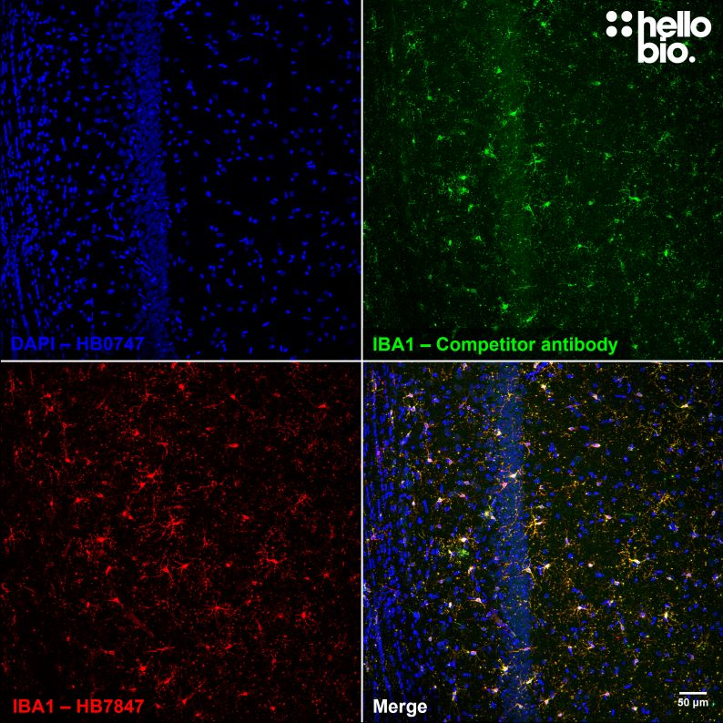
IBA1 localizes where?
Cytoplasm/membrane (common microglial marker)

DAPI identifies?
DNA-containing nuclei

DAPI localizes where?
cell nucleus (nuclear location) (DNA stain not primary antibody cell marker)
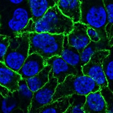
NeuN identifies?
most mature/post-mitotic neurons

NeuN localizes where?
localizes mainly cell nucleus of mature neurons

GFAP identifies?
Astrocytes

GFAP localizes where?
Cytoplasmic filaments of astrocytes in CNS
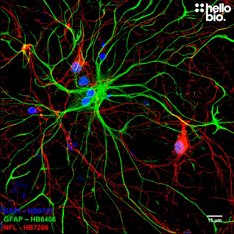
TMEM119 identifies?
specific cell marker of resident brain microglia
(specific marker for microglia)

TMEM119 localizes where?
mainly in membrane-associated structures (plasma membrane, ER, etc)
(helps distinguish residential microglia from infiltrating macrophages)

Olig2 identifies?
Oligodendrocyte lineage cells
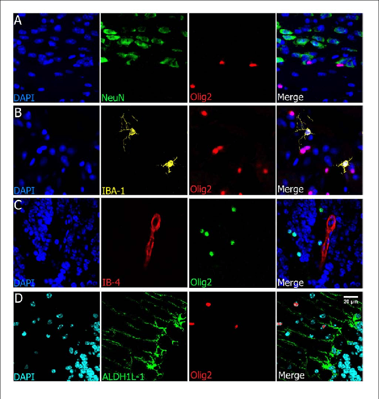
Olig2+ localizes where?
nuclear (cell nucleus)

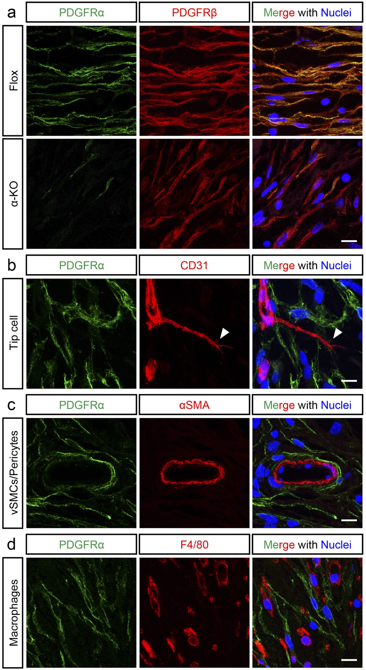
PDGFRα (NG2) identifies what?
oligodendrocyte progenitor cells

PDGFRα localizes where?
membrane
CC1 (APC) identifies what?
mature oligodendrocytes
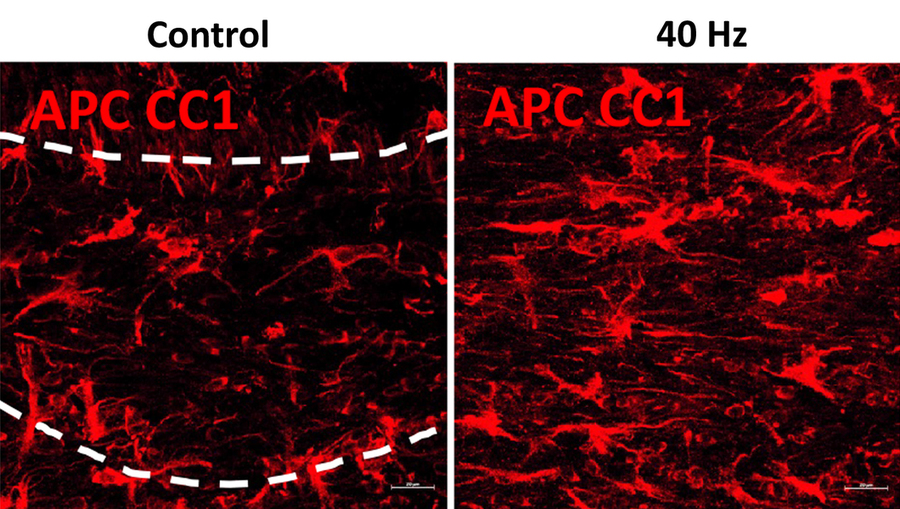
CC1 (APC) localizes where?
in cell body (useful for mature oligodendrocyte identification)

MBP identifies what?
myelin and mature oligodendrocyte-associated structures

MBP localizes where?
in myelin (stains sheath; doesn’t directly count cell bodies)

what does Ki67 tell us?
active cell cycle is occurring (cell is proliferating/cycling)

what does BrdU/EdU tell us?
cell is undergoing DNA synthesis

What does CC3 tell us?
cells is undergoing apoptosis

Double positive mean whats?
combination of interpretations
(EX: IBA+ and CC3+ means that the cell is an apoptotic microglia)

Antibodies
y-shaped proteins that target molecules called antigens

Primary antibody
targets & binds to target antigen directly

Secondary antibody
binds to primary antibody and carries fluorescent label
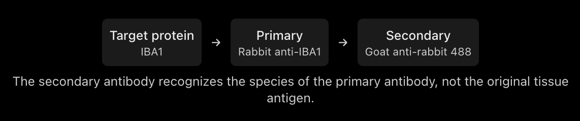
primary host and secondary target must?
Match between each other
(EX: rabbit anti-IBA1/goat anti-rabbit)
3 Things to look out for in two-marker experiments
primary antibodies cannot be same species (makes it difficult to distinguish targets in ordinary indirect staining)
using the same species antibodies on its tissue (can cause issues)
using two fluorophores that have overlapping emission spectra
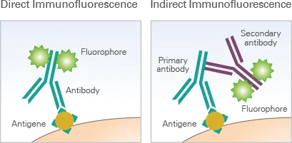
Direct immunofluorescence
fluorophore is attacked to primary antibody
fewer staining steps
often has lower signal amplification
less flexibility in secondary-antibody selection

Indirect immunofluorescence
fluorophore is attached to secondary antibody
more staining steps
can amplify fluroscent signal
many fluorophores can be used with one primary
Immunohistochemistry
antibody staining performed on tissue (cellular organization is preserved)

Immunocytochemistry (ICC)
antibody staining of isolated cells (EX: neurons grown in culture)

FFPE (Formalin-Fixed Paraffin Embedded-Tissue(
prep: fixation and paraffin embedding
sections: 3-5 µm
Tissue morphology: excellent
antigen retrieval: often required
lipid preservation: poorer
storage: stable paraffin blocks

Frozen Tissue
prep: freezing/fixation with cyroprotection
sections: 5- 50 µm
tissue morphology: good (can have freezing artifacts however)
antigen retrieval: unnecessary
lipid preservation: better than FFPE
storage: usually cold storage

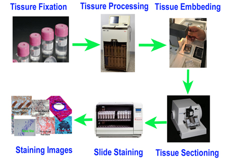
Histology staining preparation
collect & prepare tissue
dissect tissue (fix/freeze as appropriate)
pretreat specimen
remove paraffin and retrieve antigens (if FFPE protocol requires it)
block nonspecific binding
reduce unwanted antibody
apply primary antibodies
allows target-specific antibodies to recognize antigens
apply secondary antibodies
bind fluoroscent/other detection labels to primary antibodies
counterstain and image
use DAPI (if needed), mount specimen, & visualize labeled cells
Microscopy
allows researchers to examine biological samples at scale that cannot be seen/resolved by naked eye

Magnification Formula
M_total = M_objective x M_eyepiece
(63x objective & 20x eyepiece = 1260x resolution)
Resolution
how closely spaced two points can be while still appearing separate
Simplified resolution equation

Poor resolution means?
two points appear merged
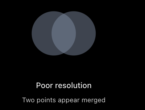
Better resolution means?
two points appear separate

Brightfield microscopy
uses transmitter light to observe cells and tissue structures
(useful for naturally colored or stained specimen)

Widefield fluoroscence
illuminates entire field simultaneously
(fast & effective for thin samples/out-of-focus light creates blur in thick samples)

Laser-scanning confocal microscope
scans sequentially to produce detailed optical sections
(excellent for fixed, thicker, multicolor tissues, but relatively slow)

Spinning-disk confocal microscope
uses many pinholes to image quickly with optical sectioning
(good for fast live-cell imaging in samples within its practical depth range)

When to use Widefield?
thin culture & fast fluoroscent imaging
When to use laser-scanning confocal microscope?
thick fixed brain tissue & fine detail
When to use spinning-disk confocal microscope?
Fast imaging requiring optical sections
When to use Brightfield?
standard visible tissue staining
Voltage
electrical potential difference
Current
flow of electrical charge
Resistance
opposition to current
Conductance
how easily current flows
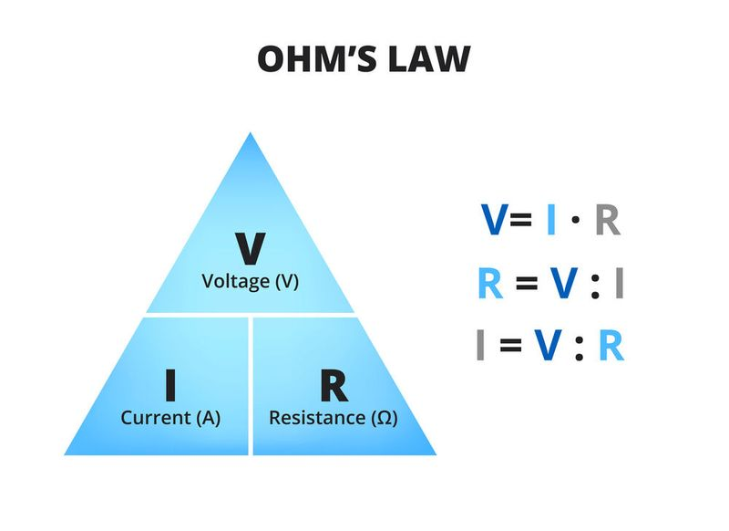
Ohm’s Law
V = I R
g = 1/ R
( Voltage (V) = pressure; current (I) = flow)
(Resistance (R) = restriction; conductance (g) = ease of flow)