Orthopaedic Examination of the ankle and foot
 Call Kai
 Learn
 Practice Test
 Spaced Repetition
 Match
 Flashcards
 Knowt Play
1/30

Earn XP
Description and Tags
Description of the steps carried during this examination of the ankle and foot, using the look, feel, move structure (special tests are interspersed).
31 Terms
What is the first step of the examination?
WINDEC
WINDEC
Wash hands
Introduce yourself
Name
DOB
Explain the examination to the patient
Consent
After WINDEC, what else should you do before starting the examination?
Expose the patient’s legs from the knee down, and position them standing.
Ask them if they have any pain before proceeding.
After the patient has been positioned correctly, what is the first section of the examination?
Look:
General inspection
Closer inspection:
Anterior
Lateral
Posterior
What are you looking for in the general inspection?
Body habitus (obesity)
Scars
Muscle atrophy
Objects and equipment (walking aids, prescriptions)
What are you looking for in the closer inspection?
Scars
Bruising
Swelling
Psoriasis plaques
Fixed flexion deformity of the toes
Big toe misalignment
Calluses
Foot arch - abnormally flat or raised
Heel misalignment
Achilles tendon
While the patient is standing, what should you assess before moving on to the next part of the examination after the inspection?
Gait assessment.
What should you be looking for in the gait assessment?
Gait cycle
Range of movement
Limping
Leg length
Turning
Height of steps
After the gait assessment, what is the next section of the examination?
Feel:
Temperature
Pulses
Metatarsophalangeal joint squeeze
Palpation
Get the patient to lay on the clinical examination couch for this section of the exam.
How do you assess the temperature of the ankle joint?
Using the back of your hand, assess and compare the temperature of each joint?

What pulses do you need to palpate?
Posterior tibial pulse
Dorsalis pedis pulse
How do you palpate the posterior tibial pulse?
It can be located posterior to the medial malleolus of the tibia.
Palpate the pulse to confirm its presence and then compare pulse strength between the feet.

How do you palpate the dorsalis pedis pulse?
It can be located over the dorsum of the foot, lateral to the extensor hallucis longus tendon, over the second and third cuneiform bones.
Palpate the pulse to confirm its presence and then compare pulse strength between the feet.
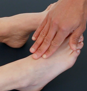
How do you perform the metatarsophalangeal joint squeeze?
Gently squeeze across the metatarsophalangeal (MTP) joints and observe for verbal and non-verbal signs of discomfort.
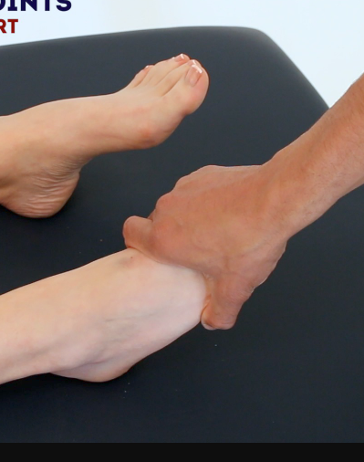
How is palpation of the ankle and foot performed?
Press firmly with one or two fingers on these areas, noting swelling, irregularity, or discomfort:
Metatarsal and tarsal bones
Tarsal joint
Ankle joint
Subtalar joint
Calcaneum
Medial / lateral malleoli
Distal fibula
Gastrocnemius muscle
Achilles tendon
While palpating the Achilles tendon and the gastrocnemius muscle, what special test can you perform?
Simmond’s test
How do you perform the Simmond’s test?
In healthy individuals, the foot should plantarflex when you squeeze the calf due to the contraction of the gastrocnemius muscle and the subsequent pulling force transmitted via the Achilles tendon.
There will be no movement of the foot if the Achilles tendon is ruptured due to loss of continuity between the gastrocnemius and the foot.

After palpation and the special test, what is the next section of the examination?
Active and passive movements.
What active movements should you ask the patient to perform?
Foot plantarflexion
Foot dorsiflexion
Toe flexion
Toe extension
Ankle / foot inversion
Ankle / foot eversion
How do you get the patient to perform active foot plantarflexion?
“Push your feet downwards, like you are pressing a car pedal.”

How do you get the patient to perform active foot dorsiflexion?
“Extend your feet backwards, like you’re trying to point them up towards your head.”

How do you get the patient to perform active toe flexion?
“Curl up your toes as tight as you can.”

How do you get the patient to perform active toe extension?
“Extend your toes backwards, like you’re trying to point them towards your head.”

How do you get the patient to perform active ankle / foot inversion?
“Try to touch the soles of your feet together.”
OR
Hold your finger medial to the foot and ask them to touch it with their foot - without moving the rest of their leg
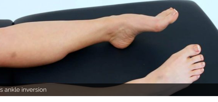
How do you get the patient to perform active ankle / foot eversion?
“Try to angle the soles of your feet outwards as far as you are able to.”
OR
Hold your finger lateral to the foot and ask them to touch it with their foot - without moving the rest of their leg.
What passive movements do you perform on the patients?
The same movements you asked the patient to perform actively, in addition to movement of the subtalar and midtarsal joint.
How do you passively move the midtarsal joint?
Wrap one hand round the ankle to stabilise it, then wrap the other hand round the tarsal area of the foot (just below the toes).
Twist the hand holding the tarsal area to assess the movement of the joint.

How do you passively move the subtalar joint?
Wrap one hand round the ankle to stabilise it, then hold the heel of the foot with your other hand.
Invert and evert the foot with the hand holding the heel of the foot.

After performing all the passive movements on the patient, what is the next step?
Finish up the examination:
Explain to the patient that the examination is now finished.
Thank the patient for their time.
Dispose of PPE appropriately and wash your hands.
Summarise your findings.
Example Summary
“Today I examined Mrs Smith, a 32-year-old female. On general inspection, the patient appeared comfortable at rest, with no stigmata of musculoskeletal disease. There were no objects or medical equipment around the bed of relevance.
“The ankles and feet appeared normal on inspection and the patient’s gait was unremarkable. The range of movement throughout the ankles and feet was normal.”
“In summary, these findings are consistent with a normal ankle and foot examination.”
“For completeness, I would like to perform the following further assessments and investigations.”
What further assessments and investigations could you suggest?
Neurovascular examination of both lower limbs.
Examination of the knee and hip joint.
Further imaging if indicated (e.g. X-ray and MRI).