Systems Lab Exam 1

0.0(0)
Studied by 0 people
call kaiCall Kai
learnLearn
examPractice Test
spaced repetitionSpaced Repetition
heart puzzleMatch
flashcardsFlashcards
GameKnowt Play
Card Sorting

1/310

encourage image

There's no tags or description

Looks like no tags are added yet.

Last updated 2:33 PM on 10/13/26
Name
Mastery
Learn
Test
Matching
Spaced
Call with Kai
Chat

No analytics yet

Send a link to your students to track their progress

311 Terms
1
New cards
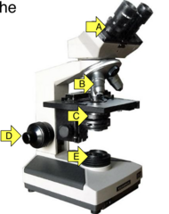
<p>What is arrow B pointing at?</p>

What is arrow B pointing at?

objective lenses

2
New cards
<p>What knob is arrow D pointing at?</p>

What knob is arrow D pointing at?

focus

3
New cards
<p>Which lens is controlled by the knob at the arrow?</p>

Which lens is controlled by the knob at the arrow?

auxillary

4
New cards
<p>What is the function of the circled lenses?</p>

What is the function of the circled lenses?

magnification change

5
New cards
<p>What is this lens called?</p>

What is this lens called?

condenser

6
New cards
<p>Choose all components that you can/should use to adjust the focus of the image (not the magnification).</p>

Choose all components that you can/should use to adjust the focus of the image (not the magnification).

A and D

7
New cards
<p>What should you do if there is too much light on the slide for your eyes?</p><p>A. Adjust the light source</p><p>B. Change the objective lens</p><p>C. Close one eye</p><p>D. Move the condenser lens farther from the stage</p><p>E. Cry</p>

What should you do if there is too much light on the slide for your eyes?

A. Adjust the light source

B. Change the objective lens

C. Close one eye

D. Move the condenser lens farther from the stage

E. Cry

A. Adjust the light source

8
New cards
<p>What does this knob do?</p>

What does this knob do?

stage control

9
New cards
<p>What happens if the condenser lens is moved too far from the microscope stage?</p><p>A. Refraction increases; slide looks fuzzy</p><p>B. Slide goes out of focus</p><p>C. Too much light on the slide</p>

What happens if the condenser lens is moved too far from the microscope stage?

A. Refraction increases; slide looks fuzzy

B. Slide goes out of focus

C. Too much light on the slide

A. Refraction increases; slide looks fuzzy

10
New cards
<p>Which part of this cell is more eosinophilic?</p><p>A. A - apical part with lots of secretory vesicles</p><p>B. B - basal part with lots of ribosomes on RER</p>

Which part of this cell is more eosinophilic?

A. A - apical part with lots of secretory vesicles

B. B - basal part with lots of ribosomes on RER

A. A - apical part with lots of secretory vesicles

11
New cards
<p>Which cell has the most heterochromatin in its nucleus?</p>

Which cell has the most heterochromatin in its nucleus?

C

12
New cards
<p>What type of staining is displayed in these images?</p>

What type of staining is displayed in these images?

eosinophilic structures

13
New cards
<p>What type of staining is displayed by the nuclei in these images?</p>

What type of staining is displayed by the nuclei in these images?

basophilic
14
New cards
<p>What is a cytoplasmic structure that usually stains eosinophilically?</p>

What is a cytoplasmic structure that usually stains eosinophilically?

mitochondria, ECM proteins, secretory granules, cytoskeleton

15
New cards
<p>What is a cytoplasmic structure that usually stains basophilically?</p>

What is a cytoplasmic structure that usually stains basophilically?

RER, ribosomes

16
New cards
<p>Identify the structure at the arrow</p>

Identify the structure at the arrow

Nucleolus

17
New cards
<p>Identify the structure at the arrow</p>

Identify the structure at the arrow

nucleolus

18
New cards
<p>What is the chromatin pattern of this nuclei?</p>

What is the chromatin pattern of this nuclei?

euchromatic

19
New cards
<p>What is the chromatin pattern of this nuclei? (red arrow)</p>

What is the chromatin pattern of this nuclei? (red arrow)

euchromatic

20
New cards
<p>What is the chromatin pattern of this nuclei?</p>

What is the chromatin pattern of this nuclei?

heterochromatic

21
New cards
<p>What is the chromatin pattern of this nuclei? (yellow arrow)</p>

What is the chromatin pattern of this nuclei? (yellow arrow)

heterochromatic

22
New cards
<p>What is this plane of section?</p>

What is this plane of section?

transverse/cross section

23
New cards
<p>What is this plane of section?</p>

What is this plane of section?

transverse/cross section
24
New cards
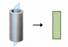
<p>What is this plane of section?</p>

What is this plane of section?

longitudinal section

25
New cards
<p>What is this plane of section?</p>

What is this plane of section?

longitudinal section

26
New cards
<p>What is this plane of section?</p>

What is this plane of section?

oblique section

27
New cards
<p>What is this plane of section?</p>

What is this plane of section?

oblique section

28
New cards
<p>What is this plane of section?</p>

What is this plane of section?

tangential section

29
New cards
<p>Which labeled structure is best describe as being in cross section?</p>

Which labeled structure is best describe as being in cross section?

C

30
New cards
<p>Which cell demonstrates prophase?</p>

Which cell demonstrates prophase?

D

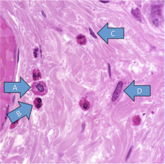
31
New cards
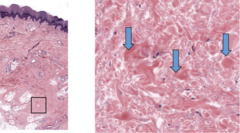
<p>Identify the eosinophilic structures at the arrows.</p>

Identify the eosinophilic structures at the arrows.

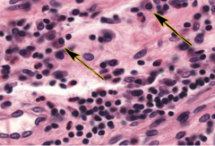
Collagen in dense irregular tissue
32
New cards
<p>Which of these cells is an eosinophil?</p>

Which of these cells is an eosinophil?

B

33
New cards
<p>What is this plane of section?</p>

What is this plane of section?

tangential section

34
New cards
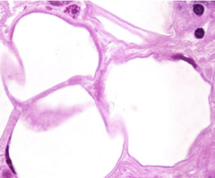
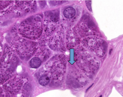
<p>Identify this</p>

Identify this

secretory granules

35
New cards
<p>Identify this</p>

Identify this

secretory vesicles

36
New cards
<p>What stage of mitosis is this?</p>

What stage of mitosis is this?

prophase

37
New cards
<p>What stage of mitosis is this?</p>

What stage of mitosis is this?

metaphase

38
New cards
<p>What image depicts the anaphase stage of mitosis?</p>

What image depicts the anaphase stage of mitosis?

A

39
New cards
<p>What image depicts the telophase stage of mitosis?</p>

What image depicts the telophase stage of mitosis?

B

40
New cards
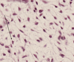
<p>Classify this connective tissue</p>

Classify this connective tissue

mesenchyme
41
New cards
<p>Identify the mesenchymal cell at the arrow.</p>

Identify the mesenchymal cell at the arrow.

stellate

42
New cards
<p>Classify this connective tissue</p>

Classify this connective tissue

loose connective tissue

43
New cards
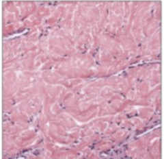
<p>Classify this connective tissue</p>

Classify this connective tissue

dense irregular connective tissue
44
New cards
<p>Classify this connective tissue</p>

Classify this connective tissue

dense irregular connective tissue

45
New cards
<p>Classify this connective tissue</p>

Classify this connective tissue

dense irregular connective tissue

46
New cards
<p>Classify this connective tissue</p>

Classify this connective tissue

dense regular connective tissue

47
New cards
<p>Classify this connective tissue</p>

Classify this connective tissue

dense regular connective tissue

48
New cards
<p>Classify this connective tissue</p>

Classify this connective tissue

adipose tissue

49
New cards
<p>Classify this connective tissue.</p>

Classify this connective tissue.

adipose tissue

50
New cards
<p>Which sample is elastic cartilage?</p>

Which sample is elastic cartilage?

B

51
New cards
<p>Identify the eosinophilic structure at the arrow.</p>

Identify the eosinophilic structure at the arrow.

collagen

52
New cards
<p>Identify this</p>

Identify this

collagen fibers in loose connective tissue

53
New cards
<p>Identify the structure at the arrows</p>

Identify the structure at the arrows

elastic lamellae

54
New cards
<p>Identify this blood vessel found in the wall of the aorta</p>

Identify this blood vessel found in the wall of the aorta

elastic artery

55
New cards
<p>What causes the blue staining of the extracellular matrix of this tissue?</p>

What causes the blue staining of the extracellular matrix of this tissue?

GAGs

56
New cards
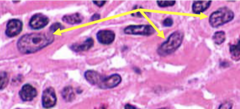
<p>Identify this cell</p>

Identify this cell

fibroblasts

57
New cards
<p>Identify this cell</p>

Identify this cell

fibroblasts

58
New cards
<p>Identify this cell</p>

Identify this cell

adipocytes

59
New cards
<p>Identify this cell</p>

Identify this cell

macrophage

60
New cards
<p>Identify this cell</p>

Identify this cell

macrophage

61
New cards
<p>What organelle causes the clear area next to the nucleus in this plasma cell?</p>

What organelle causes the clear area next to the nucleus in this plasma cell?

Golgi

62
New cards
<p>Identify this cell</p>

Identify this cell

eosinophil

63
New cards
<p>Identify this cell</p>

Identify this cell

neutrophil

64
New cards
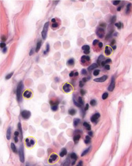
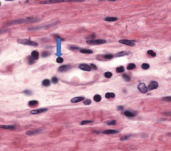
<p>Identify the circled cells</p>

Identify the circled cells

neutrophils
65
New cards
<p>Identify this cell</p>

Identify this cell

lymphocyte

66
New cards
<p>Identify this cell</p>

Identify this cell

mast cell

67
New cards
<p>Identify this cell</p>

Identify this cell

plasma cell

68
New cards
<p>Identify the cell at the arrow.</p>

Identify the cell at the arrow.

Hypertrophied chondrocytes (in fetal long bone formation)

69
New cards
<p>Addition of extracellular matrix at the level of the arrow is an example of what type of growth?</p>

Addition of extracellular matrix at the level of the arrow is an example of what type of growth?

appositional

70
New cards
<p>Identify the cells at the arrow.</p>

Identify the cells at the arrow.

Osteoblasts

71
New cards
<p>Identify this epithelia shape</p>

Identify this epithelia shape

cuboidal

72
New cards
<p>Classify the epithelium</p>

Classify the epithelium

Simple columnar non-keratinized

73
New cards
<p>Identify this epithelia shape</p>

Identify this epithelia shape

columnar

74
New cards
<p>Identify the indicated space in this bone unit.</p>

Identify the indicated space in this bone unit.

lacunae

75
New cards
<p>Identify the cells at the arrow.</p>

Identify the cells at the arrow.

Goblet cells
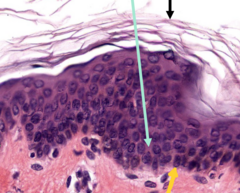
76
New cards
<p>Identify this area (yellow arrow)</p>

Identify this area (yellow arrow)

basement membrane

77
New cards
<p>Specifically identify the layer indicated by the bracket.</p>

Specifically identify the layer indicated by the bracket.

Dermis papillary layer
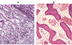
78
New cards
<p>Which bone tissue is developing directly from mesenchyme?</p>

Which bone tissue is developing directly from mesenchyme?

B

79
New cards
<p>Identify this epithelial feature</p>

Identify this epithelial feature

goblet cell
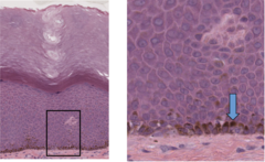
80
New cards
<p>What is the function of the brown granules in the cell at the arrow?</p>

What is the function of the brown granules in the cell at the arrow?

melanin - protection of nucleus from UV

81
New cards
<p>Identify the makeup of this brush border</p>

Identify the makeup of this brush border

microvilli

82
New cards
<p>Identify the skin structure circled in the right image</p>

Identify the skin structure circled in the right image

sebaceous gland

83
New cards
<p>Identify the eosinophilic tissue at the arrow.</p>

Identify the eosinophilic tissue at the arrow.

Bone matrix/bone ECM/osteoid

84
New cards
<p>Identify this epithelial feature</p>

Identify this epithelial feature

cilia

85
New cards
<p>What type of surface structure contains microtubules?</p>

What type of surface structure contains microtubules?

cilia

86
New cards
<p>Identify this surface feature</p>

Identify this surface feature

stereocilia

87
New cards
<p>What is the classification of the epithelium?</p>

What is the classification of the epithelium?

simple columnar with cilia

88
New cards
<p>Which is the apical surface of this duct cell?</p>

Which is the apical surface of this duct cell?

C

89
New cards
<p>Where is the basement membrane?</p>

Where is the basement membrane?

A

90
New cards
<p>Which skin gland shows evidence of apocrine secretion?</p>

Which skin gland shows evidence of apocrine secretion?

A

91
New cards
<p>Identify the structure circled in the right image, seen in the skin.</p>

Identify the structure circled in the right image, seen in the skin.

Merocrine gland

92
New cards
<p>Specifically name this outer layer of the heart (a serous membrane sitting on a layer of fat).</p>

Specifically name this outer layer of the heart (a serous membrane sitting on a layer of fat).

epicardium

93
New cards
<p>Identify this layer of the periosteum in this developing bone.</p>

Identify this layer of the periosteum in this developing bone.

periosteum

94
New cards
<p>Identify the nucleus at the arrow</p>

Identify the nucleus at the arrow

satellite
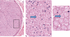
95
New cards
<p>Identify the structures at the arrows, seen here in cross section.</p>

Identify the structures at the arrows, seen here in cross section.

axon

96
New cards
<p>Classify the cell layers</p>

Classify the cell layers

simple

97
New cards
<p>Classify this epithelium</p>

Classify this epithelium

stratified squamous non-keratinized

98
New cards
<p>Classify this epithelium</p>

Classify this epithelium

simple cuboidal
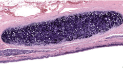
99
New cards
<p>Classify this epithelium</p>

Classify this epithelium

pseudostratified columnar with cilia and goblet cells

100
New cards
<p>Classify this epithelium</p>

Classify this epithelium

stratified cuboidal