Axial Skeleton, Histology, and Skull Bone Features w pictures
 Call Kai
 Learn
Learn Practice Test
 Spaced Repetition
 Match
Match Flashcards
 Knowt Play
1/85

Earn XP
Description and Tags
Vocabulary flashcards covering axial bones, vertebral sections, sternum, skull bone features, sutures, fetal skull structures, and histology.
Name | Mastery | Learn | Test | Matching | Spaced | Call with Kai | Chat |
|---|
No analytics yet
Send a link to your students to track their progress
86 Terms

Cervical Vertebrae
Bone group comprising everything above the ribs (7 vertebrae).

Atlas Vertebra
Bone that holds the skull; described as Yoda/syd the sloth.

Axis Vertebra
Bone where skull rotates; described as thumbs up.

Dens (Axis)
Sticky up part structure of axis (thumb).

Transverse Foramen (Cervical)
Hole described as 'eye holes' in cervical vertebra.

Thoracic Vertebrae
Bone section where ribs start to ribs end (giraffe).

Lumbar Vertebrae
Bone section comprising everything below ribs (moose).

Body (Vertebra)
Structure that holds weight; solid portion of vertebra.

Spinous Process
Point structure of vertebra.

Vertebral Foramen
Hole where the spinal cord passes through.

Transverse Process
Structure consisting of two parts that stick off sides of vertebra.

Sacrum Bone
Bone located at the base of spine.

Lateral Sacral Crest
Structure on the sides of sacrum.

Median Sacral Crest
Structure down the middle of sacrum.

Auricular Surface of the Sacrum
Sacral structure where hips connect.

Sacral Foramina
Holes in sacrum.

Coccyx Bone
Bone where pelvis connects.

Sternum
Bone where ribs meet at the front.
Jugular Notch
Notch structure at the top of manubrium.

Clavicular Notch
Structure where the collarbone meets sternum.

Manubrium
Top structure of sternum.

Body (Sternum)
Middle structure of sternum where ribs meet.

Xiphoid Process
Tail structure of sternum.

True Rib
Rib bone connected to sternum.

False Rib
Rib bone connected by cartilage.

Floating (False) Rib
Rib bone that is not connected.

Hyoid
Bone that protects esophagus.

Frontal
Forehead bone.

Parietal
Bone at middle/top of skull.

Occipital
Bone at back of skull.

Temporal
Temple bone.

Sphenoid
Bone between temporal and frontal (butterfly).

Ethmoid
Flat bone in nose and inside eyes.

Nasal
Bridge of nose bone.

Lacrimal
Bone right inside eyes (tear duct).

Zygomatic
Cheek bones.

Inferior Nasal Concha
Bone described as 'C' in nose.

Mandible
Bottom of jaw bone.

Palatine
Hard palate bone inside mouth.

Vomer
Bottom of flat bone in nose.

Maxilla
Top jaw bone.

Bone (Tissue)
Tissue found in bones that supports and protects.

Osteocyte
Cell in bone tissue appearing as a small black dot that creates boney tissue.

Hyaline Cartilage
Tissue found in embryonic skeleton that supports and reinforces.

Elastic Cartilage
Tissue found in external ear that maintains shape for structure.

Fibrocartilage
Tissue in intervertebral discs that provides tensile strength and absorbs shock.

Chondrocyte
Cell found in all cartilage.

Supraorbital Foramen
Hole in Frontal bone located above eye.

Glabella
Frontal bone structure located between eyes.

Foramen Magnum
Hole in Occipital bone.

Occipital Condyles
Occipital bone structure described as side neck hole.

Hypoglossal Canal
Hole in Occipital bone running through condyle.

Zygomatic Process
Structure located on Temporal bone.

External Acoustic Meatus
Hole in Temporal bone located behind zygomatic process.

Internal Acoustic Meatus
Hole in Temporal bone located inside on ridge.

Foramen Lacerum
Hole in Temporal bone.

Mastoid Process
Structure located on Temporal bone.

Styloid Process
Temporal bone structure that looks like tooth; located behind jaw.

Jugular Foramen
Hole in Temporal bone described as skinny spick.

Carotid Foramen/Canal
Hole in Temporal bone described as 'J' shaped hole.

Mandibular Fossa
Temporal bone structure located in front of jugular.

Sella Turcica
Sphenoid bone structure where mandible connects.

Optic Foramen/Canal
Hole in Sphenoid bone with saddle structure.

Superior Orbital Fissure
Hole in Sphenoid bone inside eye.

Inferior Orbital Fissure
Hole in Sphenoid bone appearing as slash at top of eye socket.

Foramen Rotundum
Hole in Sphenoid bone at bottom of eye socket.

Foramen Ovale
Hole in Sphenoid bone described as front hole between saddle.

Foramen Spinosum
Hole in Sphenoid bone shaped like oval.

Pterygoid Process
Sphenoid structure located on slide of side of nasal cavity.
Olfactory/Cribriform Foramina
Tiny hole(s) in Ethmoid bone for smell.

Perpendicular Plate
Ethmoid bone structure running top down in nose.
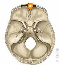
Crista Galli
Ethmoid bone structure described as bulb in front of olfactory.
Lacrimal Fossa
Hole in Lacrimal bone located in inner corner of eye.

Condylar Process
Mandible structure for back of jaw attachment.

Mandibular Foramen
Hole in Mandible inside the back of the jaw.

Mental Foramen
Hole in Mandible known as chin hole.

Mandibular Ramus
Structure on Mandible described as back of jaw think.

Coronoid Process
Structure on Mandible for front of jaw connection.

Infraorbital Foramen
Hole in Maxilla located at bottom of eyes.

Intermaxillary Suture
Suture in Maxilla forming a line in middle both inside and out.

Coronal Suture
Suture between Frontal and Parietal bones (F/P) that separates frontal and parietal.

Sagittal Suture
Suture between Parietal bones (P/P) that separates two halves of parietal.

Squamous Suture
Suture between Parietal and Temporal bones (P/T) that separates temporal and parietal.

Lambdoid Suture
Suture between Parietal and Occipital bones (P/O) that separates parietal from occipital.

Occipitomastoid Suture
Suture between Occipital and Temporal bones (O/T) that separates occipital from temporal.

Fontanelle
Fetal skull structure described as a soft spot.