Genetics
 Call Kai
 Learn
Learn Practice Test
 Spaced Repetition
 Match
Match Flashcards
 Knowt Play
1/55

There's no tags or description
Looks like no tags are added yet.
Name | Mastery | Learn | Test | Matching | Spaced | Call with Kai | Chat |
|---|
No analytics yet
Send a link to your students to track their progress
56 Terms
The human genome
-Humans are diploid
-233 pairs (46 total) of chromosomes, which are long, leaner chains of DNA
- Sizes range from 50,000,000 to 250,000,000 base pairs
- Each set of 23 chromosomes has 3.1 billion base pairs
-A complete human genome has 6.2 billion base pairs
The human genome has 20000 genes
this is not a lot flys have around 20,000 as well
Genes are small sections of chromosomes that are the basic unit of heredity

Human genetic experiment
-We are all descendants from a very small group of people who lives in East Africa 200,000 ears ago
-in The 10,000 generations since the time we have expanded across the planet and expanded from a few thousand individuals to over 8 billion individuals
- Our shared genetic material- the "human genomes" has been passed on- remixed, recombined and altered- through each generation- subject to the simples laws of genetic and the complexities of human history
What is genetics?
Genetics is the study of variation within a species
-Where does variation among individuals within a species come from?
-How is variation transmitted across generations?
-What are the consequences of variation to the organicm?
Your two copies of each chromosome are nearly identical
Your 2 copies of chromosomes differ in approximately 1 in 1,000 base pairs
When looking at it in an image that may seem like very little but there are 3 billion base pairs so there are approximately 3,000,000 differences between the 2 sets of chromosomes
allele
the specific DNA base found at a specific pace in a specific genome is usually referred to as an allele.
CFTR -Cystic Fibrosis Transmembrane Conductance Regulator
a membrane protein/channel that mainly moves Cl⁻ ions across cell membranes.
people with a mutation in CFTR the protein does not fold normally and is more quickly degraded leading to a loss of ion transport
Types of known CFTR mutations
no protein production
defective processing
defecting regulation/gating
reduced channel conductance
reduces protein quantity due to splicing defects
decreased protein stability
Where does genetic variation come from?
Genetic variation begins with error in DNA replication
DNA polymerases (the enzymes that make new copies of DNA) are really good at what they do, which is to pair Adenine- Thymine and Guanine- Cytosine
Every once in a while they make mistakes, about 1 in every 100,000 they simply make a mistake, pairing the wrong bases, matching wrong such as A-C and G-T
An error rate of 1 in 100,00 would mean 62,000 new mutations every time the human genome is copied which is simply too many
Systems that correct mistakes of DNA polymerase
Polymerases correct their own mistakes
One of the reasons that the process is so accurate is that polymerase “proofreads” its work, looking for small physical signs of an improper base pair and, when it finds it has made a mistake, correcting it. This catches most, but not all, improperly inserted basis.
Uncorrected errors create mismatches in the double strand (A-C, G-T)
Human mismatch repair (MMR) is the backup system that fixes DNA replication mistakes that DNA polymerase proofreading missed.
Think of it like:
DNA polymerase proofreading = first check
Mismatch repair = second check
MMR:
finds a mismatched base
identifies the newly made strand
removes the section containing the error
DNA polymerase fills in the correct bases
DNA ligase seals the strand
Most DNA damage can be recognized and repaired before DNA replication. Mutations are what happens in the rare cases when these error correction and repair systems fail.
In addition to simple copying errors, damage to DNA damage confuses the polymerases
UV damage creating abnormal DNA structures
chemicals modifying bases
oxidation damaging bases
That can cause polymerase to:
stall
insert the wrong nucleotide
skip damaged DNA
Common forms of DNA damage
The main ones to know are:
Depurination: a purine base like guanine falls off the DNA, leaving an empty site.
Deamination: 5-methylcytosine → thymine, which can create the wrong base pair.
Alkylation by EMS: adds ethyl groups to bases.
damaged guanine can pair with thymine → G·C → A·T mutation
damaged thymine can pair with guanine → T·A → C·G mutation
Oxidation: can produce damaged bases like thymidine glycol and 8-oxo-dG, which can also cause replication errors.

Mutations
mutations can happen anytime cells divide, we have to go through at least 35 trillion cell divisions, in our lifetime it is probably closer to 100 trillion
Humans contain somewhere between 10 and 50 trillion cells, each one a product of cell division so there are lots of opportunities for mutations to occur
Only mutations that occur in the gremlin (the lineages that make egg and sperm) can be transmitted to the next generation
Early in development, cells separate into somatic and germline lineages.
Somatic cells make the body; mutations here stay in that individual and usually are not inherited.
Germline cells make sperm/eggs; mutations here can be passed to offspring.
If a mutation happens before germline and somatic cells separate, it can end up in both lineages.
If a mutated sperm/egg forms a zygote, that mutation can be copied into the offspring’s cells.
A gremlin mutation can be inheritable a mutation confined to somatic cells cannot
A mutation before lineage separation can enter both soma and gremlin
Mutation si random
mutations is random in the sense that you can not predict what types of mutations will happen or where they will occur
but it is not truly random because there is extensive variation in the susceptibility of different parts of the genome to different types of mutation
Such as base subsititons are not equal likely transitions (purine to purine or pyrimidine to pryimidine) are roughly 2 times as common as trans versions (purine to pyrimidine or pyrimidine to purine
Cytosine and Thymine are pyridine (1 ring)
Adeline and Guanine are purine (2 rings)
base substitutions are not equally likely - changes from strong to Weak base pairs are more common than changes from weak to strong so G-C is strong A-T is week
Rates of base substations are not equal as well such as GC-AT has the highest rate where AT-TA has the lowest rate

De nova mutation
a de novo germline mutation is a new mutation that arises in one of the parent’s germline cells or in a sperm/egg precursor cell.
Types of Genova mutations and rarity
Single base-pair change: one DNA base is changed to another.
~75 per birthSmall deletion (<50 bp): a small piece of DNA is removed.
~4 per birthSmall insertion (<50 bp): a small piece of DNA is added.
~2 per birthTransposable element transposition: a mobile DNA sequence, or “jumping gene,” moves/inserts into a new place in the genome.
~1 in 25 birthsLarge insertion or deletion: a larger chunk of DNA is added or removed.
~1 in 50 birthsChromosome duplication or major rearrangement: a large chromosome region is copied, flipped, moved, or reorganized.
~1 in 500 births
Facts
Smaller deletion is more common than a long deletions
most children inherit more de nova mutations from their father than their mother
the other the parent the more mutations
Strand slippage
Strand slippage happens during DNA replication when the new strand and template strand briefly misalign, usually in areas with repeated DNA sequences.
Repition screws DNA
This can cause:
Insertion if the newly made strand loops out → extra bases get added.
Deletion if the template strand loops out → bases get skipped.

Other sources of variation in mutation rate
Local sequence context- GC rich regions mutate more rapidly
Non-unique regions of genome (repeats) are unstable
Mutation rate different at telomeres and centromeres
Transcription and recombination are mutagenic(capable of causing mutations in DNA)
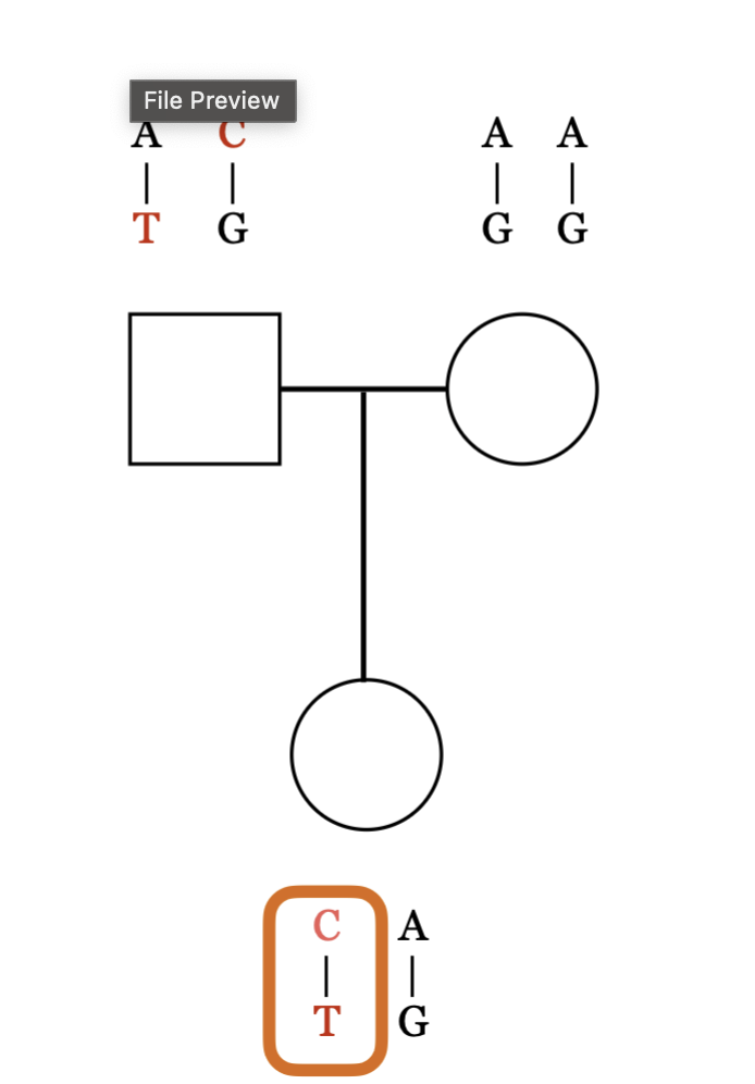
Pedigree key
if parent AA 100 percent to get A allele
if parent AG 50 percent to get A 50 percent to get G
Children get 1 allele from both parents
what one child gets has nothing to do with what the next child would get

25-50-25 rule


so it is ¾ because AA will 100 percent give AA so that’s ½ and then the 1 parent will have ½ chance to be AA and ½ to be AG but we have to keep in mind that there’s only ½ chance for 1 to be AA so we have to multiply ½ with 1/2

So each child, including #1, has:
1/2 chance AA
1/2 chance AG
Now ask: what is the chance that neither 3 nor 4 gets G?
There are two possibilities for parent #1:
If #1 = AA (probability 1/2), then #1 has no G to pass on, so both 3 and 4 definitely do not get G. Contribution = 1/2 × 1 = 1/2.
If #1 = AG (probability 1/2), then each child has a 1/2 chance of getting A instead of G. For both 3 and 4 to avoid G: 1/2 × 1/2 = 1/4. Contribution = 1/2 × 1/4 = 1/8.
Add them:
1/2 + 1/8 = 5/8
So:
P(neither 3 nor 4 has G) = 5/8
The exact same reasoning applies to 5 and 6, so:
P(neither 5 nor 6 has G) = 5/8
For the G allele to disappear from all four grandchildren, it has to disappear on both family branches:
5/8 × 5/8 = 25/64
Why you can't just do 3/4 × 3/4
Individually, each grandchild does have a 3/4 chance of being AA. But 3 and 4 are not completely independent, because they share parent #1.
If #1 happens to be AA, then both automatically cannot get G. That's why you first condition on whether #1 is AA or AG.
Fates of New Mutations
• New alleles continuously enter the population via mutation
• Most new mutations do not last very long in the population
• Some mutations reach higher frequencies in the population, but even most of these eventually disappear
• A very small fraction of new mutations get fixed in the population
• This is all the result of the random transmission of alleles from parents to their offspring
• This process is generally referred to as “genetic drift” (random change in genetic frequency)
• When we study genetic variation we are mostly looking at
alleles at different points along the path from having been
created by new mutation and disappearing
Independent Events: The occurrence of one event does not
affect the occurrence of another event
• Multiply independent event probabilities
• Mutually Exclusive Events: The occurrence of one event prevents
the occurrence of another event
• Add mutually exclusive event probabilities
• Complement Rule:
2. Lecture review
Probability of an event occurring = 1 – (probability of event not occurring)
If a question asks how likely a mutation that arrives today would be in x years the answer is just 1/x
What happens when we introduce new mutations in every generation
we can see that the most common mutations carry 1 copy and it is definitely skewed to the right
the numbers are not fixed but the shape will be consistent the size is determined by size of genome, size of population, and rate of mutation
the percent of a population having 2 of the alleles is exactly half of what the amount of populations that had 1 of the allele
we can assume that a rare variant is a older one
sites where new Allee is rare in the population are common
sites where new allele is common in the population are rare
Alleles at high frequency in population tend to be older this is very closely linked to
surviving mutations that occurred a long time ago are likely to be found in many genomes today
surviving mutations that occurred recently are generally found in fewer genomes
surviving mutations that occurred recently are generally found in fewer genomes

Classic recessive and classic dominant
A new allele G is “recessive” if heterozygotes AG has same phenotype as AA
It is “dominant” if heterozygote AG has same phenotype as GG\
In reality for most traits heterozygotes have distinct phenotypes there is usually a significant in heterozygotes and homozygotes even if it is a dominant trait.
Dominant/recessive describes the relationship between alleles for a particular phenotype — it does not mean the “recessive” allele does nothing in a heterozygote.
Normal / normal → normal β-globin production
Normal / β-thal allele → usually no severe β-thalassemia disease, but the person can still have reduced hemoglobin production, smaller red blood cells, and mild anemia
β-thal / β-thal → much more severe reduction in β-globin and can cause β-thalassemia major/intermedia

Not all mutations are loss of function
People homozygous for HbBs have sickle cell anemia which is superficially recessive, but there is a clear heterozygous phenotype with a few sickled cells but with HbBS it resists malaria. The middle column is heterozygous
Big idea- mutations in same gene can be “dominant” or “recessive” HbS allele can look dominant, recessive, or codominant depending on which phenotype you measure.

X and Y chromosomes are very different
The X chromosome looks more or less like a normal chromosome (an autosome) and contains 1400 genes, most of which function in both sexes
The Y chromosome, in contrast, is smaller and is a mostly functionless mess of receptive DNA and contains relatively few (200) genes that are almost all male- specific

SRY gene
The SRY gene is a key gene that usually starts the pathway toward male sexual development.
It is usually located on the Y chromosome and makes a transcription factor called SRY protein. Early in embryonic development, the gonads are initially bipotential, meaning they can develop along either the testis or ovary pathway.
SRY helps turn on genes such as SOX9, which pushes the gonad to develop into testes. The developing testes then produce hormones that guide further sexual development.

A single X chromosome is sufficient for Viability
You need a X chromosome to survive you don’t need a Y chromosome to survive.
You can have multiple X chromosomes for any other chromosome this can be lethal except chromosome 21 which is downsyndrome. The effects of having multiple X chromosomes are trivial.

Dosage compensation
Because males have only one X chromosome while females have two, and because there are a lot of important genes on X that do not have a copy on Y, there needs to be a system of dosage compensation to equalize the levels of expression of X chromosomal genes between the sexes.
In humans (and other non- marsupial mammals), dosage compensation occurs by the random inactivation of one X chromosome in females, so that all cells (whether male or female) have only one active X chromosome
X inactivation occurs around implantation, after many cell divisions have already occurred
The key idea is that an XX embryo starts with both X chromosomes active. The embryo then goes through several rounds of cell division first. Around the time of implantation, cells begin inactivating one X chromosome.
In each cell, usually one X is chosen for inactivation:
one cell might inactivate the maternal X
another might inactivate the paternal X
Once a cell chooses which X to shut off, its daughter cells generally keep the same X inactive. That inactive X becomes highly condensed and can be seen as a Barr body.
So this creates a mosaic female:
early embryo → many cells → random X inactivation → different groups of cells express different X chromosomes
This is why an XX individual can have patches of cells expressing genes from different X chromosomes.
The purpose is dosage compensation: XX individuals have two X chromosomes, while XY individuals typically have one, so X inactivation prevents most X-linked genes from being expressed at roughly double the level in XX cells.
One important detail: not every gene on the inactive X is completely shut off—some genes escape X inactivation.
Timing of X- inactivation creates mosaicism (one person has groups of cells that are genetically or functionally different from each other, even though all the cells came from the same fertilized egg.)
Early in development, different cells randomly inactivate one X chromosome. For example:
one cell may keep the maternal X active and inactivate the paternal X
another cell may do the opposite
Once a cell makes that choice, all of its descendants usually keep the same X inactive.
This creates patches of tissue with different activated X chromosomes.

different species use different systems to determine sex.

There are different dosage compensation systems between animals

autosome
any chromosome that is not a sex chromosome
X chromosome transmission genetics
In females the X chromosome behaves just like an autosome. There is
recombination between the two X’s and eggs get one (sometimes
recombined) X chromosome. (two X chromosomes can swap matching pieces of DNA during meiosis before they separate into eggs.)
In males there is effectively no recombination between X and Y, and each
gamete (sperm) gets either an X or a Y, thereby determining the sex of
the child.
Females always get their father’s X chromosome allele and one of their mother’s X chromosome alleles.
Males get one of their mother’s X chromosome alleles and no X chromosome alleles from their father.

this disease typically manifests in males whose single X carries a F8 mutation (h). Affected males typically do not have affected children because they only pass the disease causing allele to daughters. Daughters of affected males are always carriers.
So females usually have a 50/50 chance on if they are affected because it depends on which X chromosome is deactivated
But sex related diseases are not always because of the X chromosome such as testicular cancer
Recombination
The exchange of DNA from homologous chromosomes during meiosis
humans have countable number of recombination not many at all -Averaged over human generations, roughly
one recombination event is observed per
million bases per hundred generations.
4 chromosomes will have no recombination 2 from mom 2 from dad
Recombination hotspots-
At fine scale we see “recombination hotspots” - sites in
genome that are >10x more likely to have recombination
events. Roughly 1 hotspot/50 kb, with ~80% of recombination
events occurring in clear hotspots. They evolve very rapidly the “kill” themselves
average of one additional crossover per 100 Mb (males) or 50 Mb (million of base pairs) (females) the protein that is in charge of cuts is more prominent in males and remember there is always 1 crossover around 1.6x more recombination in females than males

Genes on seperate chromosomes

Transmission of multiple genes on same chromosome
The two genes are on the same chromosome and are extremely close together.
The mother has the alleles arranged like this:
[ AC / GT ]
Meaning one chromosome carries A and C together, and the homolog carries G and T together.
Because the genes are immediately adjacent, crossing over between them is essentially assumed to be 0% in this example.
So the mother makes only two gamete types:
[ AC ]
or
[ GT ]
each with probability:
1/2
She does not make:
[ AT or GC ]
because that would require recombination between the two genes.
The father is AA CC, so he always contributes:
[ AC ]
Therefore the child can be:
[ AACC ]
or
[ AGCT ]
each with probability \(1/2\).
So the key comparison is:
Different chromosomes → AC, AT, GC, GT each 1/4
Same chromosome and very close → only parental combinations AC and GT, each 1/2
![<p><strong>The two genes are on the same chromosome and are extremely close together</strong>.</p><p>The mother has the alleles arranged like this:</p><p><span>[ AC / GT ]</span></p><p>Meaning one chromosome carries <strong>A and C together</strong>, and the homolog carries <strong>G and T together</strong>.</p><p>Because the genes are <strong>immediately adjacent</strong>, crossing over between them is essentially assumed to be <strong>0%</strong> in this example.</p><p>So the mother makes only two gamete types:</p><p><span>[ AC ]</span></p><p>or</p><p><span>[ GT ]</span></p><p>each with probability:</p><p><span>1/2</span></p><p>She does <strong>not</strong> make:</p><p><span>[ AT or GC ]</span></p><p>because that would require recombination between the two genes.</p><p>The father is <strong>AA CC</strong>, so he always contributes:</p><p><span>[ AC ]</span></p><p>Therefore the child can be:</p><p><span>[ AACC ]</span></p><p>or</p><p><span>[ AGCT ]</span></p><p>each with probability \(1/2\).</p><p>So the key comparison is:</p><p><strong>Different chromosomes → AC, AT, GC, GT each 1/4</strong></p><p><strong>Same chromosome and very close → only parental combinations AC and GT, each 1/2</strong></p>](https://assets.knowt.com/user-attachments/a8034163-19c1-4d86-9173-b9e73cf24138.png)
Genes on separate chromosomes vs genes next to each other on same chromosome vs genes separated on same chromosome

Recombination can bring together alleles that emerged on separate chromosomes
recombination happens so often across generations, most genetic variants in humans are not strongly linked to each other at the population level.
The black horizontal lines represent chromosomes, and the colored dots are different genetic variants.
The key idea is:
Variants that are very close together on the same chromosome are more likely to stay inherited together.
Variants that are farther apart are more likely to get separated by crossing over.
Over many generations, repeated recombination breaks up old combinations of alleles.
So even if two variants started out together on the same chromosome, after enough generations they may become effectively independent.
That is what “effectively unlinked” means here: knowing which allele someone has at one location often tells you very little about what allele they have at a distant location.
A short way to think about it is:
close together → more linkage
far apart + lots of generations of recombination → less linkage / effectively unlinked
Recombination allows us to estimate how old a new allele is which helps us understand population history and evolution
Alleles that rise in frequency because of drift will have taken longer to get there, have experienced more recombination, and therefore have smaller shared haplotypes
Alleles that rise in frequency because of positive selection will have taken less time to get there, have experienced less recombination, and therefore have larger shared haplotypes
closer orange dots mean they are more strongly linked, so recombination between them happens less often.
Common alleles are usually older because they've been passed down through many generations, allowing recombination to scramble their haplotype backgrounds.

Measuring recombination event
the unit is centimorgan if you multiply by 100

Relationship between distance between alleles and recombination rate
In humans 1 centimorgan= 1MB
1 Mb apart → about 1% recombination
5 Mb apart → about 5% recombination
10 Mb apart → about 10% recombination
And remember:
1% recombination = 1cM

Genes are 25 Mb apart.
1 Mb ≈ 1 cM, so:
25 Mb = 25 cM = 25% recombination
Therefore:
Recombinant gametes = 25% total
Parental gametes = 75% total
Person 1: AG / CT
AG is a parental gamete.
P(AG from person 1) = 75% / 2 = 37.5% = 0.375
Person 2: AT / CG
AG is a recombinant gamete.
P(AG from person 2) = 25% / 2 = 12.5% = 0.125
For child 3 to be AA at Gene 1 and GG at Gene 2, child 3 must receive AG from both parents.
P(AAGG) = 0.375 × 0.125
P(AAGG) = 0.046875
Final answer = 4.6875%
How do you measure crossovers
males 1 extra crossover for 100Mb, females 1 extra crossover for 50Mb
Look at distance for example if if total distance is 100Mb and in between your target loci is 25
Probability a crossover between loci is ¼
Probability a crossover outside loci is ¾
Probability N crossovers all outside loci is (3/4)^N and Probability 1 or more crossovers between loci is 1-(3/4)^N
First do the probability of inside lets say for the father there is 2 crossover so that’s .44 we divide that by 4 because there are 4 different possibilities if there is more than one crossover inside the loci we can infer that all 4 options would happen
Then 1-.44=0.56 so that is the parental gene possibility divide that by 2 but don’t forget to add the values we got in step 3
Do the same for the parent then that makes a chart where you just add the values together

Mitochondria lineages carry unique set of mutations
Mitochondrial DNA (mtDNA) is usually inherited from the mother.
Mutations accumulate in mtDNA over generations.
Different maternal lineages can accumulate different mutations, giving each lineage a unique set.
No independent assortment occurs like it does with nuclear chromosomes during meiosis because mtDNA is not inherited as pairs of homologous chromosomes.
At some point it descents into one ancestor converging about 200,000 years ago
Best current guess is that mitochondrial EVE lived 150-200,000 years ago in sub–Saharan Africa
There is also a Y chromosomal Adam who lived roughly at the same time
Human migration history can also be inferred from mitochondrial DNA a map could be made
So on top of mutation and genetic drift we also have to account for migration
Splitting populations creates population level differeneces in allele frequencies
When a small group separates from a larger population, it carries only a subset of the original genetic variation. By chance, some alleles are lost and others become more common.
The more uneven the split, the greater the founder effect on the smaller population.
50/50 split: Both populations retain relatively more genetic variation.
90/10 split: The smaller 10% population is more likely to lose alleles due to random sampling.
99/1 split: The smaller 1% population experiences an even stronger founder effect.
Bottleneck effect: A population shrinks dramatically → genetic drift causes loss of genetic diversity.
Generally, the farther apart two human populations are geographically, the greater their genetic differences tend to be.
Closer populations: Tend to be more genetically similar because of greater gene flow.
Distant populations: Tend to be more genetically different because of reduced gene flow and accumulated genetic drift.
Exception: Migration can connect distant populations, so distance doesn't always predict genetic differences.
How to calculate the allele frequency
rare alleles are more common than high-frequency alleles.
Sₙ: Number of sites where a derived allele appears in exactly \(n\) sampled genomes.
m: Mutation rate.
n: Number of copies carrying that allele.
S1=C(m/1)
n increases → Sₙ decreases: Fewer alleles are found in many individuals.
n decreases → Sₙ increases: More alleles are found in only a few individuals.
m increases → Sₙ increases: More mutations create more genetic variants.

Rare variants are common Common alleles are rare
Rare variants are numerous, but each is unlikely to appear in your genome. Common variants are fewer, but each is more likely to appear in your genome.
C: Proportionality constant.
m: Mutation rate.
n: Number of copies carrying the derived allele.
2N: Total number of gene copies in a diploid population.

Classification of human allels by geographic distribution
We can see taht when allels are common they are usually common everywhere
And derived alleles that are rare are usually region specific
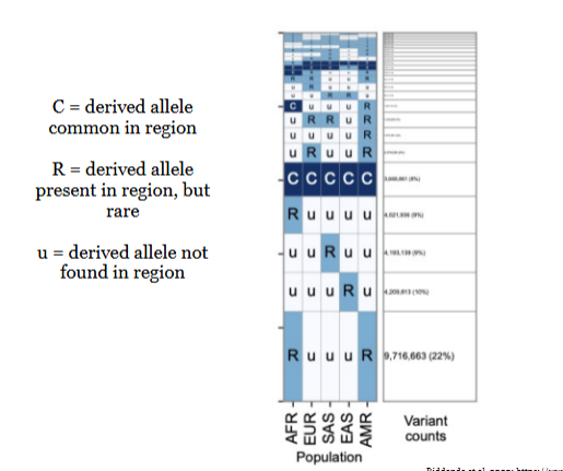

99 percent
Understanding the graph
X-axis: How common a derived allele is in the population.
0.01 = 1% of alleles in the population
0.50 = 50%
1.00 = 100%
Y-axis: How many derived alleles in your genome have that population frequency.
Why are the bars taller on the left?
A new mutation initially appears in just one individual.
Most new mutations remain rare or disappear because of genetic drift.
Some mutations spread through the population over many generations.
Therefore, your genome contains many rare derived alleles and fewer high-frequency derived alleles.
Important
Rare derived alleles: Many exist because new mutations continuously occur.
Common derived alleles: Fewer exist because most new mutations disappear through genetic drift before becoming common.
Genetic drift: Randomly causes alleles to increase or decrease in frequency, sometimes leading to fixation or disappearance.
How do we know which variants in the human genome matter and how they might affect you?
compare people who have one allele to people who have another and see if/how they differ
Linkage mapping needs to see a lot of families and get a lot of datas so it has been replaced by association mapping

Association mapping
we sort by the trait and see if people have the disease have something that’s different from people without the disease in this case its the red allele
this works best when both alleys you are comparing are resonably common because you need to observe the allele enough to see how it is associated with the trait

Is genotype associated with phenotype
Most genetic variants tested are not significantly associated with the phenotype, while a small number show strong associations.
The lines going up are the associated variants

GWAS (Genome-Wide Association Study)
a study that looks for associations between genetic variants and a particular phenotype (trait or disease)
what does it mean when a lot of traits have genetic components and a lot of traits have non genetic components
It means most traits are influenced by both genetics and the environment, not just one or the other.
Genetic components: Differences in traits caused by differences in DNA (genotypes).
Non-genetic components: Differences in traits caused by factors other than DNA, such as environment, nutrition, lifestyle, or experiences.
Example:Height
Genetic: Genes inherited from your parents influence your height.
Non-genetic: Nutrition and childhood health can also influence your height.
Example:Academicperformance
Genetic: Genetic differences can influence traits related to learning.
Non-genetic: Education, studying, sleep, and environment also affect performance.
Additive model
This is also why dominant and recessive is not necessarily true
SNPs (Single Nucleotide Polymorphisms) are variations in a single DNA nucleotide (A, T, C, or G) at a specific position in the genome.
the beta value is something we can add up depending on how much of the target allele they have
The black shaded lines are like error bars that take environment into account
