LIFE 212 Exam 2
 Call Kai
 Learn
Learn Practice Test
 Spaced Repetition
 Match
Match Flashcards
 Knowt Play
1/84

Earn XP
Description and Tags
Vocabulary flashcards covering biological membrane composition, characteristics, and solute diffusion/transport mechanisms based on LIFE210 Exam 2 lectures.
Name | Mastery | Learn | Test | Matching | Spaced | Call with Kai | Chat |
|---|
No analytics yet
Send a link to your students to track their progress
85 Terms
Plasma membrane
A selective lipid bilayer boundary that encloses the cytoplasm, protecting cell contents and regulating solute entry and exit.
Phospholipid
An amphipathic lipid consisting of a hydrophilic head group containing phosphate and hydrophobic fatty acyl tails.
Phosphoglyceride
A class of phospholipids built on a glycerol backbone esterified to two fatty acid tails and a phosphorylated head group.
Glycerol
A three-carbon alcohol molecule that forms the backbone of phosphoglycerides and triglycerides.
Amphipathic
Describing a molecule that contains both distinct hydrophilic (water-loving) and hydrophobic (water-fearing) regions.
Head group
The polar, hydrophilic region of a membrane lipid that interacts directly with aqueous cellular environments.
Fatty acid tail
The nonpolar, hydrophobic hydrocarbon chain component of a lipid molecule.
Triglyceride
A non-membrane lipid composed of three fatty acid tails attached to a glycerol backbone used primarily for energy storage.
Sphingosine
An amino alcohol containing a long hydrocarbon chain that serves as the backbone for sphingolipids.
Sphingolipid
A membrane lipid derived from sphingosine rather than glycerol, featuring fatty acid attachment via an amide linkage.
Glycolipid
An amphipathic membrane lipid containing one or more covalently attached sugar molecules.
Cholesterol
A membrane sterol that stabilizes lipid bilayer integrity across both high and low temperature extremes.
Integral transmembrane protein
An amphipathic protein embedded directly within the lipid bilayer that spans across the hydrophobic core of the membrane.
Peripheral transmembrane protein
A protein associated with the membrane surface via non-covalent interactions with integral proteins or lipid head groups without penetrating the hydrophobic core.
Single-pass vs multi-pass transmembrane protein
Classification of integral membrane proteins based on whether their polypeptide chain crosses the lipid bilayer once or multiple times.
Hydropathy plot
A quantitative graph displaying amino acid hydrophobicities across a protein sequence to identify potential membrane-spanning regions.
Self-assembly
The spontaneous formation of structured lipid arrangements (micelles or bilayers) driven by hydrophobic interactions in water.
Micelles vs bilayers
Micelles are spherical structures formed by single-tailed lipids, whereas bilayers are two-layered lipid sheets formed by double-tailed lipids.
Lateral diffusion
The rapid unhindered translational movement of lipids and proteins side-to-side within the same monolayer of a membrane.
Bilayers as “2D liquids”
Concept referring to the dynamic movement of lipid molecules diffusing freely across a single layer plane while rarely flipping across monolayers.
Lipoproteins
Water-soluble lipid-protein complexes designed to transport hydrophobic lipid molecules through circulation.
Selective permeability
The property of biological membranes that allows uncharged small molecules to cross while restricting ions, polar molecules, and large macromolecules.
Phosphatidylserine
An negatively charged phosphoglyceride asymmetric concentrated on the cytosolic monolayer of the plasma membrane.
Electrochemical gradient
The combined effect of a concentration gradient and an electrical charge difference across a membrane that drives ion movement.
Glycoprotein
A membrane protein containing covalently attached oligosaccharide chains located on the extracellular domain.
Carbohydrate layer
A protective, recognition-focused sugar coating on the outer cell surface formed by glycolipids and glycoproteins.
Lipid rafts
Thicker, specialized membrane microdomains enriched in sphingolipids, cholesterol, and specific functional proteins.
Solute vs solvent
A solute is a substance dissolved within a liquid medium, whereas a solvent is the liquid medium (such as water) doing the dissolving.
Concentration gradient
A differential density of solute molecules present between two connected regions or across a biological membrane.
Simple diffusion
Passive, unassisted transport of solutes directly across a lipid bilayer down their concentration gradient.
Passive transport
Spontaneous solute movement across a membrane down its electrochemical gradient without requiring energy (ΔG<0).
Active transport
Energy-requiring movement of solutes across a membrane against their concentration or electrochemical gradient (ΔG≥0).

Activation Energy (EA) in Membrane Transport
The energetic barrier associated with passing a solute through a membrane; lowered by facilitated transport proteins relative to non-facilitated transport without altering overall ΔG.
Osmosis
The passive net movement of solvent water molecules across a selectively permeable membrane toward higher solute concentrations.
Inorganic ion
Charged atomic species (such as Na+, K+, Ca2+, or Cl−) requiring transport proteins to cross the hydrophobic membrane core.
Electrical gradient
A potential difference in electrical voltage across a membrane generated by an unequal distribution of charge.
Transporter
A membrane protein that undergoes conformational changes to bind and translocate specific solutes across a membrane.
Channel
A membrane protein forming a hydrophilic transmembrane pore that permits rapid, selective solute diffusion down a gradient.
Gated channels
Channels that fluctuate between open and closed states in response to specific chemical, electrical, or physical signals.
Aquaporins
Selective transmembrane channel proteins designed for high-speed passage of water molecules across membranes.
Primary active transport
Active transport powered directly by cellular energy sources such as ATP hydrolysis.
Secondary active transport
Cotransport driving solute movement against its gradient powered by energy stored in an ion gradient set up by primary transport.
Antiport
A cotransporter that translocates two different solute species across a membrane in opposite directions.
Symport
A cotransporter that moves two different solute species across a membrane in the same direction.
Uniport
A carrier protein that moves a single specific solute across a biological membrane.

Epithelial Glucose Transport System
A system utilizing an apical Na+/glucose symport (Transporter #1), a basolateral glucose uniport (Transporter #2), and a basolateral Na+−K+ pump (Transporter #3) to transport glucose from the gut lumen to the bloodstream.

CFTR (Cystic Fibrosis Transmembrane Conductance Regulator)
An ABC protein functioning as a chloride ion channel regulated by nucleotide-binding domains and phosphorylation at its regulatory domain.
Describe the hydrophobic and hydrophilic components of phosphoglycerides and sphingolipids, and which regions of the plasma membrane are occupied by each.
Phosphoglycerides consist of a glycerol backbone esterified to two nonpolar, hydrophobic fatty acyl tails and a polar, hydrophilic head group containing a phosphate.
Sphingolipids are built on a sphingosine (amino alcohol) backbone, featuring a single long hydrocarbon tail attached via an amide linkage and a polar head group.
Membrane positioning: The polar hydrophilic head groups face outward, interacting directly with the aqueous extracellular fluid and intracellular cytoplasm. The nonpolar hydrophobic tails face inward, packed tightly together to form the interior core of the lipid bilayer.
Compare and contrast the structure and function of phosphoglycerides and sphingolipids.
Similarities: Both are amphipathic membrane lipids that play essential roles in forming the structural matrix of cellular membranes and protecting the cell.
Differences: Phosphoglycerides use a three-carbon glycerol backbone with two fatty acid chains. Sphingolipids use a sphingosine backbone with one primary hydrocarbon tail linked via an amide bond. Structurally, sphingolipids tend to have longer, straighter, more saturated hydrocarbon chains, which allows them to pack more tightly with cholesterol into specialized microdomains like lipid rafts.
How is a triglyceride distinct from a phosphoglycerides, and are they found in membranes?
Triglycerides consist of a glycerol backbone attached to three fatty acid tails. Because they lack a polar, phosphorylated head group, they are entirely hydrophobic (nonpolar) and not amphipathic.
Membrane presence: Because they cannot form a stable bilayer on their own, triglycerides are not found in biological membranes. Instead, they are stored as neutral fat droplets in the cytoplasm for energy storage.
Glycolipids are an important class of lipids that contain covalently attached sugar molecules.
Which of the following statements are TRUE, and which are FALSE?
• All glycolipids are sphingolipids.
• All glycolipids are phosphoglycerides.
• All glycolipids are phospholipids.
• All glycolipids are amphipathic.
All glycolipids are sphingolipids: False (some glycolipids can be built on a glycerol backbone, such as glycoglycerolipids).
All glycolipids are phosphoglycerides: False.
All glycolipids are phospholipids: False (many glycolipids do not contain a phosphate group).
All glycolipids are amphipathic: True (they possess a hydrophilic sugar head group and hydrophobic tails).
What is the primary chemical difference between different types of phosphoglycerides, such as phosphatidylcholine and phosphatidylethanolamine?
The primary difference lies in the chemical structure of the polar head group attached to the phosphate. Phosphatidylcholine has a choline group attached (carrying a quaternary amine), while phosphatidylethanolamine has an ethanolamine group attached. These differences change the net charge, size, and shape of the head group, influencing membrane curvature and protein-lipid interactions.
Compare and contrast integral and peripheral transmembrane proteins.
Integral transmembrane proteins span completely across the hydrophobic core of the lipid bilayer. Peripheral transmembrane proteins are bound loosely to the membrane surface via non-covalent interactions with integral proteins or lipid head groups without penetrating the core.
Why must integral transmembrane proteins be amphipathic?
Integral proteins must be amphipathic because they have hydrophobic amino acid stretches that interact with the fatty acid tails inside the membrane core, alongside hydrophilic domains exposed to the aqueous environments on either side of the membrane.
How can a hydropathy plot be interpreted to predict regions of a protein that pass through a membrane by a single transmembrane domain?
A hydropathy plot graphs the free energy required to transfer segments of an amino acid sequence from a hydrophobic solvent into water. A single transmembrane domain typically appears as a distinct, isolated peak of high positive hydrophobicity spanning roughly 20 to 25 consecutive amino acids (the length needed to cross the hydrophobic core as an alpha-helix).
Both bilayers and micelles have a remarkable ability to self-assemble in water. What is different about the lipid molecules that self-assemble into a bilayer versus a micelle?
Micelles are formed by single-tailed lipids (like free fatty acids or detergents) that have a wedge-shaped profile (a bulky head group relative to a single tail).
Bilayers are formed by double-tailed lipids (like phosphoglycerides and sphingolipids) that have a more cylindrical shape, making flat sheet or spherical bilayer closure geometrically favored.
Why is closure of a bilayer into a spherical structure energetically favorable?
A flat, open lipid bilayer has exposed hydrophobic edges at its perimeter that come into direct contact with water, which is energetically unfavorable. By closing into a continuous, sealed spherical vesicle, the edges are eliminated, burying all hydrophobic tails completely away from water.
Why do phospholipid molecules diffuse freely within a bilayer membrane, but cannot freely flip from one monolayer to the other?
Lateral diffusion is rapid and unhindered because the lipid stays within the same fluid monolayer plane.
Transbilayer diffusion ("flip-flop") is extremely rare spontaneously because it requires forcing a bulky, charged hydrophilic head group to traverse the completely hydrophobic, nonpolar core of the membrane—a massive energetic barrier. Specialized enzymes called flippases/flopases are required to catalyze this movement.
Describe the relationship between temperature and the fluidity of a lipid bilayer. Why does a change in temperature produce a change in fluidity?
Fluidity is directly proportional to temperature. Higher temperatures increase thermal kinetic energy, causing the fatty acyl tails to vibrate and move more rapidly, transforming the membrane from a gel-like state into a more fluid, dynamic "2D liquid." Lower temperatures decrease kinetic energy, causing tails to pack tightly together and reduce fluidity.
Describe the relationship between fatty acid saturation and fluidity of a lipid bilayer. Why does a change in saturation produce a change in fluidity?
Saturated fatty acids have no double bonds, allowing them to pack tightly together in straight chains, decreasing fluidity.
Unsaturated fatty acids contain cis-double bonds that introduce "kinks" in the hydrocarbon chains. These kinks prevent tight packing, keeping the core loose and increasing membrane fluidity.
Cholesterol has the remarkable effect of stabilizing the integrity of lipid bilayers. Why is this helpful at both very low and very high temperatures?
At high temperatures: Cholesterol rigidifies the membrane by packing closely with fatty acid tails, preventing excessive fluidity and leakage.
At low temperatures: Cholesterol inserts itself between neighboring lipid tails to prevent them from crystallizing and packing too tightly, maintaining membrane flexibility.
Rank the following molecules from highest to lowest based on their ability to diffuse through a protein-free lipid bilayer.
• lactose; Ca2+; lysine; glucose; glycerol; a 20bp DNA oligonucleotide; CO2
• Which of these molecules do you think require a transporter or channel to pass across the plasma membrane, and why?
CO2 > Glycerol > Lactsoe = Glucose > Lysine = Ca2+ > 20bp oligonucleotide
Explanation: Small nonpolar molecules (CO2) cross fastest via simple diffusion. Small uncharged polar molecules (glycerol) cross slowly. Large polar molecules (glucose, lactose) and charged ions (Ca2+, lysine, large DNA polymers) are heavily restricted by the hydrophobic core and require specific transport proteins.
In class, we described the composition of the plasma membrane bilayer as asymmetric. How is the composition of negative charges asymmetrically distributed in the membrane, and how does this affect the membrane’s potential?
Specific negatively charged phospholipids, such as phosphatidylserine, are heavily concentrated on the cytosolic monolayer of the plasma membrane. This creates a resting electrical charge asymmetry, contributing to the negative electrical potential of the cell interior relative to the outside.
How is glycosylation on the plasma membrane asymmetrically distributed, and what are some functions this glycosylation has on a membrane’s function?
Glycolipids and glycoproteins are restricted exclusively to the extracellular monolayer, creating a protective sugar coating known as the carbohydrate layer (glycocalyx). Functions include cell-cell recognition, protection against mechanical and chemical damage, and binding sites for signaling molecules.
What is a lipid raft, and how are these microdomains different from the rest of the plasma membrane? Include in your answer both the physical properties of the lipid raft, and what molecules are enriched within them.
Lipid rafts are specialized, thicker, and less fluid membrane microdomains enriched with sphingolipids, cholesterol, and specific signaling proteins. Unlike the surrounding fluid phosphoglyceride matrix, rafts act as organized platforms that concentrate receptors and enzymes for signal transduction.

In class, we used the energy diagram below to describe how facilitated transport assists in solute passage through a membrane. Answer the following questions below:
Does ΔG need to be greater than or lesser than 0 for spontaneous passage through a membrane?
For a process to be spontaneous, Delta G must be less than 0 (Delta G < 0). This applies to passive transport down an electrochemical gradient.

In class, we used the energy diagram below to describe how facilitated transport assists in solute passage through a membrane. Answer the following questions below:
Why are membranes impermeable to many solutes, like ions?
The Hydrophobic Core: The interior of a lipid bilayer is made of tightly packed, nonpolar fatty acid tails that create a strong hydrophobic barrier.
Energy Cost of Stripping Hydration Shells: Charged ions and polar molecules are energetically stable in aqueous environments because they form favorable interactions (hydration shells) with water molecules. To cross the hydrophobic core, these molecules must strip away their surrounding water molecules, which requires an immense, energetically unfavorable activation energy barrier.

In class, we used the energy diagram below to describe how facilitated transport assists in solute passage through a membrane. Answer the following questions below:
How does passive transport assist in solute transport at the reaction energy levels?
Lowering Activation Energy (EA): Facilitated passive transport (using carrier proteins or channels) provides an alternative pathway that significantly lowers the activation energy required for solutes to cross the membrane.
Thermodynamic Favorability (ΔG<0): While it does not change the overall free energy change (ΔG remains negative for movement down a gradient), it speeds up the rate of transport by bypassing the high energy barrier of the pure lipid bilayer, allowing spontaneous movement to happen rapidly.
How does a concentration gradient dictate the direction of passive versus active transport?
Passive transport always moves solutes down a concentration/electrochemical gradient (from high to low concentration).
Active transport moves solutes against a concentration gradient (from low to high concentration), which requires an external energy input (delta G).
Cells attract a high concentration of inorganic ions into their cytoplasm, due to the large presence of charged molecules in a cell. Why does this cause water to rush into the cell?
High intracellular solute concentration lowers the water potential inside the cell compared to the outside. By osmosis, water molecules move down their concentration gradient, rushing into the cytoplasm toward the region of higher solute concentration.
How do cells solve the problem of too much water uptake as inorganic ions enter the cell?
Animal cells actively pump out inorganic ions (such as via the Na^+/K^+ pump) to maintain osmotic balance and prevent excessive swelling or bursting (lysis).
What are some similarities and differences between passive transporters and channels?
Similarities: Both mediate passive transport down an electrochemical gradient without requiring external energy.
Differences: Channels form open, hydrophilic pores allowing rapid ion/water flux based on gating; carriers/transporters do not form open pores and must undergo conformational changes to ferry specific solutes across.
How does the electrochemical gradient influence passive transport efficiency?
The electrochemical gradient combines both a concentration gradient and an electrical voltage gradient across the membrane. A steeper gradient increases the driving force (Delta G), enhancing the rate and efficiency of passive transport.
Why is it important that channels are gated, and what are some types of mechanisms that regulate channel opening?
Gating prevents continuous, uncontrolled leaks, allowing the cell to control electrical signals and maintain homeostasis. Channels are regulated by voltage-gating (changes in membrane potential), ligand-gating (binding of chemical molecules), or mechanically-gated mechanisms (physical stress or pressure).
In class, we discussed aquaporins and the bacterial K+ channel as examples of highly selective channels. Why must channels be selective for their solute, and how do many channels achieve this property?
hannels must be selective to ensure only specific ions (e.g., K^+ or Cl^-) pass through, preserving cellular gradients. They achieve this via a selectivity filter inside the pore—a narrow region lined with specific amino acid carbonyl oxygens that mimic the exact hydration shell of the target ion, stripping away water only for that specific ion size and charge.
How can active transport achieve solute transport that is energetically unfavorable (ΔG ≥ 0)?
Active transport couples the unfavorable movement of a solute against its gradient directly to an energy-releasing process, such as ATP hydrolysis or the simultaneous downhill movement of another ion (secondary active transport).
Compare and contrast primary and secondary active transport.
Primary active transport is directly driven by ATP hydrolysis (e.g., the Na+/K+ pump).
Secondary active transport uses energy stored in an existing electrochemical gradient (usually set up by primary transport) to cotransport another solute uphill.
About a third of a typical animal cell’s energy is devoted to fueling the Na+/K+ pump. Why is this pump so energetically costly, and how is ATP utilized to operate it?
The pump moves three Na^+ ions out and two K^+ ions in, both against their steep electrochemical gradients. ATP is utilized via autophosphorylation: the transfer of a phosphate group from ATP onto an aspartate residue on the pump forces a major conformational change that expels Na^+ and K^+.
How does active transport establish and maintain electrochemical gradients, and why are electrochemical gradients so critical for cells?
Primary pumps constantly export ions to maintain steep concentration and charge asymmetries. These gradients are critical because they power secondary active transport, drive nerve impulse propagation, and maintain cell volume.
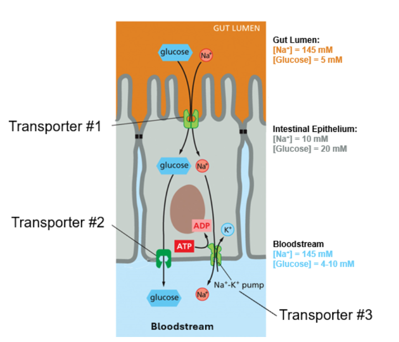
When we consume food that contains energy, often in the form of glucose, our bodies use intestinal epithelium cells to import that glucose into the bloodstream. Use the diagram below to answer the following questions about this system:
Based on the concentrations of Na+ and glucose in the gut lumen, intestinal epithelium cell cytosol, and bloodstream, which of the transporters 1-3 are for passive transport? Which are for active transport?
Transporter #1 (Na^+/glucose symport on the apical membrane) uses the Na^+ +$ gradient, making it secondary active transport.
Transporter #2 (basolateral glucose uniport) is passive transport (facilitated diffusion down a concentration gradient out to the blood).
Transporter #3 (the basolateral Na^+/K^+ pump) is primary active transport.
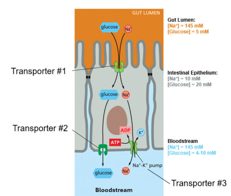
When we consume food that contains energy, often in the form of glucose, our bodies use intestinal epithelium cells to import that glucose into the bloodstream. Use the diagram below to answer the following questions about this system:
Glucose is transported into intestinal epithelium cells by transporter 1 and 2. Which transport is energetically favorable? For each transporter, is glucose transport energetically favorable or unfavorable? Explain why.
Glucose is driven into the cell via Transporter #1 by coupling its movement with the energetically favorable downhill influx of Na^+ ions established by the Na^+/K^+ pump.
When we consume food that contains energy, often in the form of glucose, our bodies use intestinal epithelium cells to import that glucose into the bloodstream. Use the diagram below to answer the following questions about this system:
Which of transporters 1–3 are uniports, symports, or antiports?
Transporter #1: Symport (Na+ and glucose in the same direction across the apical membrane).
Transporter #2: Uniport (moves a single solute, glucose, across the basolateral membrane).
Transporter #3 (Na+/K+ pump): Antiport (moves Na+ out and K+ in opposite directions).

Why is the asymmetric distribution of these transporters on the apical and basolateral membranes of intestinal epithelial cells critical for the absorption of glucose from the gut lumen into the bloodstream??
The strict polarization of the cell—placing the Na+/glucose symporter exclusively on the apical side, and the glucose uniport plus the Na+/K+ pump on the basolateral side—is what establishes a directional transepithelial flow. Without this spatial asymmetry, glucose entering from the gut would just leak back out or fail to accumulate and pass unidirectionally into the bloodstream.
CFTR (cystic fibrosis transmembrane conductance regulator) is an ABC protein that functions as a chloride ion channel in epithelial tissues.
What structural features define CFTR as a member of the ABC protein family?
CFTR contains ATP-binding cassette (ABC) domains (nucleotide-binding domains) that bind and hydrolyze ATP to regulate channel gating, making it unique among ion channels because it shares structural homology with ABC active transporters.
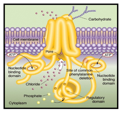

CFTR allows Cl⁻ to cross the membrane shown on the right. The arrow indicates the channel pore, not the direction of ion movement. The normal Cl⁻ concentration is approximately 120 mM outside the cell and 5–15 mM inside the cell. In which direction would Cl⁻ tend to move through CFTR? What is the role of CFTR in this process?
Given an extracellular concentration of around 120 mM and an intracellular concentration of 5-15 mM, Cl- will tend to move inward from the outside to the inside of the cell down its chemical concentration gradient when the channel is open. (Note: In native epithelial tissues, depending on local membrane potentials and regulatory chloride pumps, CFTR typically facilitates chloride secretion outward into lumens like the lungs or gut).
What is the role of CFTR in this process? CFTR acts as a gated chloride channel, providing a controlled hydrophilic pore for charged Cl- ions to cross the hydrophobic plasma membrane barrier.