chap 11
 Call Kai
 Learn
 Practice Test
 Spaced Repetition
 Match
 Flashcards
 Knowt Play
1/102

There's no tags or description
Looks like no tags are added yet.
103 Terms

Axial muscles
have both origins & insertions on axial skeleton
support & move the head & vertebral column
function in facial expression, breathing, chewing, & swallowing
support & protect abdominal & pelvic organs
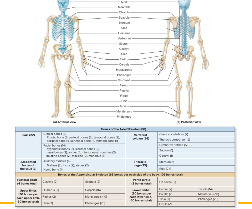
Appendicular muscles
control movements of upper & lower limbs
control movements of pectoral & pelvic girdles
organized into groups based on locations

Muscles on the front vs back
front: flex & can do other motions
back: extend & can do other motions

Tendon
attaches muscle to bone, skin, or another muscle
aponeurosis: thin, flattened, sheetlike tendon
dense regular CT
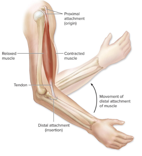
Muscle attachments
most cross at least one mobile joint
upon contraction, one bone moves while other is fixed
axial muscles
attachments
superior attachment (usually more moveable)
is pulled toward the inferior portion when the muscles contract
inferior attachment (usually less moveable)
appendicular muscles
attachments
proximal attachment (usually less moveable)
of biceps brachii are on scapula
ex. biceps brachii is on scapula
distal attachment (usually more moveable)
is moved toward the proximal attachment when muscles contract
ex. biceps brachii is on radius
contraction pulls radius toward scapula, flexing the blow
previously used
origin = less moveable attachment
insertion = more moveable attachment

Organizational Patterns of Skeletal Muscle Fibers
varied organization of fascicles (bundles of muscle fibers)
circular muscles
concentrically arranged fascicles
create a sphincter, control material passage through an opening
ex. orbicularis oris
parallel muscles
fascicles run parallel to muscles long axis
sometimes have an expanded central belly / body / gaster
high endurance & range of motion, not strong
ex. rectus abdominis, biceps brachii
convergent muscles
widespread fascicles merge toward a common attachment site
can pull in varying directions, but not as hard as parallel muscles
generates more powerful contractions
ex. pectoralis major
pennate muscles (anchor points)
fascicles organized as if part of large feather
fibers pull at an angle to the tendon
fascicles that attach obliquely to the tendon
1 or more tendons extending through their body
generate more tension/force but don’t pull their tendons as far as parallel muscles
3 subtypes “anchors”
unipennate: fascicles on same side of tendon
ex. extensor digitorum
bipennate: fascicles on both side of tendon, most common
ex. rectus femorus, interosseous muscles
multipennate: branches of tendon within muscle & fascicles arranged around both sides of each tendon branch
generates more powerful contractions
ex. deltoid
Actions of skeletal muscles
agonist
prime mover, muscle that contracts to produce a movement
ex. triceps brachii is agonist for forearm extension
antagonist
muscle whose contraction opposes that of the agonist
allows for smooth movement of controlled speed
ex. biceps brachii is antagonist for forearm extension
synergist
muscle that assists agonist by contributing tension or stabilizing point of origin / bones involved (acting as fixators, preventing movement)
most useful at start of a movement when the agonist is lengthened & cannot exert much power
ex. biceps brachii of the arm is a syngerist to the brachialis muscle (agonist), when both work synergistically to flex elbow joint
Skeletal Muscle Naming
named according to several criteria
muscle action
indicates muscles primary action
ex. flexor digitorum longus flexes digits
specific body regions
indicates muscle location
ex. rectus femoris is near the femur
muscle attachments
indicates origins and/or insertions
ex. sternocleidomastoid originates on the sternum & clavicle & inserts on the mastoid process
orientation of muscle fibers
indicates organization of muscle fascicles
ex. rectus abdominis is composed of fibers running in vertically straight (“rectus”) orientation
muscle shape
ex. deltoid is shaped like a triangular delta symbol
ex. abductor pollicis longus is a long muscle
muscle size
gluteus maximus is the largest of the buttocks muscles
magnus (big) → major (bigger) → maximus (biggest)
number of muscle heads at an attachment site
indicates # of muscle bellies or heads each contains at the superior or proximal attachment site
biceps = 2 heads / triceps = 3 heads / quadriceps = 4 heads
ex. triceps brachii has 3 heads,
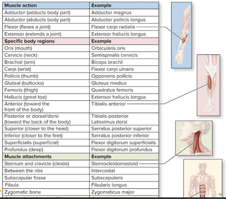
Muscle naming: Muscle action
adductor: adducts body part
ex. adductor magnus
abductor: abducts body part
ex. abductor pollicis longus
flexor: flexes a joint
ex. flexor carpi radialis
extensor: extends a joint
ex. extensor hallucis longus


Muscle naming: Specific body regions
oris (mouth)
orbicularis oris
cervis (neck)
semispinalis cervicis
brachial (arm)
biceps brachii
carpi (wrist)
flexor carpi ulnaris
pollicis (thumb)
opponens pollicis
gluteal (buttocks)
gluteal medius
femoris (thigh)
quadratus femoris
hallucis (great toe)
extensor hallucis longus
anterior (front of body)
tibialis anterior
posterior or dorsal/dorsi (back of body)
tibialis posterior
latissimus dorsi
superior (closer to the head)
serratus posterior superior
inferior (closer to the feet)
serratus posterior inferior
superficialis (superficial)
flexor digitorum super ficialis
profundus (deep)
flexor digitorum produndus

rectus
straight
deltoid
triangular
quadratus
rectangular

trapezius
trapezoidal
brevis
short
Clinical: Intramuscular injections
one route of medication administration
may be inserted into muscle w a syringe
medication to cardiovascular system thru muscles blood vessels
allows large amount of medication given at once
ensures slower, & more uniform delivery than orally or intravenously
ex. vaccines, some contraceptive medications, some antibiotics
common sites: deltoid, gluteal, quadriceps
muscle belly
the thick, fleshy, and contractile middle part of a skeletal muscle located between its origin and insertion tendons
forearm larger near elbow than it is near the wrist bc the bellies of the forearm muscles that move the wrist are located near the elbow & the tendons of these muscles extend toward the wrist
palpebrae
eyelids

Muscles of facial expression
general characteristics
arise from skull & attach to skin
generally attach to subcutaneous layer, deep to skin
cause skin to move (facial expression) during contraction
most are innervated by facial nerve (CN VII)
muscles of forehead, scalp, & eyebrows
occipitofrontalis muscle
connects w epicranial aponeurosis (galea aponeurotica) to form epicranius
frontal belly raises eyebrows, wrinkles forehead
occipital belly retracts scalp slightly
corrugator supercilii muscle (deep)
draws eyebrows together
forms vertical wrinkle lines around the nose
muscles of the eyelid
orbicularis oculi
circular muscle fibers surround orbit
closes eye (squint, blink, wink)
levator palpebrae superioris
elevates upper eyelid (open eyes)
muscles associated w the nose
nasalis
elevates corners of the nostrils for “flared” nostrils
procerus
transverse wrinkles nose in distaste
continuous w frontal belly of occipitofrontalis muscle
above nasalis
muscles associated w the mouth
orbicularis oris
closes mouth, puckers lips
depressor labii inferioris
pulls lower lip inferiorly
depressor anguli oris
pulls corners of the mouth inferiorly to frown
levator labii superioris
pulls upper lip superiorly
levator anguli oris
pulls corners of mouth superiorly & laterally
works w zygomaticus major & minor in smiling
risorius
pulls corner of the lips laterally
“why so serious” joker
mentalis
protrudes lower lip (pouting)
platysma (superficial)
tenses skin of neck & pulls lower lip inferiorly (pout)
buccinator
compresses cheek against the teeth when chewing
covered by masseter
zygomaticus major & minor
“smile” elevates the corner of the mouth

What cranial nerve mainly controls muscles in face
cranial nerve VII (7)
except levator palpebrae (CN III)
Clinical: Idiopathic Facial Nerve Paralysis (Bell palsy)
may be without known cause (idiopathic)
facial nerve inflamed & compressed
muscles on same side paralyzed
prednisone often used to reduce swelling
level & timing of recovery varies

Extrinsic Eye Muscles
are extraocular
insert onto outer / external surface (white/sclera) of the eye & move it
outside the eye, not inside
6 extrinsic muscles: 4 rectus & 2 oblique
rectus eye muscles originate from common tendinous ring
medial rectus
pulls eye medially (adducts)
lateral rectus
pulls eye laterally (abducts)
inferior rectus
pulls eye inferiorly & medially
superior rectus
pulls eye superiorly & medially
oblique eye muscles (arise from orbit)
inferior oblique
elevates & turns eye laterally
superior oblique
depresses & turns the eye laterally
passes through pulleylike loop, trochlea


Muscles of the Oral Cavity & Pharynx: Mastication
muscles of mastication
4 paired muscles move mandible at temporomandibular joint (TMJ)
all 4 are innervated by CN V (trigeminal)
temporalis
elevates & pulls the mandible posteriorly (retracts)
broad, fan shaped
when u bite u can feel it
masseter
elevates & pulls the mandible anteriorly (protracts)
most powerful & important masticatory muscle
medial & lateral pterygoid
protraction & side to side movement of mandible
medial also helps w elevation of mandible
+ buccinator is considered an accessory mastication muscle
menonmic
Must Taste Like My Burger

Muscles of the Oral Cavity & Pharynx: Tongue
intrinsic muscles
compose the tongue itself
change shape during chewing & speaking
extrinsic muscles
arise from other head & neck structures, attach to tongue
names end in -glossus (tongue)
produce movements for manipulating food (chewing/swallowing) & speaking
most are innervated by CN XII, hypoglossal nerve
except palatoglossus, CN X
are paired, & include
genioglossus: protract tongue
genie pops out of a lamp
geni = chin
styloglossus: elevate & retract tongue
imagine pulling string from styloid process
hyoglossus: depress & retract tongue
imagine pulling string from hyoid bone
palatoglossus: elevate posterior part of tongue
palate roof of mouth

Muscles of the Oral Cavity & Pharynx: Pharynx
pharynx muscles
the pharynx is a funnel shaped tube (throat) located posterior to and connects oral & nasal cavities
includes several muscles that aid in swallowing (constrictors), including superior, middle, & inferior pharyngeal constrictor muscles
innervated by CN X (vagus)
contract sequentially to push food into esophagus
palato-
relating to the palate
roof of the mouth

Muscles of the Anterior Neck: The Hyoid Muscles
suprahyoid muscles (MGSD My Gut Seems Damaged / My Gosh Suc D)
superior to hyoid bone (floor of the mouth)
elevate hyoid bone during swallowing or speaking
digastric
has 2 bellies, anterior & posterior
helps depress mandible
starts digestion (by opening mouth)
elevates hyoid bone
geniohyoid
elevates the hyoid bone
genio = chin
mylohyoid
provides muscular floor of mouth, also raises both floor & hyoid up
mylo = molar
stylohyoid
attached to styloid process of temporal bone
elevates hyoid bone, elongates oral cavity floor during swallowing
infrahyoid muscles (TOSS)
inferior to hyoid bone
depress hyoid bone or thyroid cartilage of larynx as swallowing finishes / speaking
Omohyoid
depresses hyoid by pulling it toward scapula
Sternohyoid
depresses hyoid by pulling it toward sternum
Sternothyroid
depresses thyroid cartilage by pulling it toward sternum
to return it to its og position after swallowing
deep
Thyrohyoid
extends from thyroid cartilage to hyoid
depresses hyoid bone & elevates thyroid cartilage
closes off larynx during swallowing

Muscles of the Anterolateral neck
generally act to flex neck & head
sternocleidomastoid (deep)
extends from sternum & clavicle to mastoid process
bilateral (both sides): flexes neck
unilateral (one side): lateral flexion of neck, rotates head to opposite side
if left sternocleidomastoid muscle contracts, it rotates the head to the right side of the body
anterior, middle, & posterior scalene muscles
work w sternocleidomastoid to flex neck
pull cervical vertebrae toward 1st & 2nd ribs (lateral flexion of neck)
elevate 1st & 2nd ribs during forced inhalation
“scale” the neck

Muscles of the Posterior neck
move the head & neck
generally act to extend the neck & head
trapezius
primary function is to help move pectoral girdle but also helps extend neck
medial attachment: occipital bone (superior nuchal line), ligamentum nuchae, spinous processes of C7-T12
lateral attachment: clavicle, acromion process & spine of scapula
splenius capitis, longissimus capitis
“bandage”
bilateral contraction: extend neck/head
unilateral contraction: turn / rotate head to same side (lateral flexion)
except for semispinalis capitis
bilateral contraction: extends neck/head
unilateral contraction: rotates head to opposite side
suboccipital muscles
obliquus capitis superior & inferior
turn / rotate head to same side (unilateral contraction)
rectus capitis posterior major & minor
extend neck
Clinical: Congenital Muscular Torticollis
newborn with shortened sternocleidomastoid
may persist into childhood
from birth trauma or prenatal position
head tilts to affected side & chin to unaffected side
treatments include
physical therapy
botulinum toxin, which impairs contraction of affected muscle

Muscles of the Vertebral Column: Erector spinae
maintain upright posture
bilaterally contracted: vertebral column extends
unilaterally contracted: lateral flexion toward that side
3 groups (listed lateral to medial) (ILS SLI = I Love Sushi) (all deep & paired)
iliocostalis group
comes from illiac crest to ribs
longissimus group
spinalis group

Muscles of the Vertebral Column: Other muscles
transversospinalis muscles
connect & stabilize the vertebrae (at an angle)
even deeper to erector spinae
extend vertebral column & neck
quadratus lumborum muscles
bilaterally contracted: extends vertebral column
unilaterally contracted: laterally flexes column
located primarily in lumbar region
Inspiration vs Expiration
inspiration (inhalation)
several muscles contract to increase the size of the thoracic cavity, allowing lungs to fill w air
diaphragm goes down, larger chest area, lungs fill/get bigger
expiration (exhalation)
some respiratory muscles contract & others relax, decreasing the size of the thoracic cavity & forcing air out of lungs
diaphragm goes up, smaller chest area, lungs empty/get smaller

Muscles of Respiration
found on posterior & anterior thorax
serratus posterior superior
elevates ribs during forced inspiration
increases lateral dimensions of thorax (width)
serratus posterior inferior
depresses ribs during forced expiration
scalene muscles
anterior, middle, posterior
help elevate 1st and 2nd ribs during forced inspiration
external intercostals
elevate ribs during forced & relaxed inspiration, expanding cavity
extend inferomedially (bottom to right)
internal intercostals
deep to external intercostals
fibers at right angles to external intercostals
& next to sternum (bottom to left)
depress ribs during forced expiration / pulls in
normal exhalation takes no active muscular effort
transversus thoracis
depresses ribs during forced expiration
diaphragm
internally placed dome shaped muscle that partitions thoracic & abdominal cavities
most important muscle associated with breathing
quiet, normal breathing
muscle fibers converge toward fibrous central tendon
contracts during inspiration
central tendon pulled inferiorly, increasing dimension of thoracic cavity
flattens & moves downwards
decreases pressure in the thoracic cavity & increases pressure in abdominopelvic cavity


Muscles of the abdominal wall
all 4 compress (abdominal wall) & hold abdominal organs in place
involved in forced expiration
flex vertebral column (except for transversus abdominis, at least by itself)
external oblique
located superficially, lateral abdominal wall
fibers directed inferomedially
fibers go up & right
forms aponeurosis anteriorly, that becomes inguinal ligament inferiorly
capable of rotating / flexing (+ laterally) vertebral column
internal oblique
located deep to external oblique, fibers project superomedially
fibers go down & right
forms aponeurosis anteriorly
capable of rotating / flexing (+ laterally) vertebral column
transversus abdominis
deepest abdominal muscle, fibers project transversely
forms aponeurosis anteriorly
“belt” goes straight across
provides support & stabilization
rectus abdominis
anteromedial location, fibers run vertically
partitioned into 4 segments by 3 fibrous tendinous intersections
make “six pack”
enclosed within fibrous sleeve, rectus sheath
formed from aponeuroses of external & internal oblique, & transversus abdominis
rectus sheaths of 2 sides connected by fibrous strip, linea alba
flexes vertebral column
unilateral contraction of oblique muscles & transversus abdominis helps laterally flex the vertebral column & rotate it toward the side opposite the contraction
ex. if they contracted the left side it would laterally flex to the left but also twist right
also contribute to flexion of the vertebral column when both sides contract simultaneously

Contralateral vs Ipsilateral rotator muscles
Contralateral: a muscle contracting on one side rotates the head or trunk toward the opposite side
ex.
external oblique (left contracts → rotates spine to the right)
transversospinalis (rotates vertebral column to opposite side)
sternocleidomastoid (right contract → rotates the head to the left)
semispinalis capitis
Ipsilateral: a muscle contracting on one side rotates the head or trunk toward the same side
ex.
internal oblique (left contracts → rotates spine to left)
splenius capitis,
splenius cervicis,longissimus capitis (right contracts → rotates head to right)obliquus capitis superior & inferior
*the left external oblique and right internal oblique work together to rotate your trunk to the right
Angle of fibers (might be wrong)
if its external imagine V
if its internal imagine upside down V
the point is the medial of body
Muscle Actions on Axial Skeleton
extend the neck and/or vertebral column
splenius muscles
erector spinae (ILS: illiocostals, longissimus, spinalis)
quadratus lumborum
transversospinalis group
rectus capitis posterior major & minor
flex the neck and/or vertebral column
sternocleidomastoid
scalene muscles
external oblique
internal oblique
rectus abdominis
laterally flex the vertebral column
quadratus lumborum
external oblique
internal oblique
rotate head to one side
sternocleidomastoid
splenius muscles
longissimus capitis
obliquus capitis inferior
obliquus capitis superior
elevate the ribs
serratus posterior superior
external intercostals
scalene muscles (1st & 2nd ribs only)
depress the ribs
serratus posterior inferior
internal intercostals
transversus thoracis
Clinical: Hernias
portion of viscera protrudes through weak point of abdominal wall
inguinal hernia
loop of small intestine protrudes through superficial inguinal ring
more likely to occur in males since their inguinal canals are larger to accommodate spermatic cord
high abdominal pressure (ex. straining to lift smthin heavy) can push intestine into canal
physicians test for it by palpating inguinal ring while patient coughs (cough raises abdominal pressure)

Muscles of the Pelvic floor
pelvic diaphragm
pelvic floor formed by 3 layers of muscles & fasciae
supports pelvic viscera (organs)
extends from the ischium & pubis of the ossa coxae across the pelvic outlet to the sacrum & coccyx
forms muscular wall that covers inferior pelvic opening
perineum
diamond shaped region between lower appendages
bound by pubic symphysis, coccyx, ischial tuberosities
contains 2 triangles divided by transverse line between ischial tuberosities
urogenital triangle contains external genitalia & urethra, more anterior
anal triangle contains anus, more posterior
anal triangle muscles
coccygeus
forms pelvic floor & supports pelvic viscera
right next to sacrum
external anal sphincter
constricts anal opening, must voluntarily relax to defecate
levator ani
group of muscles that form the anterior & lateral parts of pelvic diaphragm
iliococcygeus
forms pelvic floor & supports pelvic viscera
above coccygeus
pubococcygeus
forms pelvic floor & supports pelvic viscera
above iliococcygeus
puborectalis
supports anorectal junction, must relax to defecate
above pubococcygeus
urogenital triangle muscles
superficial layer
bulbospongiosus (female)
narrows vaginal opening, compresses & stiffens clitoris
bulbospongiosus (male)
ejects urine or semen, compresses base of penis & stiffens penis
ischiocavernosus
assists w erection of penis or clitoris
superficial transverse perineal muscle
supports pelvic organs
deep layer
deep transverse perineal muscle
supports pelvic organs
external urethral sphincter
constricts urethra, must voluntarily relax in order to urinate
serratus
saw
levator
raises
rhomboid
diamond shaped
trapezion
irregular 4 sided figure
-cep
head

Movements of the scapula
Elevation & Depression (Up and Down)
Elevation: Lifting the shoulder blade upward. An example of this is shrugging your shoulders.
muscle would have to be above scapula
Depression: Pulling the shoulder blade downward. An example is pushing down into your chair to lift your hips up.
muscle would have to be below scapula
Protraction & Retraction (Forward and Backward)
Protraction (Abduction): Moving the shoulder blade forward and away from the spine. This happens when you reach forward or push open a heavy door.
muscle would have to be
Retraction (Adduction): Pinching the shoulder blades backward toward the spine. This occurs when you squeeze your shoulder blades together or pull your elbows back during a row exercise.
muscle would have to be between scapula
Upward & Downward Rotation (Tilting)
Upward Rotation: Rotating the lower point (inferior angle) of the scapula outward and upward. This movement is essential for allowing you to lift your arm over your head.
imagine abducting
Downward Rotation: Rotating the scapula back down to its resting position. This happens when you pull your arms down from a high position or put your hands in your pockets
imagine adducting

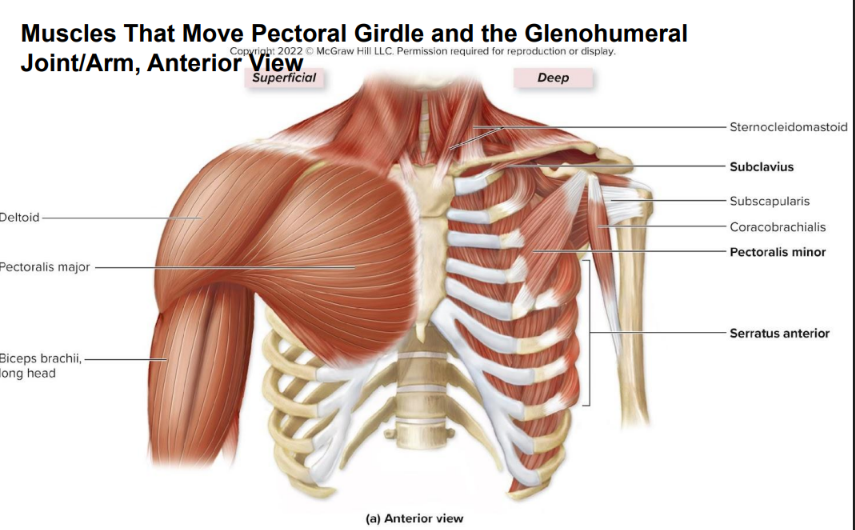
Thoracic Muscles that Move the Pectoral Girdle
muscles of the pectoral girdle originate on the axial skeleton & insert on the clavicle & scapula
anterior thoracic muscles: tend to depress / protract (abduct) scapula or clavicle
all anterior muscles that move the pectoral girdle are deep to other thoracic muscles
pectoralis minor
deep to pectoralis major
helps depress & protract (abducts) scapula
hunches shoulder
superior attachment: coracoid process of scapula
inferior attachment: ribs 3-5
serratus anterior
fan shaped muscle between anterior ribs & anterior scapula
abducts / protracts (agonist) scapula, stabilizes scapula (against the posterior side of the rib cage)
superior rotator
rotates scapula superiorly so glenoid cavity moves superiorly (imagine abducting limb)
superior attachment: medial border of scapula
inferior attachment: ribs 1-8 (anterior & superior margins)
subclavius
extends from clavicle to first rib
stabilizes & depresses clavicle
posterior thoracic muscles: tend to elevate / retract (adduct) scapula
levator scapulae
attaches to cervical vertebrae & scapula
elevates & inferiorly rotates the scapula
moves glenoid cavity inferiorly
superior attachment: transverse processes of C1-C4
inferior attachment: superior part of medial border of scapula (superior angle of scapula)
rhomboid major & rhomboid minor
runs inferolaterally from vertebrae to scapula, deep to trapezius
helps elevate, retract (adduct), inferiorly rotate the scapula
major
medial attachment: spinous processes of T2-T5
lateral attachment: medial border of scapula from spine to inferior angle
minor
medial attachment: spinous processes of C7-T1
lateral attachment: medial border of scapula superior to spine
trapezius
diamond shaped muscle extending from skull & vertebral column to pectoral girdle
superior fibers: elevate scapula, rotate scapula superiorly
middle fibers: retract (adducts) scapula
inferior fibers: depress scapula
medial attachment: occipital bone (superior nuchal line), ligamentum nuchae, spinous processes of C7-T12
lateral attachment: clavicle, acromion process & spine of scapula
in general all posterior thoracic muscles either adduct or extend the arm at the glenohumeral (shoulder) joint


Retraction (adductors) & Protraction (abductors) of the Scapula
Protractors (anteriorly located)
pectoralis minor
serratus anterior
Retractors (posteriorly located)
trapezius
rhomboids

Elevation, Depression, & Rotation of the Scapula
Elevators
rhomboid major
rhomboid minor
levator scapulae
trapezius (superior part)
Depressors
trapezius (inferior part)
pectoralis minor
Superior rotators
serratus anterior
trapezius (superior part)
Inferior rotators
rhomboid major
rhomboid minor
levator scapulae
latissimus
widest
dorsi
back
delta
triangular
teres
round
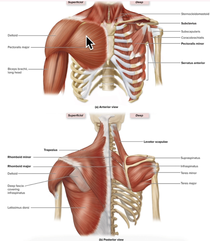
Muscles that move the arm at the glenohumeral joint
11 muscles cross the glenohumeral joint & move the humerus
agonists (prime movers) of the glenohumeral joint: latissimus dorsi & pectoralis major (handcuffs)
latissimus dorsi
broad triangular muscle on back inferiorly
agonist of arm extension
adducts & medially rotates arm
“swimmers muscle”
proximal attachment: spinous processes of T7 - T12, ribs 8-12, iliac crest
distal attachment: intertubercular sulcus of humerus
pectoralis major
thick fan shaped muscle on superior, anterior thorax
agonist of arm flexion
adducts & medially rotates arm
proximal attachment / origin: medial clavicle, costal cartilages of ribs 2-6, body of sternum
distal attachment / insertion: humerus (lateral part of intertubercular sulcus)
“the nae-nae”
triceps brachii
long head originates on scapula, spans shoulder joint
helps extend & adduct the arm
proximal attachment:
long head: infraglenoid tubercle of scapula
lateral head: posterior humerus superior to radial groove
short head: posterior humerus inferior to radial groove
distal attachment: olecranon of ulna
biceps brachii
both heads originate on scapula & span shoulder joint
assists in flexing the arm (weakly)
proximal attachment: supraglenoid tubercle & coracoid process of scapula
distal attachment: radial tuberosity & bicipital aponeurosis
deltoid
forms rounded contour of shoulder
agonist / prime abductor of the arm
anterior fibers: flex & medially rotate arm
lateral / middle fibers: abduct the arm (agonist)
posterior fibers: extend & laterally rotate arm
canNOT adduct
proximal attachment: acromial end of clavicle, acromion & spine of scapula
distal attachment: deltoid tuberosity of humerus
coracobrachialis
flexes & adducts the arm (+ medial rotation)
synergist to the pectoralis major
proximal attachment: coracoid process of scapula
distal attachment: middle medial shaft of humerus
teres major
extends, adducts, medially rotates arm
synergist to the latissimus dorsi
rotator cuff muscles (on scapula but move humerus)
subscapularis
supraspinatus
infraspinatus & teres minor
teres minor is below infraspinatus
mnemonic
“2 Majors and a Mis get cuffed Groove”
All 3 attach to intertubercular groove of humerus
all adduct & medially rotate the humerus
arm movements when your in handcuffs
if handcuffed infront: pec major
if handcuffed in back: teres major, latismiss dorsi
Pec Major
+ flexes
Teres Major
+ extends
latisMISS dorsi
+ extends
mnemonic
Deltoid is Dumb
he cant add (no adduction)
but he can flex, extend, med & lat rotation, abduction

Rotator cuff muscles
(on scapula but move humerus, not scapula)
reinforce the glenohumeral joint & secure the head of humerus in the glenoid cavity (provide strength + stability)
subscapularis
medially rotates arm (agonist)
ex. when u wind up for a pitch
connects scapula to lesser tubercle
proximal attachment: subscapular fossa of scapula
distal attachment: lesser tubercle of humerus
supraspinatus
abducts the arm
ex. when you start to execute the pitch
proximal attachment: supraspinous fossa of scapula
distal attachment: greater tubercle of humerus
infraspinatus & teres minor
laterally rotate arm (agonist) & adducts
teres minor is below infraspinatus
ex. help slow down the pitching arm upon completion of the pitch
connect scapula to greater tubercle
proximal attachment: infraspinous fossa of scapula
distal attachment: greater tubercle of humerus
mnemonic
SITS
SIT goes to great tubercle
S goes to lesser tubercle
S: Supraspinatus
abducts
I: Infraspinatus
lateral / external rotation
T: Teres minor
lateral / external rotation
S: Subscapularis
medial / internal rotation

Clinical: Rotator Cuff Injuries
result of trauma or disease
can be caused by repetitive use
can be caused by falling on the shoulder or lifting too heavy of an object
supraspinatus most commonly involved
most superficial
symptoms are swelling, tenderness, & pain w movement
especially common in baseball players
may require physical therapy or surgical repair
septa / septum
a dividing wall, fence, or partition in the body
intermuscular septa is dense CT and subdivides deep fascia
Arm & Forearm Muscles (that move the forearm at elbow joint)
limbs are organized into compartments surrounded by deep fascia & subdivided by intermuscular septa
a compartment houses functionally related muscles
anterior compartment of the arm has flexor muscles (elbow flexors)
supplied blood by deep brachial artery
innervated by musculocutaneous nerve
ex. biceps brachii, brachialis
posterior compartment of the arm has extensor muscles (elbow extensors)
supplied blood by deep brachial artery
innervated by radial nerve
ex. triceps brachii

Muscles of the arms anterior compartment
biceps brachii
proximal attachment: supraglenoid tubercle & coracoid process of scapula
distal attachment: radial tuberosity & bicipital aponeurosis
2 headed muscle on anterior humerus
short / medial head
long / lateral head, wraps around
flexes (shoulder & elbow)
weak flexor of humerus
long head only
supinates forearm
when elbow flexed (agonist)
brachialis
deep to biceps brachii
most powerful elbow flexor of forearm
proximal attachment: distal anterior surface of humerus
distal attachment: tuberosity & coronoid process of ulna
u can see it lateral view
brachioradialis
located on anterolateral forearm
actually in posterior
involved in elbow flexion
synergist to the biceps brachii
mnemonic
bicep brachii 2 heads, 3 actions
flex shoulder, flex elbow, supination

Muscles of the arms posterior compartment
triceps brachii
large 3 headed muscle on posterior arm
lateral head, long head, medial head
long head extends & adducts arm
major elbow extensor of forearm (agonist) + helps extend humerus
extend shoulder, elbow
proximal attachment:
long head: infraglenoid tubercle of scapula
lateral head: posterior humerus superior to radial groove
short head: posterior humerus inferior to radial groove
distal attachment: olecranon of ulna
anconeus
weak elbow (forearm) extensor
crosses posterolateral region of elbow
mnemonic
triceps brachii 3 heads, 2 actions
extend shoulder, extend elbow
General rules (can be changed/revised)
LOTS of muscles end in -alis
minor above major muscle
posterior (back)
extends
laterally rotate
adducts (?)
anterior (front)
flexes
medially rotate (except latissimus dorsi & teres major)
abducts (?)
abduction
radius / lateral side
adduction
ulna / medial side
for arm
biceps brachii (anterior)
long head is lateral
short head is medial
triceps brachii (posterior)
lateral head is lateral
long head is medial
for leg
biceps femoris (posterior)
short head is lateral
long head is more medial
key note: a group’s functions aren’t necessarily shared by every individual muscle!
distinguish what the group accomplishes together from what each individual muscle contributes
being active during a movement doesn’t necessarily mean a muscle directly produces that movement.
muscles are like ropes
pull one attachment toward another attachment
only move areas they attach to
muscles moving straight up & down spine (spinalis) only extend
muscles running at angles to spine (iliocostalis, SCM, QL) move the spine at angles (rotates or laterally flexes)
origin is the more stable or fixed bone, while the insertion is attached to the bone that moves
as the muscle shortens, it pulls on its insertion bone, moving it relative to the origin bone.
muscles that run in diff directions (angled) can do diff things
pay attention to orgin, thats not gonna move. also direction of fibers indicate how insertion will be pulled toward orgin

Muscles in opposite compartments tend to be
antagonists
ex. anterior forearm muscles are primarily flexors & pronators / posterior forearm muscles are primarily extensors & supinators
ex. muscles in the anterior compartment of the thigh are knee extensors / muscles in the posterior compartment of the thigh are knee flexors
ex. muscles in medial compartment of thigh are hip adductors / muscle in lateral side of thigh is hip abductor

Muscles that Pronate or Supinate the Forearm
pronator teres & pronator quadratus
rotate the radius across the ulna to pronate forearm
located in anterior compartment of forearm
teres closer to elbow (rounded)
quadratus closer to wrist (rectangle like)
more distal
supinator
supinates forearm
located in posterior compartment of forearm
more proximal
+ biceps brachii when elbow flexed (agonist)
anterior, upper arm
Clinical: Lateral Epicondylitis
also known as tennis elbow
from trauma or overuse of common extensor tendon of posterior forearm muscles
pain at lateral epicondyle of humerus, tendons attachment site
often results from repeated forceful contraction of forearm extensors
Extrinsic Forearm Muscles that move the Wrist Joint, Hand, & Fingers
forearm muscles are extrinsic to wrist & hand
intermuscular septa partition muscles into anterior & posterior compartments
anterior forearm compartment muscles (flexors of wrist)
generally flex the wrist, MP, some flex IP joint
most arise from medial epicondyle of humerus
posterior forearm compartment muscles (extensors of wrist)
generally extend the wrist, some MP & IP
most arise from lateral epicondyle of humerus
retinacula of the forearm
fibrous bands at the wrist formed from deep fascia (made of dense regular CT)
hold tendons close to bone
flexor retinaculum
covers palmar (anterior) surface of carpal bones
carpal tunnel: tight space between carpal bones & flexor retinaculum through which flexor tendons pass
extensor retinaculum
superficial to dorsal (posterior) surface of carpal bones
extensor tendons of wrist & digits pass between the bones & extensor retinaculum

Clinical: Carpal Tunnel Syndrome
space between carpal bones & flexor retinaculum
flexor tendons extending through through tunnel
median nerve extending through tunnel
syndrome caused by compression of nerve
characterized by pain and “pins and needles” (paresthesia)

Extrinsic Anterior Forearm Muscles (wrist flexors)
anterior compartment muscles
generally flex the wrist (some flex interphalangeal (IP) joint)
most arise from medial epicondyle of humerus (proximal attachment)
F ME (F for flexors, ME for medial epicondyle)
except for flexor pollicis longus & flexor digitorum profundus
superficial layer
flexor carpi radialis
prominent muscle on lateral forearm
flexes wrist & abducts hand
palmaris longus
narrow muscle on anterior forearm (between flexor carpi radialis & ulnaris)
weakly assists in wrist flexion
creates palmar aponeurosis (spiderweb on hand)
flexor carpi ulnaris
flexes wrist & adducts hand
intermediate layer
flexor digitorum superficialis
4 tendons attach distally to middle phalanges of fingers 2 to 5
flexes wrist, MP joints, & proximal interphalangeal (PIP) joints of fingers 2 to 5
deep layer
flexor pollicis longus
lateral side, attaches to distal phalanx of thumb
pollex = thumb
flexes MP & IP joints of the thumb, weak flexor of wrist
proximal attachment: anterior shaft of radius, interosseous membrane
flexor digitorum profundus
medial side, lies deep to flexor digitorum superficialis (intermediate)
its 4 tendons attach distally to distal phalanges of finger 2-5
flexes wrist, MP joints, proximal interphalangeal (PIP) joints, & distal interphalangeal (DIP) joints of fingers 2 to 5
proximal attachment: anteromedial surface of ulna, interosseous membrane
mnemonic (medial → lateral)
“Pass Fail Pass Fail”
superficial layer (from lateral to medial)
pronator teres → flexor carpi radialis → palmaris longus → flexor carpi ulnaris
deep layer (from lateral to medial)
flexor pollicis longus, flexor digitorum profundus
pronator quadratus is deep


Extrinsic Posterior Forearm Muscles (wrist extensors)
posterior compartment muscles
generally extend the wrist (some extend metacarpophalangeal (MP) & interphalangeal (IP) joints)
most arise from lateral epicondyle of humerus (proximal attachment)
superficial layer
extensor carpi radialis longus
extends & abducts hand at wrist
extensor carpi radialis brevis
extends & abducts hand at wrist
works synergistically w longus
extensor digitorum
inserts on distal phalanges of fingers 2-5
extends hand at wrist, extends MP joints, PIP joints, & DIP joints of finger 2-5
extensor digit minimi
attaches to distal phalanx of finger 5 (pinky)
extends MP, PIP, DIP joints
weakly extends wrist
extensor carpi ulnaris
distal attachment on 5th metacarpal bone (below pinky)
extends & adducts hand at wrist
deep layer
extensor pollicis brevis
helps extend MP joint of the thumb
weakly extends wrist
extensor pollicis longus
extends MP & IP joints of the thumb
weakly extends wrist
extensor indicis
extends MP, PIP, & DIP joints of index finger (2)
weakly extends wrist
abductor pollicis longus (not in slides)
abducts the thumb
weakly extends hand at wrist
superficial layer (lateral to medial)
brachioradialis → extensor carpi radialis longus → extensor carpi radialis brevis → extensor digitorum → extensor digit minimi → extensor carpi ulnaris

indicis
of the index finger / pointer
eminence
circumscribed area raised above the general level of the surrounding surface

Intrinsic Muscles of the Hand
both proximal & distal attachments are in hand
thenar group: form fleshy mass at base of thumb (thenar eminence)
flexor pollicis brevis
flexes thumb
superficial / anterior
abductor pollicis brevis
abducts thumb
superficial / anterior
opponens pollicis
assists in opposition of thumb
deep / anterior
hypothenar group: fleshy mass at base of little finger (pinky) (hypothenar eminence)
flexor digiti minimi brevis
flexes little finger
superficial / anterior
abductor digiti minimi
abducts little finger
superficial / anterior
opponens digiti minimi
assists in opposition of little finger
deep / anterior
midpalmar group: moves fingers 2-5
occupies space between thenar & hypothenar groups
lumbricals (worm shaped)
originate anteriorly but pass posteriorly
flex the MP joints
extend PIP & DIP joints of fingers 2-5
dorsal interossei (between metacarpals)
posterior
flex MP joints
extend PIP & DIP joints of fingers 2-5
abducts finger 2-5
palmar interossei (between metacarpals)
anterior
flex the MP joints
extend the PIP & DIP joints of fingers 2-5
adducts the fingers
adductor pollicis
anterior
adducts thumb
tranverse + oblique head
mnenomic for palmar / dorsal interossei
PAD DAB
Palmar ADuct
Dorsal ABduct


Summary of Muscle Actions at the Wrist & Hand
iliotibial tract
a thick, strong band of fibrous connective tissue (fascia lata) that runs along the outside of the thigh, from the hip down to the shinbone
latae
wide / broad
tensor
any muscle that stretches a part of the body or makes it tense
“to stretch”

Muscles that move the thigh at the hip joint
organization of thigh muscles (muscles that move the thigh at the hip joint)
bound by the fascia lata
deep fascia (of thigh) that partitions them into compartments
encircles the thigh muscles like a supportive stocking & tightly binds them
anterior compartment muscles
extend the knee / flex thigh
medial compartment muscles
adduct the thigh
posterior compartment muscles
most flex knee & extend thigh
some abduct
in addition to the compartments
gluteal group & deep muscles of the gluteal region
gluteus medius + minimus + tensor fascia latae (lateral)
abduct the thigh
Muscle of lateral thigh
tensor fasciae latae
attaches to iliotibial tract (lateral thickening of fascia lata)
abducts & medially rotates the thigh


Muscles of the medial compartment of thigh
most adduct & flex thigh (still kinda face anteriorly)
adductor longus, adductor brevis, gracilis, pectineus
adduct & flex the thigh (at hip joint)
gracilis: + flexes leg at knee joints
adductor magnus
adducts thigh, flexes thigh (adductor part), extends thigh (hamstring part) & laterally rotates the thigh (hip joint)
obturator externus
laterally rotates the thigh (hip joint)
mnemonic
“PAAGAm” (for all muscles) or “PAG” for more superficial deep ones
Pectineus, Adductor brevis, Adductor longus, Gracilis, Adductor magnus
help u do pee pee dance (except adductor magnus, that also helps extend & laterally rotate)

Muscles of anterior compartment of thigh
psoas major & iliacus (collectively iliopsoas)
run from lumbar vertebrae & illum to femur
flex thigh (hip joint)
quadriceps femoris
composite muscle w 4 heads
consists of: rectus femoris, vastus lateralis, vastus medialis, vastus intermedius
if it has a V it only extends knee
rectus femoris
goes up to pelvis (ASIS) / crosses hip joint, so it also flexes hip
proximal attachment: anterior inferior iliac spine
distal attachment: quadriceps tendon to patella & then patellar ligament to tibial tuberosity
vastus intermedius
underneath rectus femoris
proximal attachment: anterolateral surface of femur
distal attachment: quadriceps tendon to patella & then patellar ligament to tibial tuberosity
vastus lateralis
proximal attachment: greater trochanter & linea aspera
distal attachment: quadriceps tendon to patella & then patellar ligament to tibial tuberosity
vastus medialis
proximal attachment: intertrochanteric line & linea aspera of femur
distal attachment: quadriceps tendon to patella & then patellar ligament to tibial tuberosity
agonist of knee extension
imagine kicking soccer ball, knee extended (vastus) & hip flexed (rectus femoris)
pulls on quadriceps tendon, which becomes patellar ligament to tibia
sartorius
acts on both hip & knee joints
longest in the body
flexes & laterally rotates thigh
flexes & medially rotates leg
“seatbelt”


Muscles of posterior compartment of thigh
contraction of gluteal muscle results in extension of the thigh
contraction of deep muscles of gluteal region results in lateral rotation of the thigh
superficial muscles in gluteal region
gluteus maximus
agonist of thigh extension
laterally rotates the thigh
gluteus medius & gluteus minimus
deep to gluteus maximus
abduct & medially rotate the thigh
hamstrings (group of 3 muscles)
(lateral → medial) biceps femoris short → long head, semitendinosus, semimembranosus
bicep femoris: by itself laterally
has 2 heads
long head: extends thigh
long & short: flexes leg
laterally rotates leg when leg is flexed
proximal attachment:
long head: ischial tuberosity
short head: linea aspera of femur
distal attachment: head of fibula
semitendinosus:
medially rotate leg
proximal attachment: ischial tuberosity
distal attachment: proximal medial surface of tibia
semimembranosus:
medially rotate leg
beneath & more medial to semitendinosus
proximal attachment: ischial tuberosity
distal attachment: posterior surface of medial condyle of tibia
extend thigh (hip joint)
flex leg (knee joint)
mnemonic: imagine kicking leg up kawaii / getting ready to kick
group of muscles deep to gluteal muscles
piriformis, superior gemellus, obturator internus, inferior gemellus, & quadratus femoris (small muscle that rotates hips outward)
laterally rotate the thigh

Clinical: Thigh Muscle Injuries
groin strain (groin pull, pulled groin)
• Tearing, stretching, straining proximal attachments of medial muscles
of thigh (for example, adductor muscles, gracilis)
• Typically occurs if muscles contract too suddenly, forcefully
• ex. jumping, kicking, change direction while running
Strained or pulled hamstring
• Hamstring tendons avulsed (torn away) during quick starts/stops, fast
running, sudden lateral/medial stress to knee joint
Prevention by doing dynamic warm-up prior to exercise, static
stretching after exercise
Leg Muscles that move the Ankle, Foot, Toes
crural muscles
muscles located in leg that move ankle, foot, toes
partitioned into anterior, lateral, & posterior compartments
evert = lateral / fibula
invert = medial / tibia
anterior
dorsiflex (extends toes up)
extend
tibialis anterior inverts
posterior
plantarflex (tippy toes)
flex
tibialis posterior inverts
lateral
evert foot
weakly plantar flex
extensor retinaculum
thickening of fascia at ankle that holds tendons of the muscles within the anterior compartment close to bones

Legs anterior compartment
mainly dorsiflexors (foot) & extensors (toes)
imagine after toes grab marble it trying to let go of it
extensor digitorum longus
dorsiflexes the foot & extends toes 2-5
extensor hallucis longus
dorsiflexes the foot & extends the great toe
fibularis tertius
dorsiflexes & weakly everts foot
tibialis anterior
primary dorsiflexor & inverts foot
proximal attachment: lateral condyle & proximal shaft of tibia, interosseous membrane
distal attachment: metatarsal I & 1st cuneiform

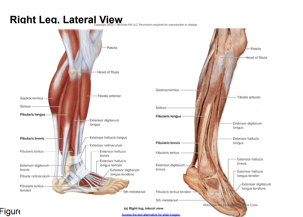
Legs lateral compartment
2 synergistic muscles, both are powerful foot evertors, weak plantar flexors (foot)
fibularis longus
attaches to plantar side of foot
fibularis brevis
lies deep to fibularis longus
attaches to base of 5th metatarsal (below pinky)


Legs posterior compartment
mainly plantarflexors & flexors (leg & toes)
imagine toes trying to grab marble / dig into ground
superficial layer
gastrocnemius
2 bellies (lat & medial head), forms calf
crosses knee & ankle joint
“more gas so it crosses 2 joints”
flexes the leg (knee joint) & plantar flexes the foot
proximal attachment: superior & posterior surfaces of lateral & medial condyles of femur
distal attachment: calcaneus
soleus
broad muscle deep to gastrocnemius
“so low” only covers 1 ankle joint
plantar flexes the foot
proximal attachment: head & proximal shaft of fibula, medial border of tibia
distal attachment: calcaneus
triceps surae
gastrocnemius + soleus
attach to heel (calcaneus) w calcaneal tendon
plantaris
weak leg flexor & plantar flexor of the foot
deep layer
flexor digitorum longus
plantar / flexes foot & MP, PIP, DIP joints of toes 2-5
medial side
flexor hallucis longus
plantar / flexes foot & great toe (MP & IP)
lateral but crosses over to medial
tibialis posterior
plantar flexes & inverts foot (agonist)
in the middle kinda hidden behind
popliteus
flexes leg & medially rotates tibia (to unlock knee)
no action at ankle or foot

Clinical: Shin Splints & Compartment Syndrome
shin splints
soreness along length of tibia
often occur in new poorly conditioned runners
may be considered a type of compartment syndrome
compartment syndrome
compression of blood vessels within a limb compartment
due to inflammation & swelling secondary to strain or trauma
increased pressure in compartment since deep fascia cannot stretch
in severe cases, fascia cut to relieve pressure

Muscles that extend or flex the leg at the knee joint

Intrinsic Muscles of Foot
have both proximal & distal attachments within foot
support arches, flex/extend toes, abduct/adduct toes
rarely perform all the precise movements their names suggest
dorsal group (top of foot): extends
extensor hallucis brevis
extends the MP joint of the great toe (1)
extensor digitorum brevis
extends the MP & PIP joints of toes 2-4
plantar group (bottom of foot): potentially flex, extend, abduct, adduct
supported by plantar aponeurosis formed by deep fascia
extends between phalanges of toes & calcaneus, encloses plantar muscles
muscles grouped into 4 layers from superficial → deep
flexor digitorum brevis, abductor hallucis, abductor digiti minimi
flexor digitorum brevis: flexes MP & PIP joints of toes 2-5
abductor hallucis: abducts great toe (1)
abductor minimi: abducts little toe (5)
quadratus plantae, lumbricals
quadratus plantae: pulls on flexor digitorum longus tendon to flex toes 2-5
lumbricals: flexes MP joints & extends PIP & DIP joints of toes 2-5
adductor hallucis, flexor hallucis brevis, flexor digiti minimi brevis
adductor hallucis: adducts great toe (1)
flexor hallucis brevis: flexes MP joint of great toe (1)
flexor digiti minimi brevis: flexes MP joint of little toe (5)
dorsal interossei, plantar interossei
dorsal interossei: abducts toes
plantar interossei: adducts toes

Clinical: Plantar Fasciitis
inflammation of the plantar aponeurosis
associated with overexertion that stresses the fascia
ex. weight bearing activities, excessive body weight, poor shoes, poor biomechanics
brachii
of the arm

Multifidus
a group of deep, short muscles that run along both sides of the spinal column from the base of the skull down to the sacrum,
largest role in lower lumbar back
extends and rotates the vertebral column toward the opposite side of the body. Its origin is on the sacrum and transverse processes of each vertebra and its insertion is the spinous processes of more superior vertebrae
What correctly describe compartments of the thigh
are anatomically defined by the fascia latae
include muscles w similar actions
include blood vessels & nerves for a single compartment
fasciae latae
deep fascia (of thigh) that partitions them into compartments
encircles the thigh muscles like a supportive stocking & tightly binds them
The anatomical & functional criteria for defining compartments of muscles that move the hip & knee joints are identical
true
bc compartments are defined anatomically by partitions of deep fascia, muscles within each compartment create similar movements at each joint
Innervation of Muscles of the Head & Neck
muscles of facial expression: CN VII (facial nerve)
except
levator palpebrae superioris: CN III (oculomotor nerve)
extrinsic eye muscles: CN III (occulomotor nerve)
except
lateral rectus: CN VI (abducens nerve)
superior oblique: CN IV (trochlear nerve)
muscles of mastication: CN V3
muscles that move the tongue: CN XII
except
palatoglossus: CN X (vagus nerve)
muscles of the pharynx: mostly CN X
muscles of the anterior neck:
muscles of the posterior neck: either cervical spinal nerves or suboccipital nerve
except sternocleidomastoid: CN XI (accessory nerve)

Innervation of Vertebral Column
either cervical, thoracic, & lumbar spinal nerves (or a combination of them)
Innervation of Muscles of Respiration / thoracic
thoracic spinal nerves
except
diaphragm: phrenic nerves (C3-C5)

Innervation of Muscles of the Abdominal Wall
spinal nerves T8-T-12
except
rectus abdominis: spinal nerves T7-T12
Innervation of

Innervation of Muscles of the Pelvic Floor
pudendal nerve (S2-S4)
except
coccygeus: spinal nerves S4-S5
Innervation of Thoracic Muscles that Move the Pectoral Girdle