Module 4.2 Eukaryotic
 Call Kai
 Learn
Learn Practice Test
 Spaced Repetition
 Match
Match Flashcards
 Knowt Play
1/65

There's no tags or description
Looks like no tags are added yet.
Name | Mastery | Learn | Test | Matching | Spaced | Call with Kai | Chat |
|---|
No analytics yet
Send a link to your students to track their progress
66 Terms
membrane fluidity
refers to the physical state of the lipids in the bilayer
2 physical states
2D liquid crystal in which lipids can move within the plane of one leaflet of the bilayer
frozen crystalline gel in which the movement of the lipids is greatly restricted

transition temperature
the temperature in which membrane states change
lipids may exist in either state, and the temperature at which they pass from one state to the other
very quick change from one state to the other
liquid crystal state
individual lipid molecules in the membrane
can rotate and move laterally in the plane of the leaflet
frozen crystalline gel state
all movement of lipids is greatly restricted
determination of transition temperature
transition temperature of a lipid bilayer depends on the particular lipids of which it is constructed
proteins do not contribute much to transition temperature, mainly lipids contribute
degree of unsaturation of the fatty acid: fewer double bonds, higher transition temperature
length of the fatty acid chain: longer chains need higher transition temperature
cholesterol presence: cholesterol disrupts close packing of fatty acyl chains and interferes with their mobility.
abolishes sharp transition temperature, becomes more gradual
creates intermediate fluidity, a state in between crystalline gel and liquid crystal (created due to the gradual change)
biological significance of membrane fluidity
2D liquid crystal is a compromise between a rigid, ordered structure that would lack mobility and a completely fluid, nonviscous liquid in which components of the membrane could not be oriented and structural organization and mechanical support would be lacking
most important reason why membrane needs 2D liquid crystal
fluidity allows for interactions to take place within the membrane
fluidity is essential to membrane assembly
the growth of membrane is accomplished by the insertion of lipid and protein components into the fluid matrix of the membranous sheet
many of the most basic cellular processes, including cell movement, cell growth, cell division, and secretion, etc. depend on the movement of membrane components
maintenance of membrane fluidity
cell wants to stay in liquid crystal state, not gel crystalline
cells adapt to temperature changes
maintenance of membrane fluidity - cold response
Desaturating single bonds in fatty acyl chains to form double bonds
reshuffling the chains between different phospholipid molecules to produce ones that contain two unsaturated fatty acids, which lowers the melting temperature of the bilayer
desaturating: desaturases
reshuffling: phospholipases and acyltransferases
increase amount of double bonds or have the same amount of double bonds but concentrate them
reduces melting temperature
phospholipases vs acyltransferases
phospholipases will cut the ester bond between fatty acids and glycerol
acyltransferases: transfers fatty acid from one lipid to another
lipid rafts
detergent: triton x-100 (nonionic)
extracts lipids from membrane by dissolving them, but patches of lipids are left behind
tritone x-100 cannot dissolve/break interactions between these lipids
cholesterol, lipids with long saturated fatty acyl tails (particularly sphingolipids)
pack very tightly, which forms patches since they are very well organized
packed in a microdomain
SDS can break up lipid rafts/microdomains since it is ionic (dissolves entire membrane)
function of lipid rafts
semi-permanent structure that can concentrate functional units
functional proteins like receptors or single-end proteins
keeps protein complexes in place as one functional unit
dynamic nature of membrane
movement of membrane lipids and proteins in the 2D lipid crystal bilayer gives membrane its dynamic nature
not dynamic in the crystalline gel state
if cells stay in this state for too long, they will die from lack of exchange
no carbohydrate movement because they are attached to protein and lipids, so the movement of the latter will move the carbohydrates
movement of lipids in membranes
in fluid state, phospholipids are free to move in the plane of membrane bilayers (laterally in their leaflet)
movement between bilayers (leaflets) is restricted
rotation/flex: lipids turn around, very quick (10^-9 sec)
lateral shift: lipids move in same leaflet, quick, (10^-6 sec)
Flip-flop: movement from one leaflet to another, very slow (10^5 sec), requiring enzyme flippase

confined diffusion of phospholipids in plasma membrane
track movement of single lipid by labeling it and use high speed microscope to watch it
ex: tagging
records movement of one lipid
movement is not random, it is restricted
movement of membrane proteins
the degree of protein movement in the plane of the membrane varies with individual proteins
some proteins move more than others
some proteins are free to move within plane of membrame, but movement is very slow compared to lipids
proteins are bigger than lipids
other protein movement is restricted by their attachment ot other proteins or structures in the cell, like attachment to cytoskeleton
no transmembrane movement of proteins
membrane has sideness because proteins cannot move from one leaflet to another
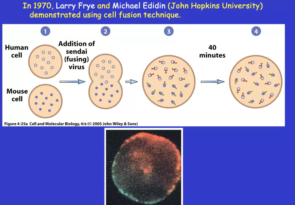
cell fusion technique
larry frye and michael edidin in 1970
movements of proteins demonstrated
fuse two cells (human and mouse cell) since they contain different proteins
labeled antibodies
look at their fluorescence change before and after fusion
i cannot understand his accent but he says there’s a name for the cell that is the human and mouse fusion something like “sick cell” ?????????????????????????
cell starts out with proteins of human and mouse separated on both sides of the cell then they will fuse because there is movement
fluidity of membrane determines movement of proteins
presence of mosaic cells increases sharply as the temperature passes transition temperature
movement of proteins has been enhanced
fluorescence recovery after photobleaching (FRAP)
experiement to measure membrane fluidity and test if proteins can move laterally within the cell membrane.
label: Tag membrane proteins with a fluorescent marker.
bleach: Use a intense laser beam to destroy fluorescence in a tiny spot (photobleaching).
recover: Monitor how fast fluorescence returns to the bleached area.
unbleached, glowing proteins from adjacent (surrounding) areas diffuse laterally into the bleached spot, replacing the zapped proteins.
if the bleached spot never recovered its fluorescence, it means the tagged proteins are anchored/fixed in place and cannot move laterally through the membrane
findings of FRAP
membrane proteins move much slower in plasma membrane than in artificial bilayer
in real PM, membrane proteins are physically restricted by underlying cytoskeleton
30-70% of membrane proteins were not free to diffuse back to the irradicated circle
movement of these proteins are restricted
limitations to FRAP
can only follow the average movement of a large number of labeled molecules as they diffuse over a large distance
cannot distinguish between truly immobile proteins and ones that can only diffuse over a limited distance
single-particle tracking (SPT)
observes movement of single protein
label single protein and use high-speed computer enhanced microscope to record movement of protein
movement patterns in SPT
A: random movement
largest fraction of proteins exhibit random movement
B: no movement (immobilized)
C: highly directed movement
control of membrane protein mobility (SPT)
B: tethered or restricted by membrane skeleton
D: impeded by crowded neighbors
E: restricted by membrane skeleton fences
F: slowed by extracellular materials
membrane domains and cell polarity
a specialized, distinct region within a cell membrane or protein structure that has a specific lipid and protein composition or a distinct functional role
membrane domains and cell polarity: epithelial cells
the apical surface contains integral proteins functioning in ion transport and hydrolysis of disaccharides
the lateral surface contains integral proteins functioning in intercellular interactions.
the basal surface contains integral proteins functioning in the association of the cell with the underlying basement membrane.

differentiation of mammalian sperm plasma membrane
highest differentiated plasma membrane
protein distribution/domain is very specific per area of the sperm cell
red blood cell (erythrocyte) plasma membrane
The most studied and best understood PM
the red blood cells are inexpensive and readily available;
they are present as single cells
they are simple, lacking nuclear and cytoplasmic membranes;
purified erythrocyte PMs can be simply obtained by placing the cells in a dilute salt solution.
when proteins are extracted from membrane, they are all different sizes
SDS-PAGE extracts proteins, can show how they form membrane using a cartoon..?
carbohydrates are only on the extracellular surface
proteins form a network in the protein, which gives the red blood cells in properties
movement of band 3 protein and glycophorin A
the Band 3 proteins are immobilized in the membrane by the network of spectrin and other proteins.
glycophorin A is free to diffuse in the membrane, but because its cytoplasmic domain extends in the cytosol of the erythrocyte, its diffusion is restricted to a limited area created by the specctrin network on the inside of the erythrocyte membrane
gives erythrocyte property of resistance to shearing forces (mechanical dragging force caused by blood or fluid rushing rapidly across the surface of a cell. the cell will not be torn apart from these forces by allowing the cell to bend and squeeze through narrow spaces)

dual functions of cell membrane
retains dissolved materials of the cell so they do not leak out into the environment
must allow the necessary exchange of materials in and out of the cell
types of movement of substances across membranes
simple diffusion through the lipid bilayer
simple diffusion through an aqueous, protein-lined channel
diffusion that is facilitated by a protein transporter
active transport
movement of substances across membranes
influx: movement of substance into the cell
efflux: movement of substance out of the cell
net flux: result of influx and efflux
energetics of solute movement
diffusion is a spontaneous process in which a substance moves from a region of high to low concentration, eventually eliminating the concentration difference between the two regions
if [Ci]/[Co] < 1, delta G is negative, influx is thermodynamically favored
[Ci]/[Co] = 1, delta G is zero, at equilibrium, no net flux
Ci]/[Co] > 1, delta G is positive, efflux is thermodynamically favored
gradients that control diffusion
tendency of a charged solute (electrolyte) to diffuse between two compartments depends on two gradient
the chemical gradient: determined by concentration difference
electric potential gradient: determined by difference in charge
these gradient differences are combined to form an electrochemical gradient
formula: delta G= RT ln [Ci]/[Co] + zF(delta)Em
![<ul><li><p>tendency of a charged solute (electrolyte) to diffuse between two compartments depends on two gradient</p></li><li><p>the chemical gradient: determined by concentration difference</p></li><li><p>electric potential gradient: determined by difference in charge</p></li><li><p>these gradient differences are combined to form an electrochemical gradient</p></li><li><p>formula: delta G= RT ln [Ci]/[Co] + zF(delta)E<sub>m</sub></p></li></ul><p></p>](https://assets.knowt.com/user-attachments/5eb920cb-bef7-466e-b6b9-3678e829d0ce.png)
diffusion of water through membranes
the process of the movement of water through semipermeable membrane from a region of lower solution concentration to a region of higher solute concentration osmosis
when separated by semipermeable membrane, the compartment of higher solute concentration hypertonic (hyperosmotic) to the compartment of the lower solute concentration that is hypotonic (hypoosmotic)
hypertonic: higher solute concentration than the cell
water will flow out and cause cell to shrink
hypotonic: has a lower solute concentration than the cell
causing water to flow in and the cell to swell

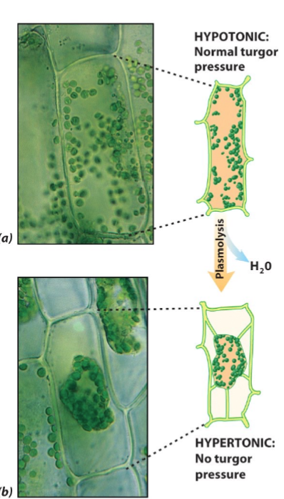
effect of osmosis on plant cells
plants are generally hypertonic compared to their fluid environment
tendency for water to enter the cell develops an internal pressure (turgor) to push the cell against its surrounding wall, which provides support for nonwoody plants
plasmolysis: the process where a plant cell loses water and shrinks, causing the cell membrane to pull away from the rigid cell wall
plant will wilt and drop
diffusion of ions through membranes
most of the ion channels identified can exist in either an open or closed conformation
closed channels are gated:
voltage-gated channels: conformational state depends on the difference in ionic charge on the two sides of the membrane.
ligand-gated channels: conformational state depends on the binding of a specific molecule (the ligand), which is usually not the solute that passes through the channel
mechano-gated channels: conformational state depends on mechanical forces (like stretch tension) that are applied to the membrane
bacterial KcsA K+ channel
diameter:
the four oxygen atom ring, 3 angstrom
dehydrated K+, 2.7 angstrom
dehydrated Na+, 1.9 angstrom
hinge-bending model for the opening of the KcsA channel
Change causes conformational change in the channel, causing M2 to bend and open the channel
Pore is closed by M1 and M2 helix
establishes the structural and mechanistic basis for potassium selectivity and conduction in all living cells
polypeptide sequence: T, V, G, Y, G (will be on test)
carbonyl groups of amino acids V, G, Y, G point into the pore, which form the oxygen ring
Per layer there is 4 oxygens. K will interact with all oxygens in all layers
hydroxyl group of T can form an oxygen ring
K+ binds to ion binding position, there are 4 positions which are all at each carbonyl O
Will go from 1-3 or 2-4 (jumps)
Pore has specificity for K+
Allows big ions to go through but not small
Ions are surrounded by water shields, which is too big for the pore
Water shield must be removed, which will be removed by the oxygen ring because it will interact with the ion, repressing the water shield
If the ion is too small, interaction is too weak so the shield cannot be removed. Potassium is big enough to have a strong interaction to repress water shield (size is optimal)
The attraction between oxygen and ion takes the ion out of the water and attracts it to the pore

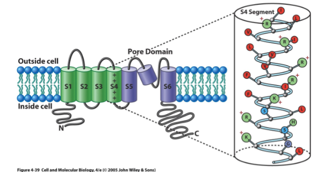
the eukaryotic voltage-gated K+ (Kv) channel
four subunit
each subunits contain six transmembrane helices (S1-S6) and pore domain (P segement) which is a partial helix
grouped into two functionally distinct domains
A pore domain: including helices S5 and S6 and the P segment. They are homologous to helices 1 and 2 and the P segment of the KcsA channel. The the inner end of S6 forms the gate.
A voltage-sensor domain: including helices S1-S4 that senses the voltage across the plasma membrane. S4 is the key element of the sensor.
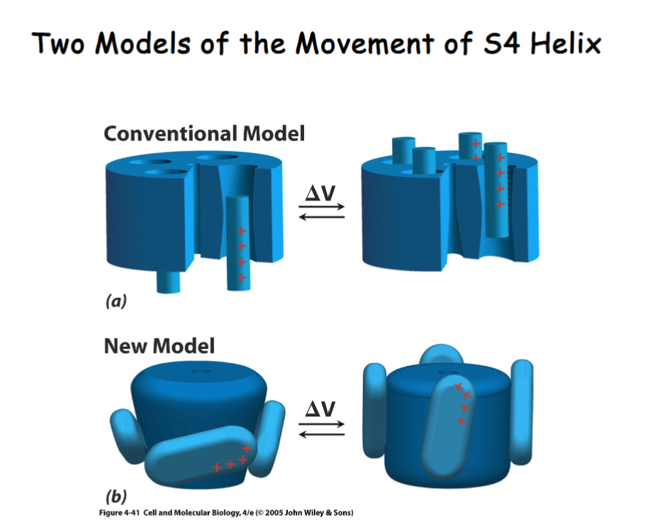
S4 is positively charged, so when voltage changes it moves the position of S4
the models of movement of S4 helix
Change conformation of S5 and S6 to open channel (moves up and down)
Change conformation of S4 by swinging it to open and close pore
inactivation of the Kv channel
Channel is open for very short time (a millisecond)
inactivation of the channel is accomplished by movement of the N-terminal cytoplasmic portion of the polypeptide through the mouth of the pore.
The inactivation peptide moves into the lateral window to inactivate the channel
After inactivation, it will go back to rest (closed) conformation
Once inactivated, it takes a while to go back to rest position, meaning it cannot be automatically or quickly reopened

facilitated diffusion
Using a protein to transport from high to low concentration across a membrane
No ATP required, it is passive movement that requires a transporter
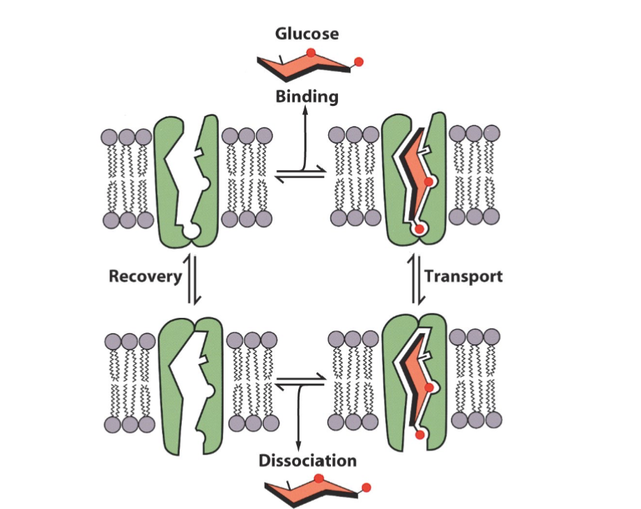
kinetics of facilitated diffusion
similar to an enzyme-catalyzed reaction
specificity- only one solute can be transported by a specific transporter
saturation-type kinetics
regulation

active transport
the gradients in the pic are generated by active transport
requires ATP
Against concentration gradient
endergonic process that is coupled to an exergonic process, such as the hydrolysis of ATP, the absorbance of light, the transport of electron, or the flow of other substances down their gradients.

comparison between facilitated and active transport

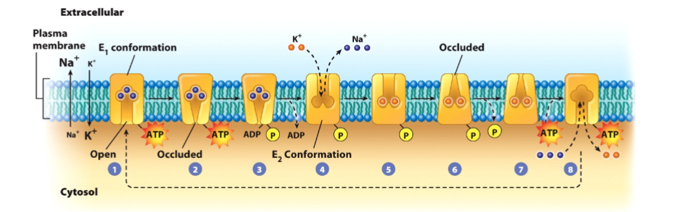
model of Na+/K+ ATPase Transport
Jens Skou in 1957 discovered an ATP hydrolyzing enzyme in the nerve cells of a crab that was active only in the presence of both Na+ and K+
proposed this protein transporting the two ions.
the enzyme was called Na+/K+ ATPase, or sodium-potassium pump.
3 sodium out, 2 potassium in
Gives cell a resting membrane potential of -70 V
phosphorylation of the transport changes the protein’s shape and affinity for the ions.
p-type pump
Phosphate group comes from ATP
p-type pump: phosphorylation of the transporter
v-type pump: Vacuolar transporter
ABC transporter: ATP-binding cassette transporter
H+/K+ ATPase Transport
epithelial lining of stomach contains this P-type pump
Pump is within vesicles that sit in the cytoplasm
Food induces secretion of histamine, which binds to a receptor to trigger fusion of the vesicle with the plasma membrane
Pump is not integrated into plasma membrane, making it active
Pumps protons out and potassium into the cell using ATP (active transport)
Transporter must be phosphorylation to work
Helps prevent heartburn

Regulation of H+/K+ ATPase
Acid-blocking Drug: Zantac is a drug that can bind to the receptor like histamine does, which prevents fusion of vesicles to the plasma membrane
Pump-Inhibiting Drug: prilosec will inhibit H+/K+ ATPase
Acid-Neutralizing Drug: neutralize pH in lumen of the stomach
Zantac, Pepcid, and Tagamet do not inhibit the ATPase directly, they block a receptor on the surface of the cells that stop activation by the hormone
coupling active transport to existing ion gradients (in Na+/glucose cotransporter)
the primary active transport system, the Na+/K+ ATPase, produces the Na+ gradient
the potential energy stores in the Na+ gradient is utilized to transport glucose into the cell
glucose is driven by secondary transport
energetics of the Na+/glucose cotransport
primary Na+/K+ ATPase keeps the external [Na+] 10 times the internal
the voltage across the membrane of a cell is typically –70 mV,
the free-energy change for the movement of a mole of Na+ into the cell under these conditions would be 3.1 kcal/mol
two moles of Na+ are utilized to cotransport 1 mole of glucose
[Ci]/[Co] = 1/23,000
the Na+/glucose cotransporter is capable to transporting
glucose against a concentration gradient > 20,000.
uniport vs cotransport
uniport: transport of a single solute across a membrane (may occur with either facilitated diffusion or active transport).
cotransport: the linked transport of more than one solute across a membrane.
symport: the transport of two solutes in the same direction.
antiport: he transport of two solutes in opposite directions.
irritability
a property that an organism responds to external stimulation
irritability in a single-celled amoeba depends on the same basic properties of membranes that lead to the formation and propagation of nerve impulses.
membrane resting potential
a voltage or electronic potential difference between the inside and outside of the plasma membrane is the membrane potential (for nonexcitable cells).
for a nerve or muscle cell, this same potential is referred to as the resting potential
watch video
if you change the membrane potential, the voltage gated channel will open or close
the picture is a voltage gated potassium channel
domains will move to open or close channel

movement of K+ across membrane
the movement of K+ across the membrane is the major factor in determining resting potential
cell is most permeable to K+
the vast majority of the ion channels that are open in the plasma membrane of a resting cell are K+ leak channels that are selective for K+.
these channels lack the S4 voltage sensor and fail to respond to changes in voltage
K+ at equilibrium
equilibrium potential using Nerst equation: -91 mV
measured resting potential: -70 mV
differ because of membrane’s slight permeability to Na+

depolarization
decrease in polarity between two sides of the membrane
creates an action potential when it reaches the threshold
Na+ comes in to decrease the polarity between inside and outside, which makes the cell reach threshold to create an action potential
action potential
Leak K+ channels keep the resting potential of the membrane (-70mV)
External stimulation opens some Na+ channels, allowing Na+ ions to move into the cell, which causes depolarization of the membrane
If the depolarization passes the threshold (-50mV), the change in voltage causes the voltage- gated Na+ channels to open, allowing Na+ ions diffuse freely into the cell, which reverse the potential to +40mV, approaching the equilibrium potential for Na+.
after 1 msec, the Na+ channel are inactivated. The change in membrane potential triggers the opening of voltage-gated K+ channels, allowing the K+ ions to diffuse freely out of the cell, which causes the potential to swing back to about –80mV, approaching the K+ equilibrium potential. The large negative potential causes the voltage-gated K+ channels to close and the membrane returns to the resting state.
the changes in membrane potential collectively are called an action potential

two characteristics of action potential
The all-or-none law: once the membrane is depolarized to the threshold value, a full-blown action potential is triggered without further stimulation.
Subthreshold depolarization is incapable of triggering action potential.
An action potential process does not require energy: It results from the flow of ions down their respective electrochemical gradients.
The Na+/K+ ATPase requires energy to generate the steep ionic gradients across the plasma membrane.
propogation of action potential
membranes enter a brief refractory period following an action potential because the Na+ channels cannot reopen for several milliseconds after their inactivation.
membranes in refractory period cannot be restimulated
this gives the propogation of an action potential a direction
once triggered, an action potentials travels down the entire length of the neuron with the same strength and arrives at its target cell with the same strength as at its initiation. Stronger stimuli cannot produce “bigger” impulse than weaker stimuli

differences in strengths of stimuli
Stronger stimuli activate more nerve cells than do weaker stimuli
Stronger stimuli activate “high-threshold” neurons that would remain at rest if the stimuli were weaker
stimulus strength is also reflected by the pattern and frequency by which action potentials are launched down a particular neuron.
the stronger the stimulus, the greater the number of impulses generated.
structure of a nerve cell
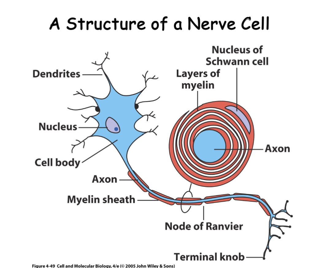
saltatory conduction
to increase speed of a nerve impulse:
increase in the diameter of the axon
wrap the axon with a myelin sheath
saltatory conduction: propagation of an impulse by a myelin sheath-wrapped axon

synapses
specialized junctions that link neurons with their target cells
synaptic cleft: a narrow gap (20-50nm) between a neuron and its target cell.
presynaptic cell: a sensory cell or a neuron that conducts impulses toward a synapse.
postsynaptic cell: a neuron, muscle, or gland cell that lie on the receiving side of a synapse.
synaptic vesicles: carry neurotransmitters into the synaptic cleft
neurotransmitters: acetylcholine, norepinephrine
neuromuscular junction
neuron whose target cell is a muscle cell
the space the vesicles release the neurotransmitters into is called the synaptic cleft
action potential goes from neuron to muscle cell

synaptic transmission
arrival of action potential to the axon terminal
depolarizes presynaptic membrane
voltage-gated calcium channels open
calcium rushes in
calcium willl trigger the fusion of synaptic vesicles and presynaptic membrane
neurotransmitters are released into the cleft (acetylcholine in the image)
diffuses across cleft to receptor on postsynaptic membrane
Voltage-gated sodium channels open (sodium rushes in) and depolarize it
nerve impulse gets sent down the axon
or Cl- channels will open (chlorine rushes in) instead of Na+ channels, which will inhibit the action potential
desensitizes the membrane
two types of responses can occur on the postsynaptic membrane depending on the voltage-gated channels opening

receptors on post-synaptic membrane
number of receptors on postsynaptic membrane can change
more receptors make target cell more sensitive since it can bind to the neurotransmitter more
less receptors make target cell less sensitive
synaptic plasticity
the physical structure of synapses are genetically encoded, whereas their dynamic quality can be affected by environment.
the responses of the dynamic quality of synapses is called synaptic plasticity
when hippocampal neurons are repeatedly stimulated over a short period of time, the synapses that connect these neurons to their neighbors become “strengthened” by a process known as long-term potentiation (LTP), which last for days, weeks, or even longer
synapses have undergone LTP are able to transmit weaker stimuli and evoke stronger responses in postsynaptic cells.
the synaptic plasticity plays a major role as newly learned information or memories are encoded in the neural circuits of the brain