SciOly Respiratory System
 Call Kai
 Learn
 Practice Test
 Spaced Repetition
 Match
 Flashcards
 Knowt Play
1/72

There's no tags or description
Looks like no tags are added yet.
73 Terms
Functions of Respiratory system
Protection, Ventilation, Respiration, Regulating blood pH, and Speech
Quiet breathing
Muscles contract involuntarily to let air in the lungs (actively), and relax when you exhale (passively)
Forced breathing
muscles contract voluntarily in inspiration and expiration
When diaphragm relaxes
Thoracic cavity decreases in size, intercostal muscles push down on ribs, increase in internal pressure, breathing out
When diaphragm contracts
Thoracic cavity increases in size, lungs gain volume and lose pressure, partial vacuum created, intercostal muscles pulled up, diaphragm pulls down, breath in
Upper respiratory system organs
nostrils through the pharynx
Lower respiratory system
top of trachea through the diaphragm
Parts of the nose
Nasal septum, nasal conchae, respiratory mucosa
Nasal conchae
provides more surface area within the nose, three bones rolled up
Respiratory muscosa
tissue lining nasal cavity, larynx, trachea, and bronchial tree. contains cilia to move mucus out of nostrils
Lacrimal glands
secretes tears, drains through the lacrimal puncta into nasolacrimal ducts and nasal cavities, causes nose to run when crying.
Sinuses
air spaces in skull to lighten weight, lined with mucous membranes
Pharynx
large muscle tube that extends from the nose and down the neck, connects respiratory and digestive systems
Choana
area between nasal cavities and pharynx
Nasopharynx
top part of the throat where the nasal cavities drain into
Hard palate
bony plate that separates the mouth cavity from the nasal cavities
Soft palate
in front of the nasopharynx, moves backwards when swallowing food to ensure nasopharynx is blocked
Oropharynx
middle part of the throat, from the uvula to the hyoid bone
Hyoid bone diagram
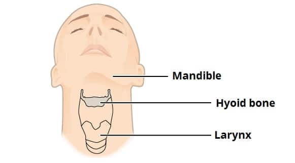
Epiglottis
cartilage structure that guides materials into either the trachea or esophagus, covers the glottis
Laryngopharynx
lower part of throat adjacent to the larynx
Larynx
voice box, when talking voice boxes closes, which is why you have to take breaths when speaking
Larynx anatomy

Glottis
top of larynx, opening when air passes through, contains vocal cords
Trachea
tube that runs from the larynx, divides into two large branches behind the sternum called primary bronchi
Heart location
cavity between the two lungs called the mediastinum
Lung lobe categories
3 for right lung, 2 for left, then segments, then lobules
Pleurae
two layers that seal off lungs from inside surface of thoracic cavity
Pleurae level names
visceral pleurae - outer surface
Pleural cavity
potential space between the two pleural and contains lubricating intrapleural fluid
Difference in pressure between lungs and atmosphere
4 mmHg
Motor phrenic nerves
nerves that contract and relax the diaphragm
Spirometer
measures pulmonary volumes
Normal breathing rate
12-20 cycles per minute
Spirogram

Tidal volume (TV)
volume of air inspired or expired in a regular breath (500 ml)
Inspiratory reserve volume (IRV)
maximum amount of air that can be inspired after breathing in normally (3000ml)
Expiratory reserve volume (ERV)
the maximum amount of air that can be expired after breathing out normally (1100ml)
Residual Volume (RV)
the amount of air still remaining in the respiratory passages and lungs are maximum expiration (1200 ml)
Functional Residual Capacity
ERV + RV
Inspiratory Capacity
TV + IRV
Vital Capacity
IRV + TV + ERV
Total Lung capacity
IRV + ERV + TV + RV (normally around 6000ml)
Pulmonary Ventilation
TV x respiratory rate
Alveolar ventilation
( TV - dead space ) x respiratory rate
Dead space ventilation
dead space x respiratory rate
Acute Rhinitis
Common cold: runny nose, teary eyes, and smell impairment. Bacterial/viral infection
Asthma triggers + reaction
Airway inflammation: muscles surrounding airways become tight and air passage swells. Common triggers are allergies, chemicals, exercise, mold, infections, stress, and smoke
Control Drugs
prevent asthma attacks, take always
Rescue drugs
tkane when having an asthma attack, usually an inhaler
COPDs
Chronic obstructive pulmonary disease
2 forms of COPD
chronic bronchitis and emphysema
COPD charactarization
gradual and irreversible decrease in ability to push air out of lungs, can be caused by smoking or genetic mutations
Chronic brochitis
bronchi become inflamed, goblet cells overproducing, increased airway resitance
Emphysema
Lung tissue and alveolar wall destroyed, causing resistance to blood flow and collapses airways, caused by long-term exposure to airborne irritants
Cystic fibrosis
thick mucus buildup in lungs, pancreas, and rest of body, must inherit recessive CF gene, no cure but treatment includes antibiotics for infections
Hypoxia
Whole body or region is deprived of oxygen, usually characterized by blue skin
Types of hypoxia
Anemic, Ischemic, histotoxic, hypoxemic
Anemic hypoxia
abnormally low RBC or too little hemoglobin
Ischemic hypoxia
abnormal blood flow due to partial or complete blockage
Histotoxic hypoxia
inability of blood cells to metabolize oxygen, caused by metabolic poisons (cyanide)
Hypoxemic hypoxia
abnormally low partial pressures of oxygen in blood, ie breathing in air with too little oxygen, (CO poisoning), does not give usually symptoms of hypoxia, usually just confusion and headache
Oxygen toxicity
result from breathing pure oxygen at high pressures, affects CNS and pulmonary functions
Lung Cancer
symptoms: constant cough, shortness of breath, and infections
Laryngitis
Inflammation of the vocal folds, caused by viruses, can be acute or chronic
Pneumonia
infections of the lungs, caused by bacteria, viruses, fungi. can be treated with IV fuilds and antibiotics, oxygen therapy, humid air, and deep breathing
Sleep apnea
breathing repeatedly stops while sleeping
Sleep apnea types
obstructive, central, and complex sleep apnea syndrome
Obstructive sleep apnea
muscles in the back of through narrowing or closing during inspiration, people sleep well at night
Central sleep apnea
when brain fails to transmit signals to breathing muscles and is usually caused by heart disease or stroke, usually wake up in the night
Complex sleep apnea syndrome
airway obstruction along with problems with breathing rhythm; symptoms: hypersomnia, loud snoring, and waking up abruptly with shortness of breath
Tuberculosis
contagious bacterial infection of the lungs, symptoms: weight loss, excessive sweating, and coughing up blood. Treated with Isoniazid and Rifampin.
How does TB work
Mycobacterium tuberculosis has a cell wall with a lot of myco. to common antibiotics and the immune system. It is also an obligate aerobic.
Alveolar macrophages engulf the TB bacteria, but the bacteria survive the insides of the macrophage. Therefore, the immune system builds a cellular wall around infected macrophages, called tubercules or granulomas. Over time, these calcified granulomas and nearby lymph nodes form what is known as a Ghon Complex.