Mycology
 Call Kai
 Learn
 Practice Test
 Spaced Repetition
 Match
 Flashcards
 Knowt Play
1/82

There's no tags or description
Looks like no tags are added yet.
83 Terms
An aseptate mold isolated from a respiratory specimen exhibits internodal rhizoids, with sporangiophores arising from stolons between the rhizoids and bearing pyriform sporangia) What is the identity of this organism?
Absidia species
Which microscopic feature is characteristic of Syncephalastrum species?
Tubular merosporangia (finger shaped) containing chains of sporangiospores radiating around a vesicle
Which structure serves as the site of origin for erect conidiophores in Aspergillus species within the vegetative hyphae?
Foot cell
A mold culture yields blue-green colonies with a white apron. Microscopic examination shows septate hyphae with erect conidiophores branching into metulae and phialides that form a brush-like structure. Most strains are inhibited at 37°C. Which organism does this represent?
Penicillium species
Zygomycota (Mucorales)
– found in soil
-immunocompromised patients
• Lichtheimia (Absidia), Mucor, Rhizomucor, Rhizopus
The most significant order
• Rapid growers
• Produce gray to white aerial mycelium with hyaline, sparsely septate hyphae
Asexual reproduction= Sporangiospores
Ascomycota
Produce sexual spores (ascospores)
• Microsporum, Trichophyton, Pseudallescheria boydii\
Sexual spores=Ascospores that are formed in a saclike structure known as an ascus
Basidiomycota
Not many clinically significant
• Cryptococcus neoformans var. neoformans
Clamp connections are sometimes key identifying feature
• Members of this order are typically sterile
• A portion of the hypha on one side of the septation grows out and connects to the hypha on the other side of the septation, thereby bypassing the septation

Deuteromycota (fungi imperfecti)
Largest class of etiologic agents of mycoses including cutaneous, subcutaneous and systemic disease
• Have no mode of sexual reproduction identified
Cutaneous, subcutaneous, and systemic diseases
Yeast
Single vegetative cell
Reproduce by budding or fission(budding produces blastoconidium)
No aerial hyphae (smooth/creamy/bacterial-like)
ID based on biochemical testing and molecular diagnostic methods
Mold
Aerial or vegetative hyphae(long filaments of cells joined together)
Aerial mycelia: fuzzy appearance
ID based reproductive structures that produce conidia (used to id different fungi)
Vegetative mycelia extend down into the media to absorb nutrients
Which special histopathological stain selectively stains fungal cell wall melanin brown to black?
Fontana-Masson stain
An India ink preparation performed on cerebrospinal fluid (CSF) reveals budding yeast cells surrounded by large, clear haloes against a dark background) This finding is characteristic of which organism?
Cryptococcus neoformans
A laboratory scientist performs a microscopic evaluation of a mold colony by gently pressing transparent tape against the surface of the aerial mycelium and transferring it to a slide containing lactophenol aniline blue (LPCB). What is this procedure called?
Cellophane tape prep
A patient presents with progressive, nonhealing skin ulcers, draining sinus tracts, and verrucous crusted nodules on the lower extremity following a puncture wound) Direct blood cultures remain negative. Which location category of mycosis is most consistent with these clinical findings?
Subcutaneous mycoses
Which of the following is a general cellular characteristic of all fungi?
Presence of a chitinous cell wall
Why is the Latin term "tinea" used in naming dermatophytic fungal infections?
Infections were historically believed to be caused by burrowing worms forming ring-shaped patterns.
A microscopic examination of a mold reveals hyphae with swollen branching tips that resemble moose antlers. This specific hyphal morphology is classified as:
Antler hyphae (favic chandeliers)
How are arthroconidia formed during asexual reproduction of fungi?
By the fragmentation of fertile hyphae rather than production from conidiogenous cells
What is the defining diagnostic microscopic structure created by certain Basidiomycota hyphae to bypass a septation?
Clamp connection
Cycloheximide is added to primary fungal isolation media (such as Sabouraud Dextrose Agar) for what specific purpose?
To inhibit saprophytic/contaminant molds while allowing recovery of dermatophytes and primary pathogens
What severe clinical manifestation does Pneumocystis spp. cause in immunocompromised patients, particularly those with AIDS?
Pneumonia
What microscopic morphology does Cryptococcus spp. exhibit when cultured on Cornmeal agar?
Blastoconidia only (no true hyphae or pseudohyphae)
What result is observed when Cryptococcus neoformans is inoculated onto a Urea agar slant?
Rapid positive reaction with a color change to pink/magenta
On HardyCHROM / CHROMagar Candida media, what colony color is typical for Candida albicans?
Green
What is the typical colony appearance of Candida krusei on CHROMagar Candida medium?
Dry, rough, pale pink colonies with spreading borders
Which feature microscopic morphology differentiates Geotrichum spp. from Trichosporon spp.?
Geotrichum spp. produces true hyphae that fragment into barrel-shaped arthroconidia but lacks blastoconidia
What characteristic arrangement of fungal elements is classically described on a KOH preparation of skin scrapings infected with Malassezia furfur?
Spaghetti and meatballs appearance (clusters of round budding yeast and short septate hyphae)
Which rapid screening test provides a presumptive identification of Candida albicans and Candida dubliniensis by detecting short hyphal projections within 2 to 3 hours?
Germ Tube test
How are true yeasts distinguished from yeast-like fungi regarding their reproductive capabilities?
True yeasts reproduce sexually by forming ascospores or basidiospores
Which clinical skin condition is caused by Malassezia furfur?
Tinea versicolor (pityriasis versicolor)
Microscopic evaluation of Sporothrix schenckii in its yeast phase reveals which characteristic cell morphology?
Small, elongated, cigar-shaped yeast cells
Histoplasma capsulatum infection is most strongly associated with exposure to soil contaminated with what material?
Chicken droppings and bat guano
What temperature and growth conditions are required to convert dimorphic fungi to their yeast phase in culture?
37°C with increased carbon dioxide (CO2)
Paracoccidioides brasiliensis is geographically restricted to which region?
Central and South America
In its mold phase, Talaromyces (Penicillium) marneffei displays which microscopic fungal structures?
Biverticillate conidiophores with terminal metulae and needle-like phialides
Coccidioides immitis is endemic to which specific ecological zone in the United States?
Lower Sonoran life zone, including San Joaquin Valley CA, Arizona, and SW Texas
What distinctive microscopic spore structure is produced by Histoplasma capsulatum in its mold phase?
Large tuberculate macroconidia with knobby surface projections
Which microscopic feature is characteristic of Blastomyces dermatitidis in tissue or yeast phase cultures?
Large spherical yeast cells with double-contoured walls and broad-based budding
How is fungal dimorphism defined in clinical mycology?
The ability of a fungus to exist in two distinct structural forms (mold and yeast) based on growth conditions
Blastomyces dermatitidis is geographically endemic to which region?
Mississippi and Ohio River basins
An aseptate mold isolated from a respiratory specimen exhibits internodal rhizoids, with sporangiophores arising from stolons between the rhizoids and bearing pyriform sporangia) What is the identity of this organism?
Absidia species
Which microscopic feature is characteristic of Syncephalastrum species?
Tubular merosporangia (finger shaped) containing chains of sporangiospores radiating around a vesicle
Which structure serves as the site of origin for erect conidiophores in Aspergillus species within the vegetative hyphae?
Foot cell
A mold culture yields blue-green colonies with a white apron. Microscopic examination shows septate hyphae with erect conidiophores branching into metulae and phialides that form a brush-like structure. Most strains are inhibited at 37°C. Which organism does this represent?
Penicillium species
Which microscopic feature distinguishes uniseriate from biseriate Aspergillus species?
Uniseriate phialides attach directly to the vesicle, whereas biseriate phialides arise from an intermediate support cell called a metula)
What is the characteristic microscopic morphology of macroconidia produced by Fusarium species?
Banana or canoe shape with multiple cells
Which Aspergillus species is most commonly isolated from clinical cases of invasive aspergillosis?
Aspergillus fumigatus
A fungal isolate shows flat, sandy-to-powdery tan colonies. Microscopic exam exhibits annellides with bowling-pin shape and rough, spiky (echinulate) conidia with flattened bases borne in chains. Which organism is this?
Scopulariopsis species
A medical laboratory scientist notes a rapid-growing mold covering an entire Sabouraud dextrose agar plate within 4 days with thick woolly mycelia) Microscopic review demonstrates wide, clear hyphae with no cross-walls and sporangiophores lacking rhizoids at their base. How should the scientist differentiate this isolate from Rhizopus?
By noting the total absence of rhizoids and stolons in Mucor, whereas Rhizopus possesses nodal rhizoids directly below sporangiophores
A skin biopsy specimen yields a septate hyaline mold with single-celled pyriform or clavate conidia featuring flat (truncate) bases, resting directly on hyphae or short pedicels. The culture resembles a dermatophyte. Which fungus is this?
Chrysosporium species
If Cryptococcus is suspected
India Ink
Colony Morphology yeast
Growth in 2-3 days
• BAP@ 25-37°C; best at 30°C
• Smooth/mucoid/butter-like
• White, cream, tan (some pink!)
• Some phaeoid (dark pigment)
• May be filamentous (FEET ON AGAR!)
Cornmeal-Tween 80 Agar yeast
Coverslip the agar after inoculation
• Growth in 1-2 days
• 25-37°C; best at 30°C
• Observe microscopically on cover slip
• Blastoconidia
• Pseudohyphae
• Chlamydoconidia
• Arthrospores
Germ Tube Production
Differentiates Candida from other yeasts
(+) Candida albicans and Candida dubliensis
Urease
Positive (pink):
• Cryptococcus sp. (rapid)
• Trichosporon sp.
• Rhodotorula sp.
• Negative (yellow):
• Candida sp.
• Geotrichum sp.
India Ink
Capsule detected surrounding the
budding yeast in CSF with India Ink
• Background is black/dark and clear, unstained halos are seen in individual yeast cells
• Positive (light halo on black background):
• Cryptococcus sp.
Lactophenol Cotton Blue (LPCB)
Used in wet mounts
• Cotton Blue
• Acidic dye that binds to chitin
• Stains hyphae, conidia and spores
• Phenol
• Disinfectant and fungicidal agent to kill live orgs
• Lactic Acid
• Preserves fungal morphology
Rapid Trehalose Assimilation
Aids in detection of Candida glabrata
• Identifies yeasts that can utilize/hydrolyze the sugar trehalose more quickly than other Candida species
• Incubate inoculated broth at 42°C for 3 hours
• Color changes from blue to yellow
when positive
Candida albicans

Candidia tropicalis

Candidia krusei

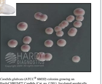
Candidia glabarata
Candidiasis Mucocutaneous
Most common Candidiasis
Thrush (oral)
Vaginitis
Bronchial/Pulmonary
Can be chronic
Cutaneous
Areas of profuse sweating
Areas of water immersion
Skin
Diaper Rash
Nails - Onychomycosis
Systemic
Most detrimental
UTI
Endocarditis
Septicemia
CANDIDA ALBICANS Identification
Gram Positive Organism
• Large
• Germ Tube (+)
• Growth at 42C
C.dublin… and c.para
no groth past 37
• C. neoforman
major cause of opportunistic infections in AIDS patients
• Commonly found in soil contaminated with pigeon droppings
• Acquired by inhalation
Surrounded by capsule which gives characteristic mucoid appearance
India Ink or latex
• C. neoforman identification
Produce blastoconidia only
• No true hyphae or pseudohyphae on
cornmeal agar
• Cryptococcal antigen assay detect
neoformans and gattii in CSF and
serum
Cryptococcus neoformans vs. gattii
Identification
Use CGB agar or PCR
Trichosporon spp
Opportunistic systemic pathogen= Rare, but fatal
• Immunocompromised host with
hematologic disorders, malignancies,
chemotherapy
• Causes white piedra
• Occurs on the hair shaft
• Characterized by soft mycelial mat
surrounding hair of scalp, face & pubic
region
An isolate from a toe web lesion (Tinea pedis) produces fluffy white colonies with a deep wine-red reverse. Microscopic prep shows tear-shaped microconidia along the hyphae ("red birds on a wire"). The hair perforation test is negative and urea is not hydrolyzed. How should the laboratory specialist classify this organism's diagnostic characteristics?
T. rubrum can infect hair, skin, and nails, but it is Wood's lamp negative, urea negative, and hair perforation negative
Which clinically significant dermatophyte species does NOT produce microconidia?
Epidermophyton floccosum
Which dermatophyte genus is known to infect skin and nails, but does NOT infect hair?
Epidermophyton
Microconidia arranged in a classic "red birds on a wire" pattern are characteristic of which species?
Trichophyton rubrum
A mold isolated from skin scrapings forms khaki-colored, suede-like colonies with a raised center. Microscopic exam reveals smooth, thin-walled macroconidia in clusters ("beaver tail") and an absence of microconidia. What is the identity of this isolate?
Epidermophyton floccosum
A diagnostic lab receives a skin scraping. Direct KOH mount displays septate hyphae. Culture on Mycosel agar yields flat, cinnamon-brown, suede-like colonies within 7 days. Microscopic mount reveals abundant thin-walled, elliptical macroconidia with 4 to 6 cells. Hair perforation is negative and rice grain media shows poor growth. How should the technologist resolve the identification?
Identify as Nannizzia gypsea (Microsporum gypseum) based on thin-walled elliptical macroconidia and brown colony morphology
A slow-growing dermatophyte isolated from a scalp infection forms greyish-white downy colonies with a reddish-brown reverse. Macroconidia and microconidia are rarely seen, but terminal chlamydospores are present. Growth on rice grain media is negative. What is the organism?
Microsporum audouinii
A specimen collected from an infected nail bed yields a mold. Microscopic evaluation demonstrates broad, thin-walled, club-shaped macroconidia produced directly from hyphae without any microconidia. Based on anatomical tropism and morphology, why is this organism categorized as Epidermophyton floccosum?
It infects skin and nails but cannot invade hair tissue
A suede-like, flat dermatophyte colony produces symmetrical, thin-walled macroconidia containing 4 to 6 cells. Rice grain media shows poor to negative growth, and hair perforation is negative. What is this organism?
Microsporum gypseum
A suspected fungal isolate fails to grow on Trichophyton Agar No. 1 (vitamin-free agar) unless thiamine is added. Microscopic examination demonstrates tear-shaped (pyriform) microconidia born at right angles to hyphae. What is the organism?
Trichophyton tonsurans