cardiac rehab exam 1 study guide
 Call Kai
 Learn
Learn Practice Test
 Spaced Repetition
 Match
Match Flashcards
 Knowt Play
1/105

There's no tags or description
Looks like no tags are added yet.
Name | Mastery | Learn | Test | Matching | Spaced | Call with Kai | Chat |
|---|
No analytics yet
Send a link to your students to track their progress
106 Terms
Cardiovascular Disease
Host of diseases that affect the heart and/or blood vessels.
Forms of Cardiovascular Disease
Coronary artery disease (CAD), cerebral vascular disease (stroke), peripheral artery disease (PAD), heart failure, hypertension.

Coronary Arteries
Supply oxygen to the heart muscle.
Coronary Artery Disease (CAD)
Plaque develops in the coronary arteries and creates blockages, leading to a heart attack.
Cerebral Vascular Disease (stroke)
Significant plaque blockages restrict blood flow to the brain, leading to a stroke.
Peripheral Artery Disease (PAD)
Plaque blockages in the legs blood vessels leading to discomfort. Pain goes away when walking begins.
Heart Failure
Heart muscle is in a weakened state and is unable to pump enough blood to meet body’s needs for blood + oxygen.
Hypertension
Where the force of blood against the artery walls is consistently too high, leading to heart disease or stroke.
Atherosclerosis
The accumulation of fat and fibrous tissue in medium sized blood vessels.
Atherosclerosis begins with…
Repeated/prolonged exposure to smoking, high LDL, high BP, obesity, diabetes, and sedentary lifestyle.
% of blockage → beginning to feel symptoms
75%
Myocardial Oxygen Consumption
Amount of oxygen that is consumed/used by the heart muscle.
Myocardial Oxygen Demand
Amount of oxygen that is required/needed by the heart muscle.
Myocardial Oxygen Supply
Amount of oxygen that is being delivered to the heart muscle.
Physiological determinants of myocardial oxygen demand and myocardial oxygen supply
MVO2 demand = MVO2 supply
Ischemia
An inadequate delivery of blood and oxygen to the heart muscle. MVO2 demand > MVO2 supply
Angina
Discomfort associated with ischemia. Includes lightheadedness, left arm pain, chest pain, neck, jaw, upper back (women), upper abdominal pain.
Myocardial Infarction
Heart attack; occurs when blood flow to a part of the heart is blocked due to blood clot in coronary artery. Prolonged period of ischemia of at least 30-60 minutes results in the death of heart muscle.
Research based benefits of cardiac rehab:
25% reduction in mortality, being able to return to work/leisure activities sooner, and higher exercise capacity.
Phase 1: Inpatient Cardiac Rehab
Events: Myocardial infarction, PCI, CABG, chronic heart failure, heart transplant.
Goals: Offset effects of bedrest, evaluate ability to return to daily activities, prepare for discharge/entry to outpatient CR.
Phase 2-3: Early Outpatient CR
3 weeks after discharge
Goals: detect changes in disease status, return to recreational activities, implement safe/ effective program, educate on risk factors.
Phase 4: Maintenance Cardiac Rehab
Supervised but not monitored, self-reliant on equipment, in program indefinitely.
Functions of the cardiovascular system:
Transport blood through arteries, veins and capillaries to deliver oxygen and nutrients to every cell while removing CO2 and waste products.
4 Chambers of the Heart
Right atrium, right ventricle, left atrium, left ventricle.
Sequence of blood flow through the heart:
Vena cava → Right Atrium → Right Ventricle → Lungs → Left Atrium → Left Ventricle → Aorta
Function of heart valves:
Ensure one-way blood flow through the heart and prevent backflow.
Impulse pathway
SA Node → AV node → AV bundle → bundle branches → Purkinje fibers
Autonomic Nervous System and HR
ANS acts on the SA node.
SNS → accelerates HR
PNS → slows HR
Endocrine system and HR
Sympathetic → releases norepinephrine
Parasympathetic → releases acetylcholine
Diastole
Phase where the heart muscle relaxes and allows the chambers to fill with blood.
Systole
Phase where the heart muscle contracts and pumps blood from the chambers into the arteries.
Stroke Volume
The volume of blood pumped from the ventricles per beat.
Ejection Fraction
How much blood the left ventricle pumps out with each contraction.
Cardiac Output
The volume of blood pumped by each ventricle of the heart over a given period of time (measured in liters/min).

Left Main Coronary Artery
Sends blood to the left side of the heart; including the left ventricle + left atrium.

Left Anterior Descending Artery
Branches off the left coronary artery; sends blood to the front left side of heart + septum.

Circumflex Artery
Branches off left coronary artery and circles the heart; sends blood to the outer + back side of the heart.

Right Main Coronary Artery
Sends blood to the right ventricle, right atrium, SA node, and AV node.

Right Acute Marginal Artery
Supplies blood to the right ventricle.

Posterior Descending Artery
Supplies the ventricles and interventricular septum.
HR + Acute Exercise
Increased HR, increased cardiac output, increased SBP.
Stroke Volume + Acute Exercise
Increased stroke volume, venous return.
Cardiac Output + Acute Exercise
Increased cardiac output (volume of blood increases).
Blood Pressure + Acute Exercise
Increases systolic blood pressure while diastolic blood pressure remains stable.

P wave
Activation/depolarization of the atrium.

PR interval
Reflects conduction through the AV node.
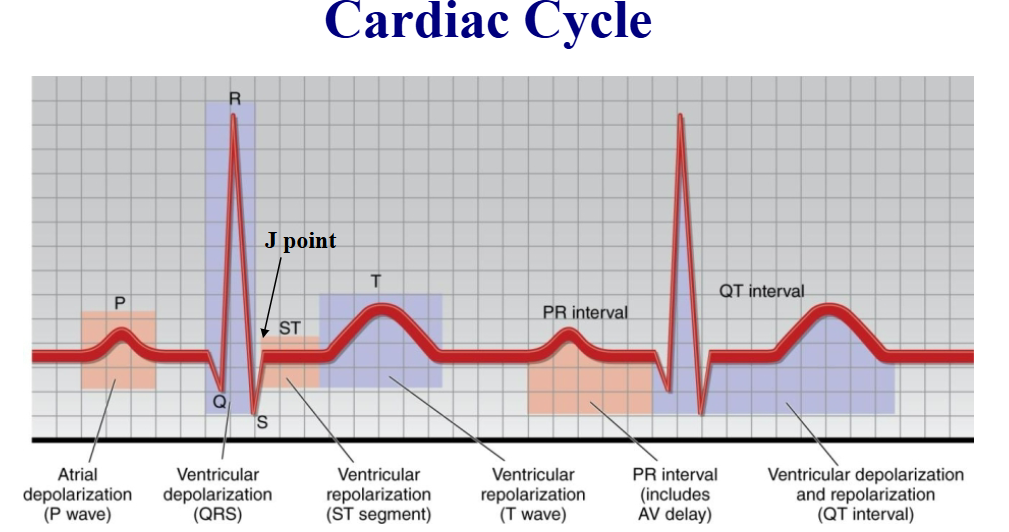
QRS duration
Activation/depolarization of the two ventricles.

ST segment
Interval between ventricular depolarization and repolarization.

T wave
Follows QRS; represents the relaxation/repolarization of the ventricles.

QT interval
Represents time taken for ventricular depolarization and repolarization.

Normal Sinus Rhythm
60-100 bpm.
Sinus Bradycardia
Less than 60 bpm.
Sinus Trachycardia
More than 100 bpm.

Sinus Arrythmia
1:1 ratio of P waves to QRS complexes, variation in heart rate (distances in QRS complexes). Based on ANS changes with beating.

PAC (premature atrial complexes)
Early beat, P wave may appear different, normal width QRS complex.

PVC (premature ventricular complexes)
Early beat, premature beat not preceded by P wave, large, upside-down QRS complex.

Ventricular Bigeminy
PVC every other beat.

Ventricular trigeminy
PVC every 3rd beat.

Ventricular Couplet
2 PVC every row.

Ventricular Triplet
3 PVC every row.

Unifocal PVCs
Multiple PVCs coming from the same ecotpic focus, identical.

Multifocal PVCs
Coming from more than 1 ectopic focus, not identical.

Sinus Tachycardia in ECG
Gradual onset, normal P waves, normal QRS.

Supraventricular Tachycardia
Sudden onset, HR 150-250, P waves abnormal/buried in T wave, normal width QRS.

Atrial Flutter
“Saw-tooth” pattern, flutter waves override P waves and T waves, normal width QRS complex.

Atrial Fibrillation
Irregular rhythm, no organized P waves, normal QRS.
Implications for atrial fibrillation:
Drop in cardiac output/symptoms, higher risk of stroke, medications.

Ventricular Tachycardia
>3 consecutive ventricular beats, HR: 100-200, wide QRS, dissociation between P waves and QRS complexes. (serious + urgent)

Ventricular Fibrillation
No organized ventricular activity, lethal rhythm.

Components of a pacemaker:
Pulse generator and placing wires (leads).
Pacemakers treat:
Severe bradycardia, SA block (idioventricular), AV block (2nd + 3rd degree).

Pacemakers changes on ECG:
Sharp vertical spikes, wide QRS complexes, abnormal/flat P wave, T waves in opposite direction.
SA Node Blocks
Junctional rhythm and Idioventricular rhythm.

Junctional Rhythm
40-60 bpm, impulses blocked + unable to leave SA node → inverted P waves, normal width QRS.

Idioventricular Rhythm
15-40 bpm, impulses coming from ventricles + can’t travel ro SA + AV node due to blockage → no P waves, wide QRS.
AV node blocks
1st degree, 2nd degree (mobitz 1 + 2), 3rd degree.

1st Degree AV Block
Delay in conduction, fixed prolonged PR interval > 0.20 sec.

2nd degree AV block - Mobitz 1 (wenckebach)
Intermittent blocked conduction — progressive lengthening of PR interval → dropped QRS complex.

2nd Degree AV Block - Mobitz 2
Intermittent blocked conduction — fixed PR interval with occasional dropped QRS complex.
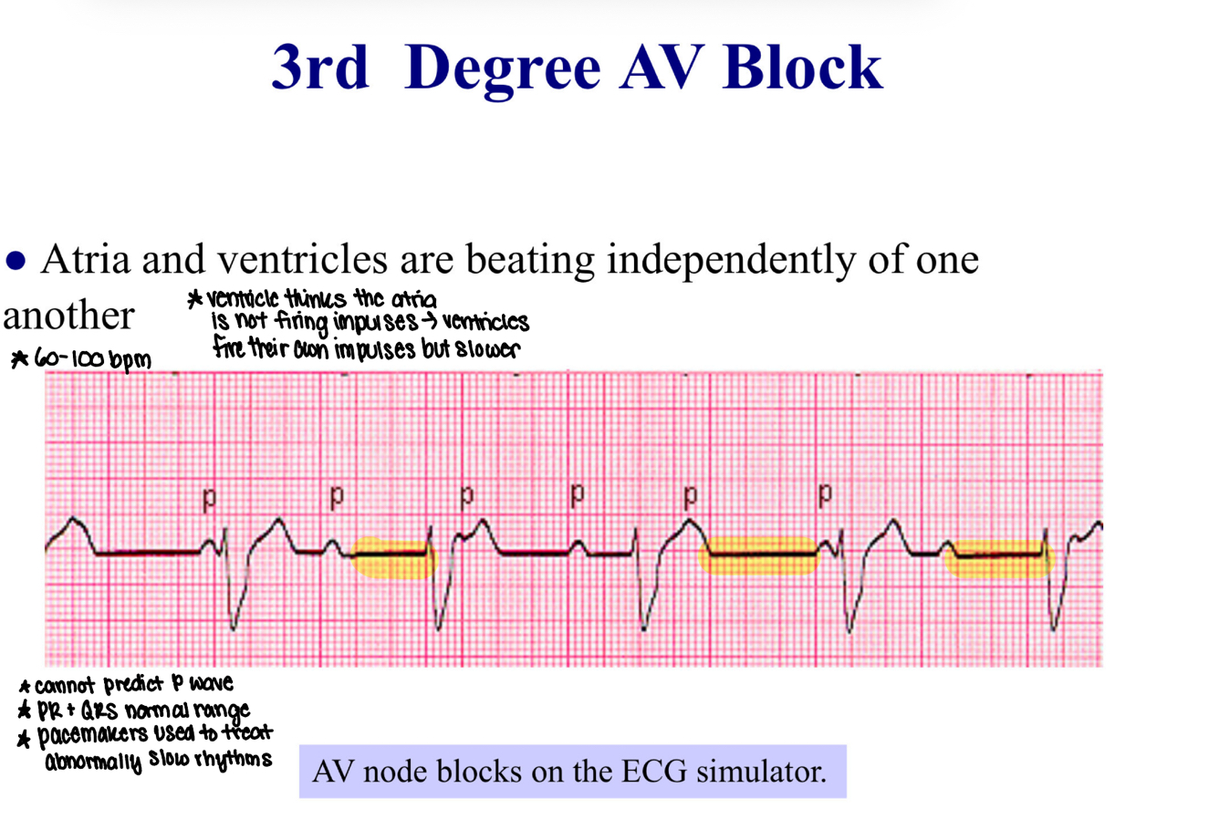
3rd Degree AV Block
Complete blocked conduction — atria and ventricles are beating independently of one another.
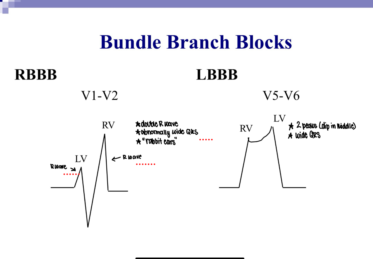
Bundle Branch Blocks
One of the bundles is blocked, sequential depolarization, wide QRS.

Bundle Branch Blocks (right) leads:
V1-V2.

Bundle Branch Blocks (left) leads:
V5-V6.
Primary Prevention of Heart Disease:
Quit smoking, heart-healthy diet, regular physical activity, weight control, manage cholesterol + BP.
Secondary Prevention of Heart Disease:
Prevents recurring cardiovascular events → cardiac rehab, anti-platelet therapy, BP control, ACE inhibitors/beta-blockers.
Smoking
Positive cardiovascular risk factor if individual is a current smoker or has quit within the past 6 months.
Smoking – Pharmacological Interventions
Nicotine replacement therapy, varenicline, bupropion.
Blood Pressure Risk for CVD
Hypertension >130/80 mmHg.
AHA/ACC lifestyle recommendations for lowering BP:
Quit smoking/drinking, limit caffeine, manage diabetes, weight, cholesterol, diet, become physically active.
LDL Cholesterol + Atherosclerosis
Primary driver → initiates plaque formation.
HDL cholesterol + Atherosclerosis
Linked to lower atherosclerosis.
Blood Glucose as a risk factor:
>250 mg/dL
Type 1 Diabetes
Autoimmune condition where the body stops making insulin.
Type 2 Diabetes
Autoimmune condition caused by insulin resistance and the body not using insulin effectively.
ACSM criteria for physical inactivity
Not participating in at least 30 mins of moderate-intensity physical activity on at least 3 days per week for at least 3 months.
MET-min weekly
1 MET = 3.5 x weight (kg) / 200
Recommendation for physical inactivity (min/week) for general population:
150 min → moderate aerobic activity
75 min → vigorous aerobic activity
Recommendation for physical activity (min/week) for heart disease patients:
240-360 min of moderate activty a week.
ACSM criteria for obesity - BMI
BMI greater than 30 kg/m2.
ACSM criteria for obesity - waist girth
Greater than 38 inches in women, 40 inches in men.