Balance Wk1 Bootcamp LG2
 Call Kai
 Learn
Learn Practice Test
 Spaced Repetition
 Match
Match Flashcards
 Knowt Play
1/95

There's no tags or description
Looks like no tags are added yet.
Name | Mastery | Learn | Test | Matching | Spaced | Call with Kai | Chat |
|---|
No analytics yet
Send a link to your students to track their progress
96 Terms
What is the purpose of renal autoregulation?
Answer: To stabilize glomerular filtration rate and renal blood flow despite fluctuations in systemic blood pressure.
Extra Information:
The kidneys can self-regulate their blood flow.
Glomerular filtration rate is affected by systemic blood pressure.

What are the two renal autoregulatory mechanisms shown on the slide?
Answer: The myogenic mechanism and tubuloglomerular feedback.
Extra Information:
Both mechanisms help maintain glomerular filtration rate and renal blood flow.
Trace the myogenic response to increased blood pressure.
Answer: Increased blood pressure → increased stretch → afferent arteriole contraction → vasoconstriction → decreased renal blood flow.
Extra Information:
This response opposes the initial increase in pressure.
The overall mechanism helps maintain glomerular filtration rate and renal blood flow.

Trace the myogenic response to decreased blood pressure.
Answer: Decreased blood pressure → decreased stretch → afferent arteriole relaxation → vasodilation → increased renal blood flow.
Extra Information:
This response opposes the initial decrease in pressure.
The overall mechanism helps maintain glomerular filtration rate and renal blood flow.

Which arteriole changes its tone during the renal myogenic mechanism?
Answer: The afferent arteriole.
Extra Information:
Increased stretch causes it to contract.
Decreased stretch causes it to relax.

What variable does the macula densa sense during tubuloglomerular feedback?
Answer: Sodium chloride.
Extra Information:
Changes in sodium chloride trigger changes in afferent arteriole tone.

Trace tubuloglomerular feedback when the macula densa senses increased sodium chloride.
Answer: Increased sodium chloride sensed by the macula densa → afferent arteriole contraction → vasoconstriction → decreased renal blood flow.
Extra Information:
This response helps maintain glomerular filtration rate and renal blood flow.

Trace tubuloglomerular feedback when the macula densa senses decreased sodium chloride.
Answer: Decreased sodium chloride sensed by the macula densa → afferent arteriole relaxation → vasodilation → increased renal blood flow.
Extra Information:
This response helps maintain glomerular filtration rate and renal blood flow.

Compare the stimulus for the myogenic mechanism with the stimulus for tubuloglomerular feedback.
Answer: The myogenic mechanism responds to changes in blood pressure and stretch, while tubuloglomerular feedback responds to sodium chloride sensed by the macula densa.
Extra Information:
Both ultimately alter afferent arteriole tone.
Both help stabilize glomerular filtration rate and renal blood flow.

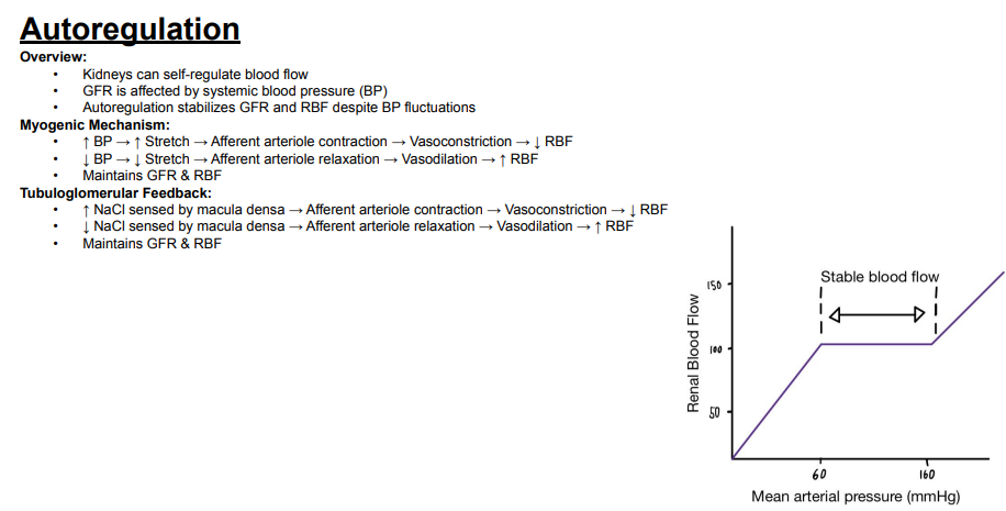
Over approximately what mean arterial pressure range does the graph show stable renal blood flow due to autoregulation?
Answer: Approximately 60–160 mmHg.
Extra Information:
Within this range, renal blood flow remains relatively stable despite changes in mean arterial pressure.
Outside this range, the graph shows renal blood flow changing with pressure.

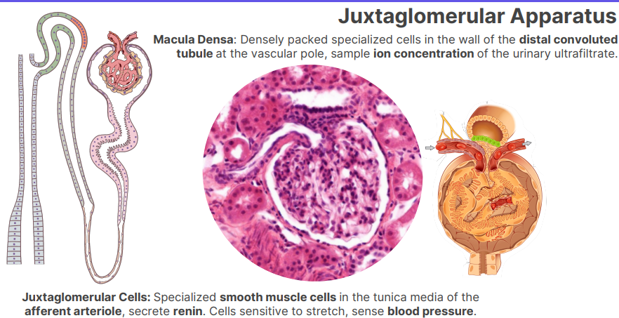
What are juxtaglomerular cells?
Answer: Specialized smooth muscle cells.
Extra Information:
They are part of the juxtaglomerular apparatus.
They are located in the afferent arteriole.

Where are juxtaglomerular cells located?
Answer: In the tunica media of the afferent arteriole.
Extra Information:
They are specialized smooth muscle cells.

What do juxtaglomerular cells secrete?
Answer: Renin.
Extra Information:
Juxtaglomerular cells are located in the tunica media of the afferent arteriole.
What do juxtaglomerular cells sense?
Answer: Blood pressure.
Extra Information:
These cells are sensitive to stretch.
This allows them to sense changes in blood pressure.

What are macula densa cells?
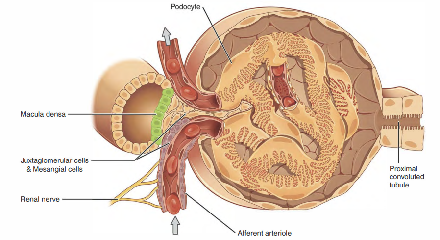
Answer: Densely packed specialized cells in the wall of the distal convoluted tubule.
Extra Information:
They are part of the juxtaglomerular apparatus.
Where is the macula densa located?
Answer: In the wall of the distal convoluted tubule at the vascular pole.
Extra Information:
The cells are densely packed and specialized.

What does the macula densa sample?
Answer: The ion concentration of the urinary ultrafiltrate.
Extra Information:
The macula densa performs this function from its location in the distal convoluted tubule.

Compare what juxtaglomerular cells and the macula densa sense.
Answer: Juxtaglomerular cells sense blood pressure through stretch, while the macula densa samples ion concentration in the urinary ultrafiltrate.
Extra Information:
Juxtaglomerular cells are in the afferent arteriole.
Macula densa cells are in the distal convoluted tubule at the vascular pole.

What are juxtaglomerular cells?
Answer: Specialized smooth muscle cells.
Extra Information:
They are part of the juxtaglomerular apparatus.
They are located in the afferent arteriole.
Where are juxtaglomerular cells located?
Answer: In the tunica media of the afferent arteriole.
Extra Information:
They are specialized smooth muscle cells.
What do juxtaglomerular cells secrete?
Answer: Renin.
Extra Information:
Juxtaglomerular cells are located in the tunica media of the afferent arteriole.
What do juxtaglomerular cells sense?
Answer: Blood pressure.
Extra Information:
These cells are sensitive to stretch.
This allows them to sense changes in blood pressure.
What are macula densa cells?
Answer: Densely packed specialized cells in the wall of the distal convoluted tubule.
Extra Information:
They are part of the juxtaglomerular apparatus.
Where is the macula densa located?
Answer: In the wall of the distal convoluted tubule at the vascular pole.
Extra Information:
The cells are densely packed and specialized.
What does the macula densa sample?
Answer: The ion concentration of the urinary ultrafiltrate.
Extra Information:
The macula densa performs this function from its location in the distal convoluted tubule.
Compare what juxtaglomerular cells and the macula densa sense.
Answer: Juxtaglomerular cells sense blood pressure through stretch, while the macula densa samples ion concentration in the urinary ultrafiltrate.
Extra Information:
Juxtaglomerular cells are in the afferent arteriole.
Macula densa cells are in the distal convoluted tubule at the vascular pole.
What are fenestrations in capillaries?
Answer: Small pores within capillary endothelial cells.
Extra Information:
Fenestrations are a characteristic feature of fenestrated capillaries.

What type of barrier do fenestrated capillaries form?
Answer: A slightly selective barrier.
Extra Information:
Their endothelial cells contain small pores called fenestrations.

Which organs are specifically associated with fenestrated capillaries on the slide?
Answer: The kidney and intestine.
Extra Information:
- Fenestrated capillaries provide a slightly selective barrier in these organs.

What specialized epithelial cells are shown surrounding glomerular capillaries?
Answer: Podocytes.
Extra Information:
- The slide illustrates podocytes extending processes around the glomerular capillaries.
- Podocytes contribute to the urinary filtration barrier.

What disease is specifically associated with podocytes on the Bootcamp slide?
Answer: Minimal change disease.
Extra Information:
The slide identifies minimal change disease as a clinical association with podocytes.
The slide does not provide additional disease mechanism or findings.
Minimal change disease is a primary kidney disorder characterized by diffuse podocyte foot process effacement that leads to heavy proteinuria and nephrotic syndrome

What is the overall function of the urinary filtration barrier?
Answer: Ultrafiltration of molecules.
Extra Information:
- The barrier determines which molecules can pass from the blood during filtration.

What two molecular properties determine ultrafiltration across the urinary filtration barrier?
Answer: Size and charge.
Extra Information:
- The filtration barrier therefore provides selective filtration rather than allowing all molecules to pass equally.

What capillary feature contributes to the urinary filtration barrier?
Answer: Fenestrated capillary endothelium.
Extra Information:
- Fenestrations are small pores in endothelial cells.
- The kidney is specifically identified as a location of fenestrated capillaries.

What structures shown across these slides contribute to filtration at the glomerulus?
Answer: Fenestrated capillary endothelium and podocytes.
Extra Information:
- The slides visually show these structures surrounding the glomerular capillary.
- The resulting urinary filtration barrier performs ultrafiltration based on molecular size and charge.

What disease is specifically associated with the urinary filtration barrier on the Bootcamp slide?
Answer: Goodpasture syndrome.
Extra Information:
- Goodpasture syndrome is labeled as a clinical association on the urinary filtration barrier slide.
- The slide does not provide additional disease mechanism or findings.

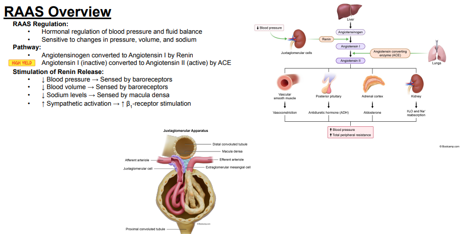
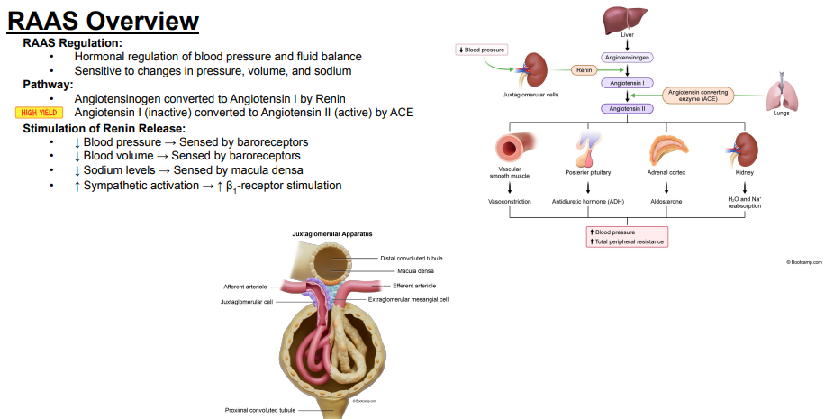
What is the overall function of the renin-angiotensin-aldosterone system?
Answer: Hormonal regulation of blood pressure and fluid balance.
Extra Information:
The system responds to changes that affect circulation and fluid status.

What three variables is the renin-angiotensin-aldosterone system sensitive to?
Answer: Pressure, volume, and sodium.
Extra Information:
Changes in these variables can contribute to activation of the system.

What converts angiotensinogen into angiotensin I?
Answer: Renin.
Extra Information:
This is the first conversion shown in the renin-angiotensin-aldosterone system pathway.

What converts angiotensin I into angiotensin II?
Answer: Angiotensin-converting enzyme.
Extra Information:
Angiotensin-converting enzyme is abbreviated ACE.
This conversion is marked as high yield on the slide.

Which form is inactive versus active: angiotensin I or angiotensin II?
Answer: Angiotensin I is inactive, while angiotensin II is active.
Extra Information:
Angiotensin-converting enzyme converts angiotensin I into angiotensin II.

Trace the pathway from angiotensinogen to active angiotensin II.
Answer: Angiotensinogen → renin → angiotensin I → angiotensin-converting enzyme → angiotensin II.
Extra Information:
Angiotensin I is inactive.
Angiotensin II is active.

How does decreased blood pressure stimulate renin release?
Answer: Decreased blood pressure is sensed by baroreceptors, which stimulates renin release.
Extra Information:
Decreased pressure is one stimulus for activation of the renin-angiotensin-aldosterone system.

How does decreased blood volume stimulate renin release?
Answer: Decreased blood volume is sensed by baroreceptors, which stimulates renin release.
Extra Information:
The renin-angiotensin-aldosterone system is sensitive to changes in volume.
How do decreased sodium levels stimulate renin release?
Answer: Decreased sodium levels are sensed by the macula densa, which stimulates renin release.
Extra Information:
The macula densa is part of the juxtaglomerular apparatus shown on the slide.
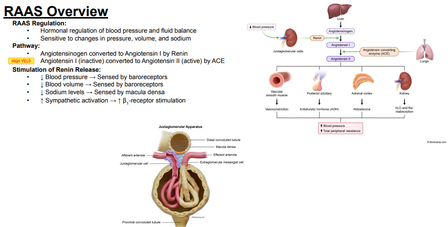
How does increased sympathetic activation stimulate renin release?
Answer: Increased sympathetic activation increases beta-1 receptor stimulation, which stimulates renin release.
Extra Information:
Beta-1 receptor stimulation is the sympathetic mechanism emphasized on the slide.
Beta-1 receptors are a type of adrenergic receptor that respond to stress hormones like adrenaline and norepinephrine to speed up heart rate and boost blood flow

What four changes stimulate renin release according to the slide?
Answer: Decreased blood pressure, decreased blood volume, decreased sodium levels, and increased sympathetic activation.
Extra Information:
Decreased blood pressure and volume are sensed by baroreceptors.
Decreased sodium is sensed by the macula densa, while sympathetic activation acts through beta-1 receptor stimulation.
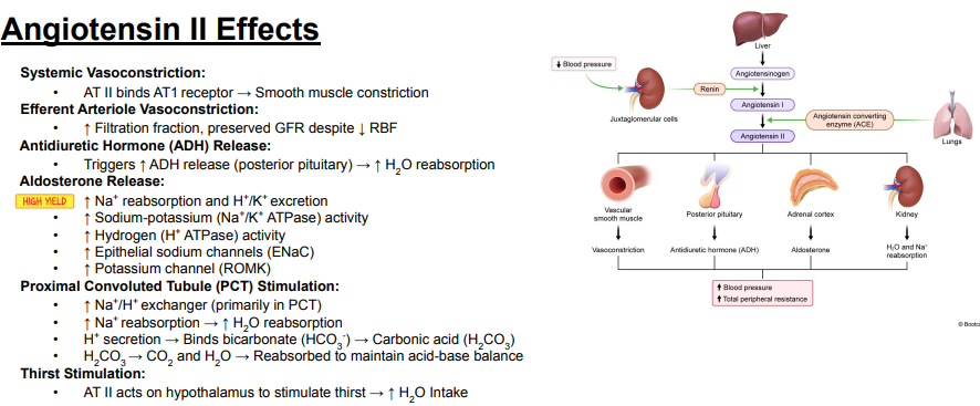
What major downstream effects of angiotensin II are shown in the RAAS diagram?
Answer: Vasoconstriction, antidiuretic hormone release, aldosterone release, and increased water and sodium reabsorption.
Extra Information:
The diagram connects these effects with increased blood pressure.
Vasoconstriction also increases total peripheral resistance.

Through which receptor does angiotensin II cause systemic vasoconstriction?
Answer: The AT1 receptor.
Extra Information:
Angiotensin II binding to the AT1 receptor causes smooth muscle constriction.
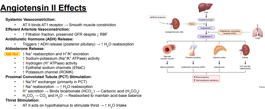
How does angiotensin II produce systemic vasoconstriction?
Answer: Angiotensin II binds the AT1 receptor → smooth muscle constriction.
Extra Information:
Systemic vasoconstriction is one of the major effects of angiotensin II.
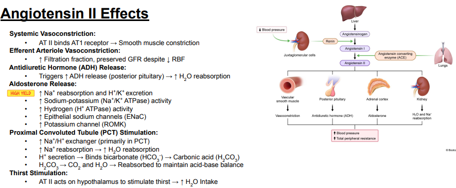
Which renal arteriole does angiotensin II constrict according to the slide?
Answer: The efferent arteriole.
Extra Information:
This affects filtration fraction, glomerular filtration rate, and renal blood flow.

How does angiotensin II-mediated efferent arteriole vasoconstriction affect filtration fraction, glomerular filtration rate, and renal blood flow?
Answer: It increases filtration fraction, preserves glomerular filtration rate, and decreases renal blood flow.
Extra Information:
The slide emphasizes preservation of glomerular filtration rate despite decreased renal blood flow.
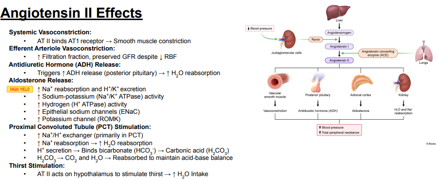
How does angiotensin II affect antidiuretic hormone release?
Answer: Angiotensin II increases antidiuretic hormone release from the posterior pituitary.
Extra Information:
Increased antidiuretic hormone increases water reabsorption.

What is the effect of angiotensin II-induced antidiuretic hormone release on water handling?
Answer: It increases water reabsorption.
Extra Information:
Angiotensin II triggers increased antidiuretic hormone release from the posterior pituitary.

How does angiotensin II-induced aldosterone release affect sodium, hydrogen, and potassium handling?
Answer: It increases sodium reabsorption and increases hydrogen and potassium excretion.
Extra Information:
These aldosterone effects are marked as high yield on the slide.

Which sodium-potassium pump is increased by aldosterone according to the slide?
Answer: The sodium-potassium ATPase.
Extra Information:
Aldosterone increases sodium-potassium ATPase activity.

Which hydrogen pump is increased by aldosterone according to the slide?
Answer: The hydrogen ATPase.
Extra Information:
Aldosterone increases hydrogen ATPase activity.

Which epithelial sodium channel is increased by aldosterone?
Answer: ENaC.
Extra Information:
ENaC stands for epithelial sodium channel.
Aldosterone increases its activity.

Which potassium channel is increased by aldosterone?
Answer: ROMK.
Extra Information:
ROMK is one of the channels increased during the aldosterone response shown on the slide.

What transporter does angiotensin II increase primarily in the proximal convoluted tubule?
Answer: The sodium/hydrogen exchanger.
Extra Information:
The slide identifies the proximal convoluted tubule as the primary location of this effect.

How does angiotensin II stimulation of the proximal convoluted tubule affect sodium and water reabsorption?
Answer: It increases sodium reabsorption, which increases water reabsorption.
Extra Information:
This is linked to increased sodium/hydrogen exchanger activity.

What happens to hydrogen secretion when angiotensin II stimulates the sodium/hydrogen exchanger in the proximal convoluted tubule?
Answer: Hydrogen secretion increases.
Extra Information:
The secreted hydrogen then participates in bicarbonate handling.
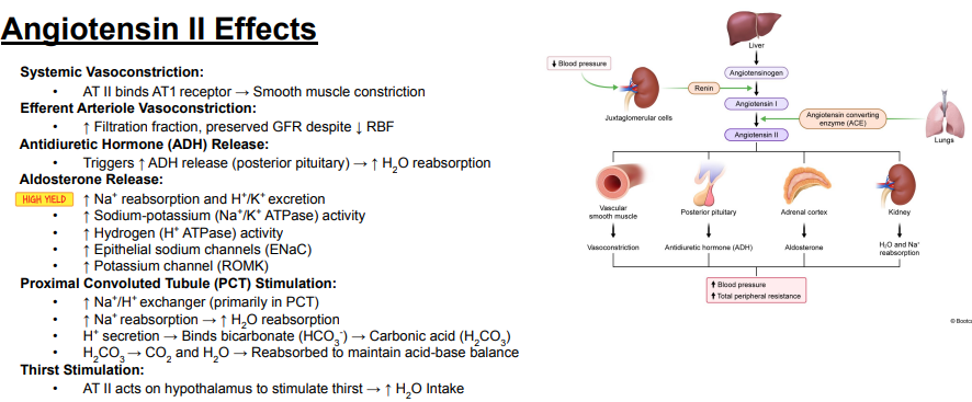
What happens when secreted hydrogen binds bicarbonate in the proximal convoluted tubule?
Answer: Hydrogen binds bicarbonate to form carbonic acid.
Extra Information:
The pathway is H+ + HCO3− → H2CO3.
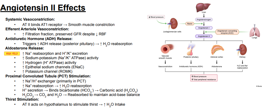
What happens to carbonic acid during proximal convoluted tubule bicarbonate handling?
Answer: Carbonic acid is converted to carbon dioxide and water.
Extra Information:
The carbon dioxide and water are then reabsorbed.
The slide connects this process with maintenance of acid-base balance.
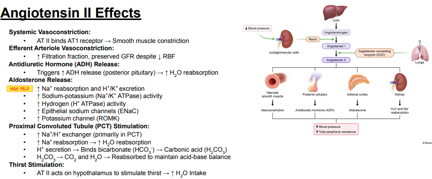
Trace the proximal convoluted tubule mechanism stimulated by angiotensin II.
Answer: Angiotensin II → increased sodium/hydrogen exchanger → increased sodium reabsorption and hydrogen secretion → increased water reabsorption, while hydrogen binds bicarbonate → carbonic acid → carbon dioxide and water → reabsorption.
Extra Information:
The slide connects this process with maintenance of acid-base balance.
How does angiotensin II stimulate thirst?
Answer: Angiotensin II acts on the hypothalamus to stimulate thirst, increasing water intake.
Extra Information:
Increased water intake is one of the mechanisms by which angiotensin II supports fluid balance.

Where are macula densa cells located?
Answer: In the wall of the distal convoluted tubule near the vascular pole.
Extra Information:
Macula densa cells are specialized cells of the juxtaglomerular apparatus.

What do macula densa cells sense according to the slide?
Answer: Low ion concentration in the urine.
Extra Information:
The slide states that low urinary ion concentration indicates a high amount of water in the urine.

What do macula densa cells do after sensing a low ion concentration in the urine?
Answer: They stimulate juxtaglomerular cells.
Extra Information:
- This connects macula densa sensing to the juxtaglomerular cell response.

Where are juxtaglomerular cells located?
Answer: In the wall of the afferent arteriole.
Extra Information:
- Juxtaglomerular cells are specialized smooth muscle cells.

What do juxtaglomerular cells sense?
Answer: Low blood pressure.
Extra Information:
- These cells participate in the juxtaglomerular apparatus response to decreased blood pressure.

What do juxtaglomerular cells secrete?
Answer: Renin.
Extra Information:
- Renin is part of the renin-angiotensin-aldosterone system.

What major effect of angiotensin II is emphasized on the slide?
Answer: Angiotensin II is a vasoconstrictor.
Extra Information:
- The slide connects angiotensin II with the response that increases blood pressure.

Which part of the pituitary gland is associated with antidiuretic hormone on the slide?
Answer: The pars nervosa.
Extra Information:
- Antidiuretic hormone is abbreviated ADH.

Which part of the pituitary gland is associated with adrenocorticotropic hormone on the slide?
Answer: The pars distalis.
Extra Information:
- Adrenocorticotropic hormone is abbreviated ACTH.

Which adrenal gland layer is associated with aldosterone?
Answer: The zona glomerulosa.
Extra Information:
- Aldosterone is shown as part of the hormonal response on the slide.

What happens to sodium ions in the distal convoluted tubule according to the slide?
Answer: Sodium ions are actively transported out of the urine.
Extra Information:
- This sodium movement creates an ion gradient.

What is created when sodium ions are actively transported out of the urine in the distal convoluted tubule?
Answer: An ion gradient.
Extra Information:
- The slide connects this gradient with subsequent water movement.

How does water move out of the thin descending Loop of Henle according to the slide?
Answer: Water passively diffuses out of the urine.
Extra Information:
- The slide connects this movement to the ion gradient.
- Water then diffuses into the blood.

What does the collecting duct do with water according to this slide?
Answer: It transports water out of the urine.
Extra Information:
- This contributes to water moving back toward the blood.

Trace the overall response shown on the slide that begins with low urinary ion concentration and contributes to increased blood pressure.
Answer: Low urinary ion concentration → macula densa cells stimulate juxtaglomerular cells → juxtaglomerular cells secrete renin → renin-angiotensin-aldosterone system response → angiotensin II vasoconstriction and increased renal water recovery → increased blood pressure.
Extra Information:
- The slide also associates antidiuretic hormone with collecting duct water transport.
- Sodium transport creates an ion gradient that promotes water movement toward the blood.

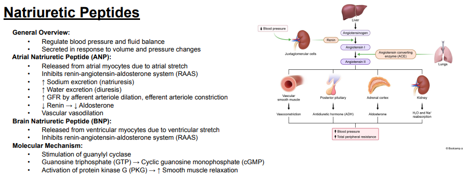
What is the overall function of natriuretic peptides?
Answer: They regulate blood pressure and fluid balance.
Extra Information:
They are secreted in response to changes in volume and pressure.

What stimulates the release of atrial natriuretic peptide?
Answer: Atrial stretch.
Extra Information:
Atrial natriuretic peptide is released when increased volume or pressure stretches the atria.
Which cells release atrial natriuretic peptide?
Answer: Atrial myocytes.
Extra Information:
Atrial stretch stimulates these cells to release atrial natriuretic peptide.
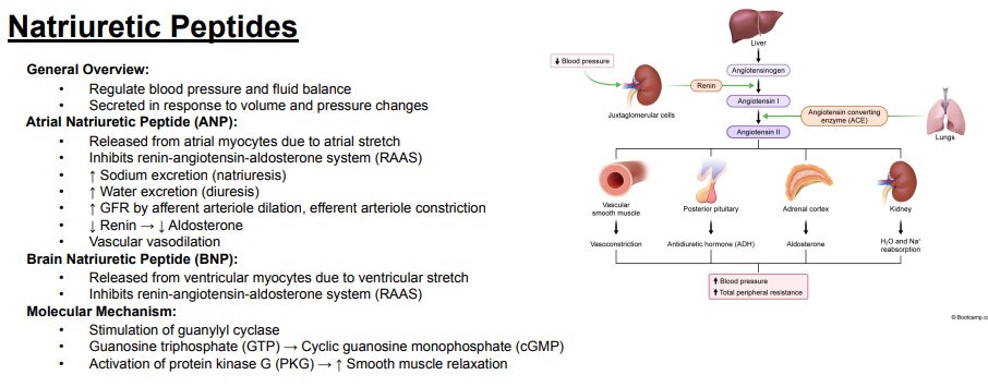
How does atrial natriuretic peptide affect the renin-angiotensin-aldosterone system?
Answer: It inhibits the renin-angiotensin-aldosterone system.
Extra Information:
Atrial natriuretic peptide decreases renin and consequently decreases aldosterone.

How does atrial natriuretic peptide affect sodium and water excretion?
Answer: It increases sodium excretion and water excretion.
Extra Information:
Increased sodium excretion is called natriuresis.
Increased water excretion is called diuresis.

What is natriuresis?
Answer: Increased sodium excretion.
Extra Information:
Atrial natriuretic peptide promotes natriuresis.

What is diuresis?
Answer: Increased water excretion.
Extra Information:
Atrial natriuretic peptide promotes diuresis.
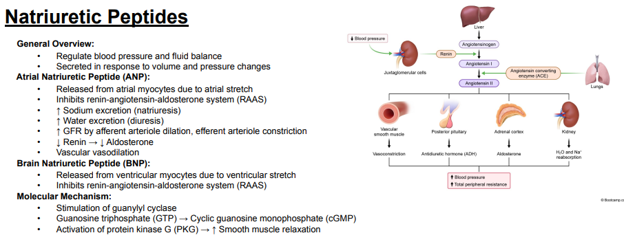
How does atrial natriuretic peptide affect the afferent and efferent arterioles?
Answer: It dilates the afferent arteriole and constricts the efferent arteriole.
Extra Information:
These vascular changes increase glomerular filtration rate.

How does atrial natriuretic peptide affect glomerular filtration rate?
Answer: It increases glomerular filtration rate.
Extra Information:
It does this through afferent arteriole dilation and efferent arteriole constriction.

How does atrial natriuretic peptide affect renin and aldosterone?
Answer: It decreases renin, which decreases aldosterone.
Extra Information:
This contributes to inhibition of the renin-angiotensin-aldosterone system.

What effect does atrial natriuretic peptide have on systemic blood vessels?
Answer: It causes vascular vasodilation.
Extra Information:
This is one of the blood-pressure-regulating effects of atrial natriuretic peptide.

What stimulates brain natriuretic peptide release, and which cells release it?
Answer: Ventricular stretch stimulates ventricular myocytes to release brain natriuretic peptide.
Extra Information:
Brain natriuretic peptide is released in response to changes in volume and pressure.

How does brain natriuretic peptide affect the renin-angiotensin-aldosterone system?
Answer: It inhibits the renin-angiotensin-aldosterone system.
Extra Information:
This is an effect shared with atrial natriuretic peptide on the slide.

What intracellular signaling pathway is stimulated by natriuretic peptides?
Answer: Guanylyl cyclase converts guanosine triphosphate into cyclic guanosine monophosphate, which activates protein kinase G.
Extra Information:
Protein kinase G promotes smooth muscle relaxation.

Trace the molecular mechanism by which natriuretic peptides cause smooth muscle relaxation.
Answer: Guanylyl cyclase stimulation → guanosine triphosphate → cyclic guanosine monophosphate → protein kinase G activation → increased smooth muscle relaxation.
Extra Information:
Cyclic guanosine monophosphate is the second messenger in this pathway.
The final vascular effect is smooth muscle relaxation.
