Chapter 4 Molecular Biology (not done)
 Call Kai
 Learn
 Practice Test
 Spaced Repetition
 Match
 Flashcards
 Knowt Play
1/3

There's no tags or description
Looks like no tags are added yet.
4 Terms
DNA Structure
General Overview
Significant detail about the structure of DNA and RNA is provided in MCAT Biochemistry Review, here, we will give a brief overview of this topic. DNA is short for deoxyribonucleic acid. DNA and RNA (ribonucleic acid) are called nucleic acids because they are found in the nucleus and possess many acidic phosphate groups.
The building block of DNA is deoxyribonucleoside 5’ triphosphate (dNTP, where N represents one of the four basic nucleosides). Deoxyadenosine 5’ triphosphate (dATP) is shown in Figure 1; the other bases are shown in Figure 2. Nucleo-tides are built from three components: a sugar (deoxyribose for DNA, ribose for RNA), an aromatic, nitrogenous base, and 1-3 phosphate groups. The bases G and A are derived from a precursor called purine, so they are referred to as the purines. C,T, and U are the pyrimidines.
A nucleoside is ribose or deoxyribose with a purine or pyramidine linked to the 1’ carbon; nucleotides are phosphate esters of nucleosides, with one, two or three phosphate groups joined to the ribose ring by the 5’hydroxy group. When nucleotides contain three phosphate residues, they are also referred to as nucleoside triphosphates, abbreviated NTP (if the usgar is deoxyribose, they are abbreviated dNTP). In individual nucleotides, N is replaced by A, G, C, T, or U.
d: deoxyribose
N: any base
TP: triphosphate.
dNTPs are the building blocks of DNA.
Put figure 1 here

Backbone of DNA
The sugar + phosphate portion of the nucleotide is refereed to as the backbone of DNA, because it is invariant. The base is the variable portion of the building block.
put the picture of the bases on page 66 (figure 2) here

Polynucleotides
Nucleotides in the DNA chain are covalently linked by phosphodiester bonds between the 3’hydroxy group of one deoxyribose and 5’ phosphate group of the next deoxyribose (Figure 3).
A polymer of several nucleotides linked together is termed an oligonucleotide, and a polymer of many nucleotides is a polynucleotide.
Since the only unique part of the nucleotide is the base, the sequence of a polynucleotide can be abbreviated by simply listing the bases attached to each nucleotide in the chain.
The end of the chain with a free 5’ phosphate group is writen first in a polynucleotide, with other nucleotides in the chain indicated in the 5’ to 3’ direction.
put figure 3 here.

The Watson Crick Model of DNA Structure
James Watson and Francis Crick (with the help of Maurice Wilkins and Rosalind Franklin) developed a model of the structure of DNA in the cell. According to the Watson-Crick model, cellular DNA is a right-handed double helix held together by hydrogen bonds between bases.
In the cell, DNA does not exist in the form of a single polynucleotide. Instead, the DNA found in the nucleus is double-stranded (ds). In ds-DNA, two very long polynucleotide chains are hydrogen-bonded together in antiparallel orientaion.
Anti-parallel means 5’end of one chain is paired with 3’ end of the other (parallel would mean 5’ end is bonded to 5’ end).
The H-bonds in ds-DNA are between the bases on adjacent chains. This H-bonding is very specific: A is always H-bonded to T, and G is always H-bonded to C (Figure 4). Note that this means that an H-bonded pair always consists of a purine plus a pyrimidine2. Thus both types of base pairs (AT or GC) take up the same amount of room in the DNA double helix. The GC pair is held together by three hydrogen bonds, the AT pair by two. Two chains or DNA sare said to be complementary if the bases in each strand can hydrogen bond when the strands are oriented in an antiparallel fashion. If we are talking about ds-DNA 100 nucleotides long, we would say it is 100 base pairs (bp) long. A kbp (kilobase pair) is ds-DNA 1000 nucleotides long.
question: This fact has a fringe benefit: we can calculate the number of purines if we know the number of pyrimidines. We can actually calculate several variables. Chargoff’s rule states that [A]=[T], and [G]=[C]; and [A] + [G]=[T]+[C]
binding of two complementary strands
The binding of two complementary strands of DNA into a double-stranded structure is termed annealing, or hybridization.
separation of strands
The separation of strands is termed melting or denaturation.
Which of the following is/are true about ds-DNA?
I. If the amount of G in a double helix is known, the amount of C can be calculated
II. If the fraction of purine nucleotide and the total molecular weight of a double helix are known, the amount of cytosine can be calculated
III. The two chains in a piece of ds-DNA containing mostly purines will be bonded together more tightly than the two chains in a piece of ds-DNA containing mostly pyrimidines.
IV. The oligonucleotide ATGTAT is complementary to the oligonucleotide ATACAT?
I and IV are true
for IV, start at the end of the sequence, in the last sequence, start at the front.
Item I: true. For every G, there is a C; and for every A there is a T. Item II: False. The ratio of purines to pyramidines is always the same (50:50) since each purine is paired with a pyrimidine. In order to calculate the amount of any one base, you have to know the ratio of AT to GC pairs. Item III: False. Again, the ratio of purines to pyrimidines is always the same; 50:50. However, two chains containing mostly GC pairs will bond more tightly than two chains containing mostly AT pairs, since GC pairs are held together by 3 H-bonds while AT pairs are held together by 2 H-bonds. Item IV is true: Remember: the strands are antiparallel, A and T pair, G and C pair, and the 5’ end is always written first.
DNA structure is double stranded and coiled. In ds-DNA, the two hydrogen bonded antiparallel DNA strands form a right-handed double helix (meaning it corkscrews in a clockwise motion). with the bases of the interior and the ribose/phosphate backbone on exterior.
The double helix is stabilized by van der Waals interactions between the bases, which are stacked upon each other.

Chromosome Structure and Packing
Genome: the sum total of an organism’s genetic information.
Eukaryotic genomes are composed of several large species of linear ds-DNA; each piece of ds-DNA is called a chromosome.
Chromosome: a thread-like structure made of protein and a single molecule of DNA that serves to carry genomic information from cell to cell.
genomes are the sum total of an organisms genetic information
chromosomes are ds-DNA that make up the genome.
Humans have 46 chromosomes, 23 of which are inherited from each parent.
Prokaryotic (bacterial) genomes are composed of single circular chromosome.
Viral genomes may be linear or circular DNA or RNA.
The human genome consists of 109 base pairs while bacterial genomes contain only 106 base pairs. Much of the size different in higher eukaryotic genomes is the result of repetitive DNA that has no known function.
If the DNA remained as a simple double helix floating free in the cell, it would be very bulky and fragile. Prokaryotes have a distinctive mechanism for making their single circular chromosome more compact and sturdy. DNA Gyrase uses the energy of ATP to twist the gigantic circular molecule. Gyrase functions by breaking the DNA and twisting the two sides of the circle around each other. The resulting structure is a twisted circle that is composed of double stranded DNA. The two strands are already coiled, forming a helix. The twists created by DNA gyrase are called supercoils, since they are coils of a structure that is already coiled.
DNA gyrase is an essential bacterial Type II topoisomerase enzyme that introduces negative supercoils into double-stranded DNA using energy from ATP hydrolysis.
DNA Gyrase is Exclusive to Prokaryotes: Found in bacteria and archaea, making it absent in humans and an ideal target for selective drug action.
From the Greek word gyros, meaning "turn”. This reflects the enzyme's primary action of twisting, looping, and supercoiling circular DNA strands.
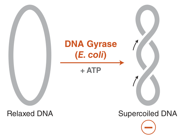
Since eukaryotes have even more DNA in their genome than prokaryotes, the eukaryotic genome requires denser packaging to fit within the cell (Figure 6). To accomplish this:
Histones: DNA is wrapped around globular proteins called histones.
Nucleosomes: After being wrapped around histones, but before being completely packed away, DNA has the microscopic appearance of beads on a string. The beads are called nucleosomes, nucleosomes are composed of DNA wrapped around an octamer of histones (a group of eight).
The string between the beads is a length of double-helical DNA called linker-DNA and is bound by a single linker histone.
Fully packed DNA is called chromatin; it is composed of closely stacked nucleosomes.

Staining chromosomes with chemicals
To look for patterns and morphology, chromosomes can be stained with chemicals. Usually, condensed metaphase chromosomes are used, as they are compact and easier to see. When chromosomes are treated, distinct light and dark regions become visible.
-the darker regions are denser and are called heterochromatin. Heterochromatin is rich in repeats
-the lighter regions are less dense and are called euchromatin. Density gives a sense of DNA coiling or compactness, and these patterns are constant and heritable. It’s now known that the lighter regions have higher transcription rates and therefore higher gene activity. The lower packing makes DNA accessible to enzymes and proteins.
Centromeres
A centromere (centro: center) is the region of a chromosome to which spindle fibers attach during cell division.
The spindle fibers attach via kinetochores, multiprotein complexes that act as anchor attachment sites for spindle fibers.
spindle fibers attach to the centromere via kinetochores.
Other protein complexes also bind to the centromere after DNA replication to keep sister chromatids attached to each other. Centromeres are made of heterochromatin, and repetitive DNA sequences. Chromosomes have p (short) and q (long) arms, and centromere position defines the ratio between the two (Figure 7).

(Meta)centric: the centromere is in the middle
(Sub)metacentric: the centromere is almost in the middle
(Acro)centric:
Telomeres
The ends of linear chromosomes are called telomeres. At the DNA level, these regions are distinguished by the presence of distinct nucleotide sequences repeated 50 to several hundred times.
-the repeated unit is usually 6-8 base pairs long and guanine rich.
-many vertebrates (including humans and mice) have the same repeated: 5’-TTAGGG-3’.
-Telomeres are composed of both single- and double-stranded DNA. Single stranded DNA is found at the very end of the chromosome and is about 300 base pairs in length. It loops around to form a knot, held together by many telomere associated proteins. This stabilizes the end of the chromosome.
-specialized telomere cap proteins distinguish telomeres from double stranded breaks and this prevents activation of repair pathways.
Telomeres function to prevent chromosome deterioration and also prevent fusion with neighboring chromosomes. They function as disposable buffers, blocking the ends of chromosomes.
DNA replication of telomeres represents a special challenge to cellular machinery. Since most prokaryotes have circular genomes, their DNA does not contain telomeres.
Prokaryotes vs Eukaryotes
-prokaryotes have DNA Gyrase, eukaryotes do not
-prokaryotes do not have telomeres, eukaryotes do.
d