Respiratory System Anatomy and Physiology - Lab 10
 Note
 Call Kai
 Learn
 Practice Test
 Spaced Repetition
 Match
 Flashcards
 Knowt Play
1/32

Earn XP
Description and Tags
Vocabulary practice flashcards covering gross anatomy, histology, respiratory tract divisions, and practical identification from Lab 10.
33 Terms
Respiratory System
The organ system that obtains oxygen from the atmosphere and removes carbon dioxide from body cells; consists of tubes that filter, warm, and moisturize incoming air, and microscopic air sacs that exchange gases.
Upper Respiratory Tract
The division of the respiratory tract that includes the nose, nasal cavity, paranasal sinuses, and pharynx.
Lower Respiratory Tract
The division of the respiratory tract that includes the larynx, trachea, bronchial tree, and lungs.
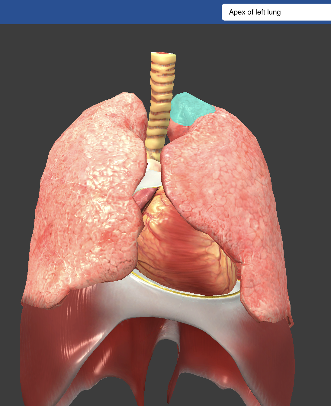
Apex
Apexis the topmost part of the lungs, extending above the first rib.


Nose - Nostrils
Openings in the nose that provide an entrance and exit for air; contain coarse internal hairs that prevent the entry of large particles.

Nasal Cavity
Hollow space posterior to the nose that conducts air to the pharynx; lined with a mucous membrane that filters, warms, and moistens incoming air.

Pseudostratified Columnar Epithelial Cells
Tall, specialized epithelial cells of the respiratory lining that appear stratified and bear cilia on their apical surfaces to move mucus.

Goblet Cells
Specialized mucus-secreting epithelial cells interspersed within the pseudostratified columnar epithelium that trap dust and foreign particles from incoming air.

Frontal Sinuses
Paranasal sinuses located in the frontal bone superior to the orbits that drain into the nasal cavity.

Sphenoidal Sinus
A paranasal sinus located deep in the body of the sphenoid bone posterior to the upper nasal cavity.
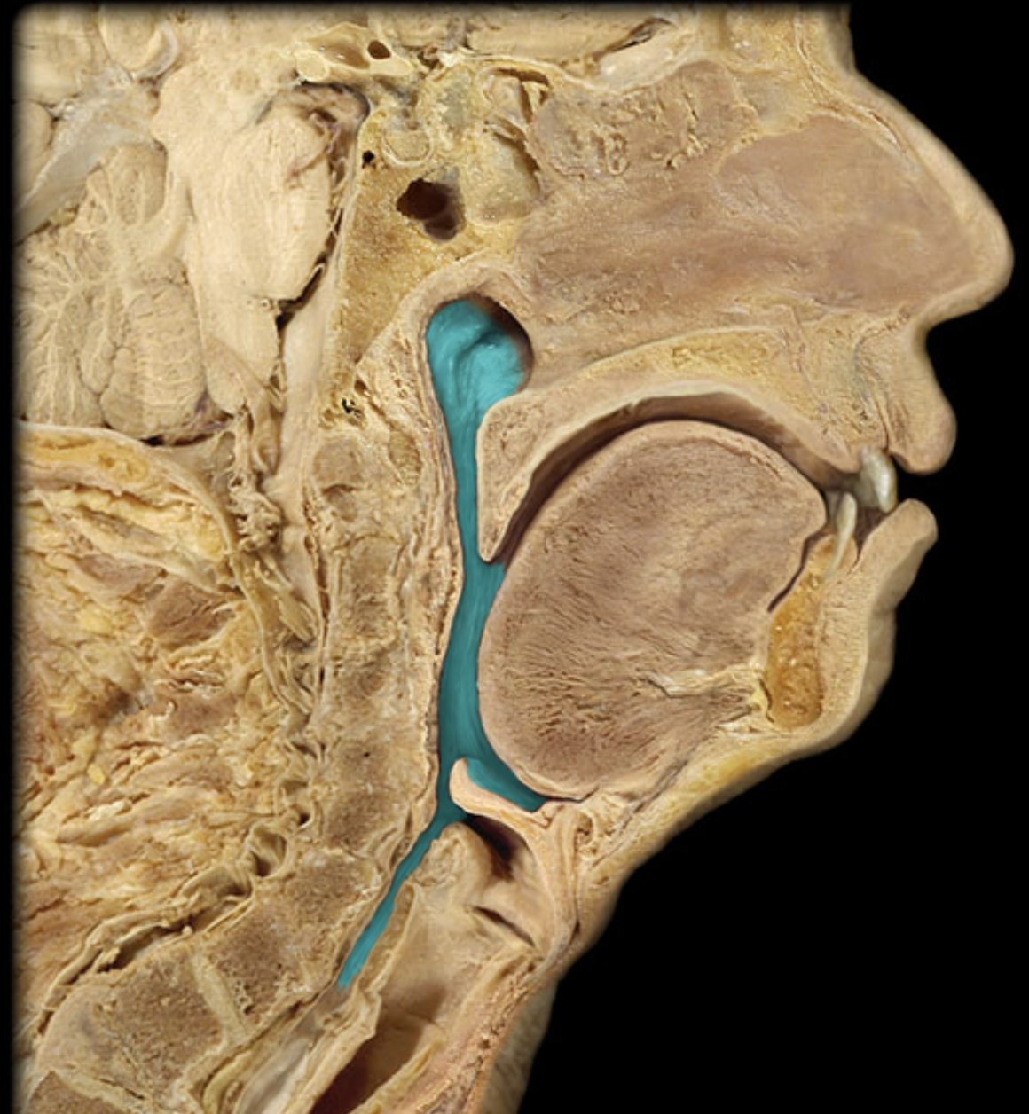
Pharynx
A chamber located behind the oral cavity, nasal cavity, and larynx that serves as a common passageway for air moving to the larynx and food moving to the esophagus; also aids in speech production.

Larynx
An enlargement in the airway superior to the trachea and inferior to the laryngopharynx; transports air, prevents foreign objects from entering the trachea, and houses the vocal cords.

Thyroid Cartilage
the largest laryngeal cartilage, shield-shaped, commonly called the Adam’s apple; forms the anterior and lateral walls of the larynx.

Epiglottis
A flaplike structure supported by epiglottic cartilage that stands upright during normal breathing and folds downward to close off the glottis during swallowing.

False Vocal Cords (Vestibular Folds)
The upper pair of mucosal folds inside the larynx that do not produce sound; help close the glottis during swallowing to prevent food from entering the lower respiratory tract.

True Vocal Cords (Vocal Folds)
The lower pair of mucosal folds in the larynx containing elastic ligaments and muscles; vibrate when air is forced past them to produce vocal sounds.

Trachea (Windpipe)
A flexible cylindrical tube extending downward anterior to the esophagus from the larynx into the thoracic cavity, where it splits into primary bronchi.


Tracheal Cartilaginous Rings
Approximately 20 incomplete, C-shaped hyaline cartilage rings supporting the tracheal wall to prevent airway collapse while allowing esophageal expansion during swallowing.
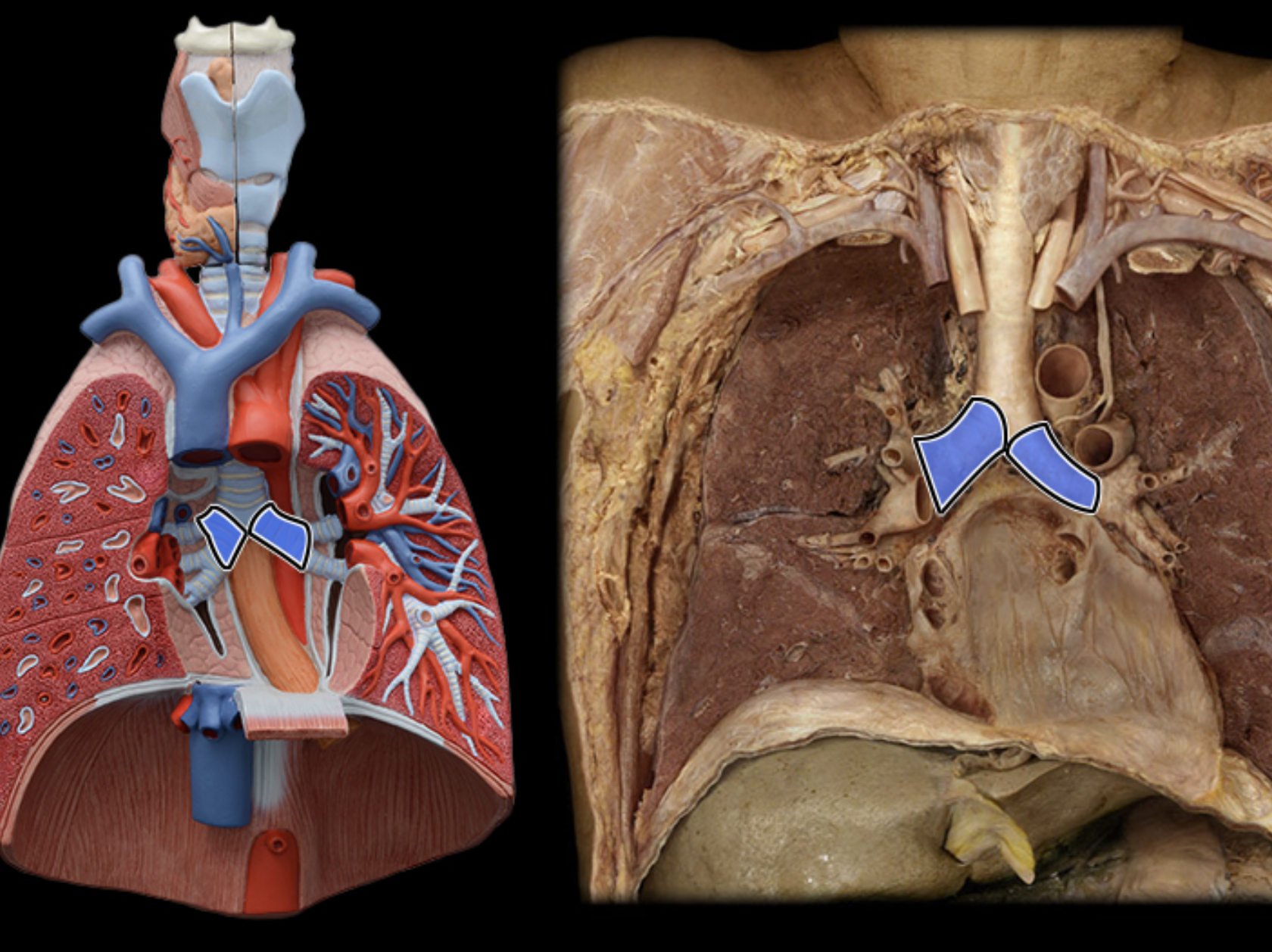
Main (Primary) Bronchus
The direct branch off the tracheal bifurcation that conducts air into the right lung.

Left Main (Primary) Bronchu
The direct branch off the tracheal bifurcation that leads laterally into the left lung.
Lobar (Secondary) Bronchi
Branches of the main bronchi that enter each lobe of the lungs; there are 3 in the right lung and 2 in the left lung (5 total).

Segmental (Tertiary) Bronchi
Airway branches derived from lobar bronchi that supply individual bronchopulmonary segments within each lung lobe.

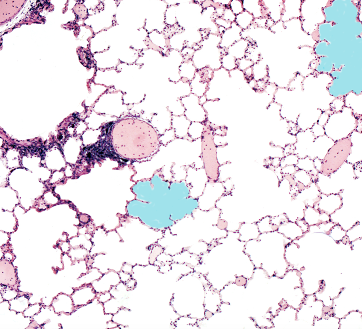
Alveoli
Microscopic air sacs clustered at the distal ends of alveolar ducts; surrounded by extensive capillary networks, serving as the sole sites of gas exchange between atmosphere and blood.

Superior Lobe of Right Lung
The upper division of the right lung, separated from the middle lobe by the horizontal fissure.

Middle Lobe of Right Lung
The intermediate wedge-shaped lobe of the right lung, bounded by the horizontal fissure superiorly and the oblique fissure inferiorly.

Inferior Lobe of Right Lung
The lower, posterior division of the right lung located beneath the oblique fissure.

Horizontal Fissure of Right Lung
The transverse groove that separates the superior lobe from the middle lobe of the right lung.

Oblique Fissure of Right Lung
The diagonal groove dividing the inferior lobe from both the middle and superior lobes of the right lung.
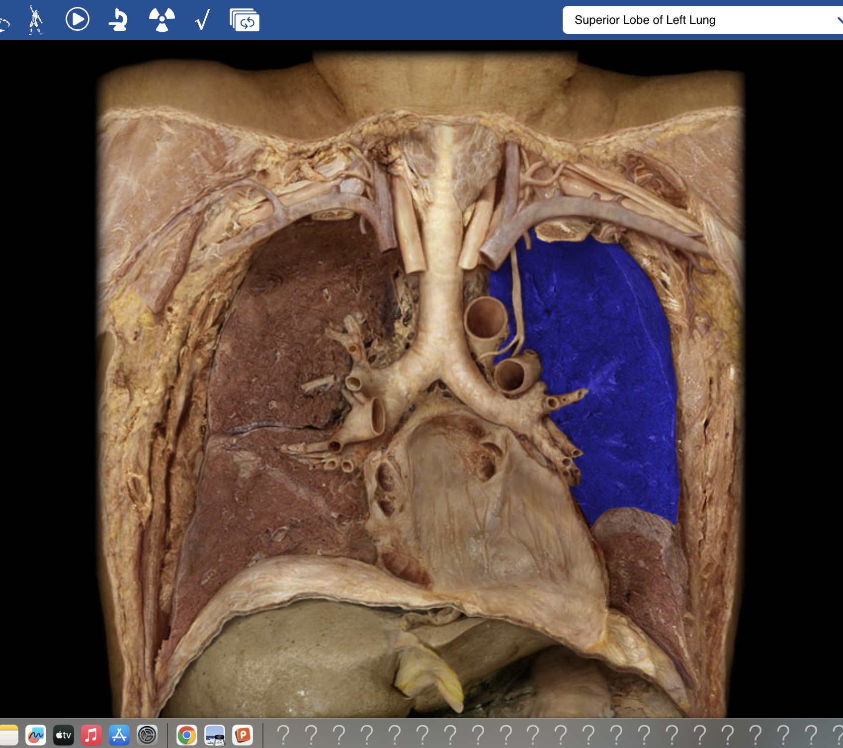
Superior Lobe of Left Lung
The upper lobe of the two-lobed left lung, situated above the oblique fissure.

Inferior Lobe of Left Lung
The posteroinferior lobe of the left lung situated below the oblique fissure.

Oblique Fissure of Left Lung
The single diagonal fissure dividing the left lung into superior and inferior lobes.

Diaphragm
The large dome-shaped primary muscle of respiration that forms the floor of the thoracic cavity; contracts downward to enlarge the thoracic cavity during inspiration.

External Intercostal Muscles
Respiratory skeletal muscles situated between adjacent ribs whose contraction elevates the rib cage to expand thoracic cavity volume.