Muscle practical
 Call Kai
 Learn
 Practice Test
 Spaced Repetition
 Match
 Flashcards
 Knowt Play
1/76

There's no tags or description
Looks like no tags are added yet.
77 Terms
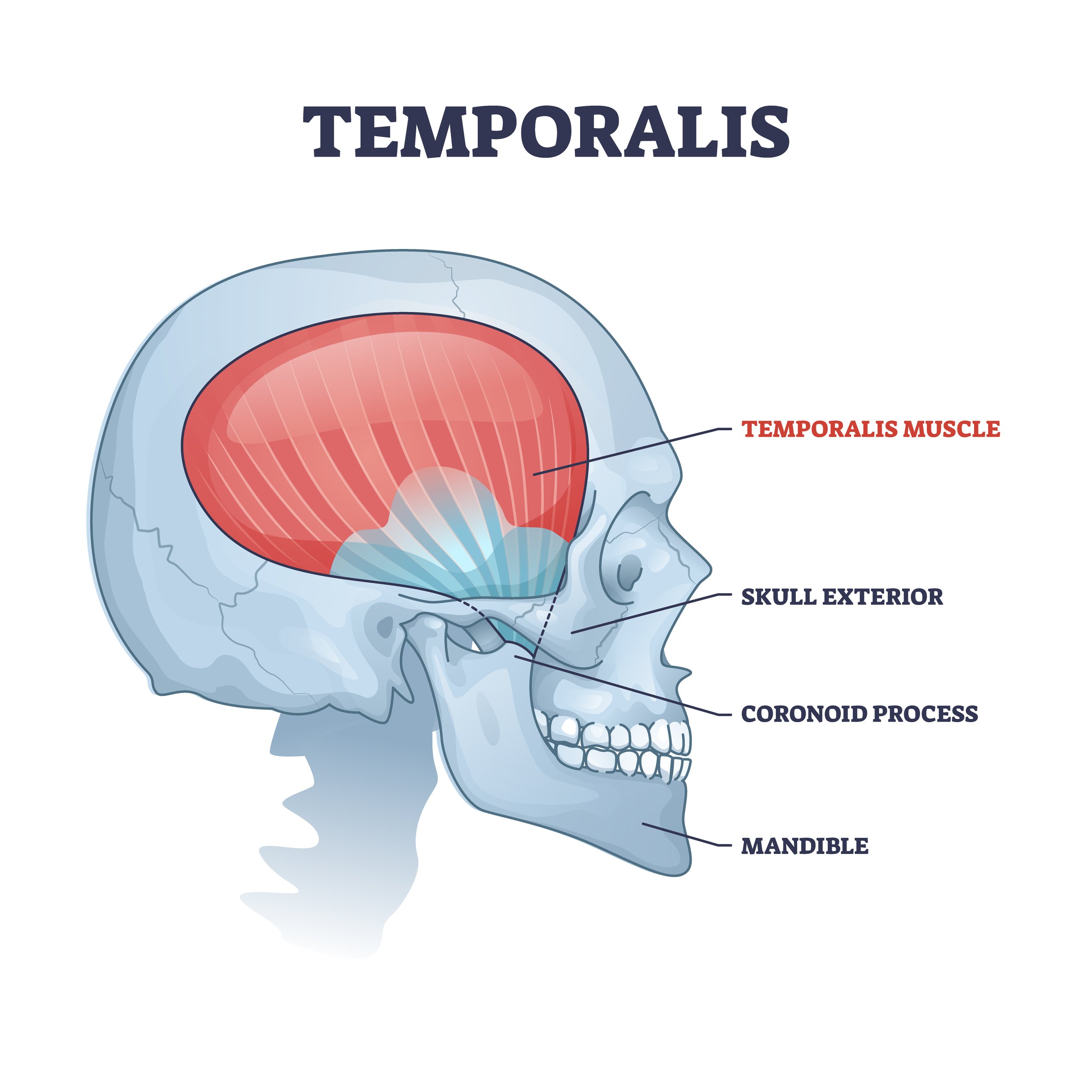
Temporalis
Location:
temporal fossa
Function:
closes jaw
synergist of the masseter
enabling jaw movement for chewing, biting, and grinding food
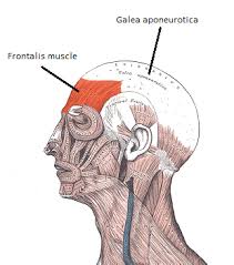
Frontalis
Location:
forehead region of the head
Function:
raise eyebrows
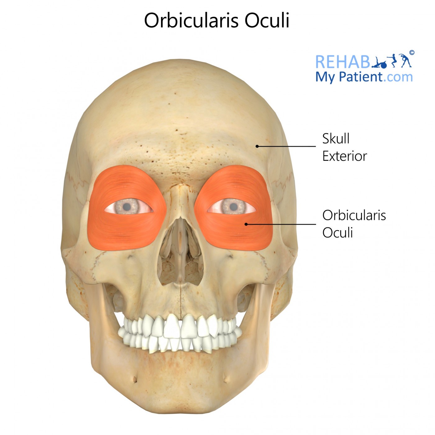
Orbicularis oculli
Location:
beneath the eyelid skin
Function:
close eyes, squints, blinks, wink
to close the upper and lower lids
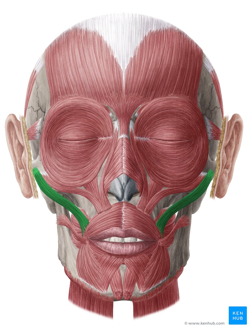
Zygomaticus
Location:
lateral surface of the zygomatic bone\
anterior to the zygomaticotemporal suture
lateral to the origin of zygomaticus minor
Function:
raises corners of the mouth
raises the upper lip to bare the upper teeth
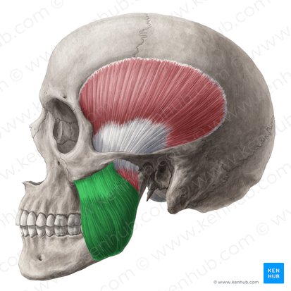
Masseter
Location:
below the cheekbone
the side of the face
Function:
closes jaw
elevate the mandible
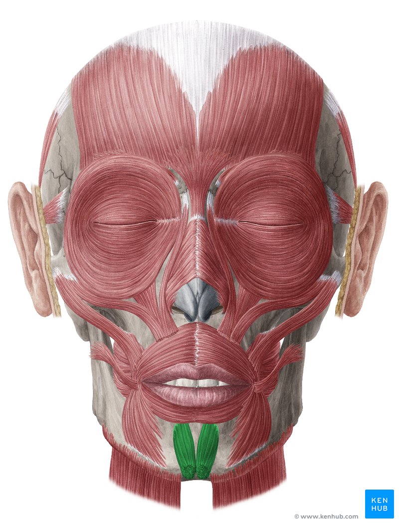
Mentalis
Location:
on the mandible
each side of the frenulum of the lower lip
Function:
raise the skin of the chin and elevate
protrude the lower lip
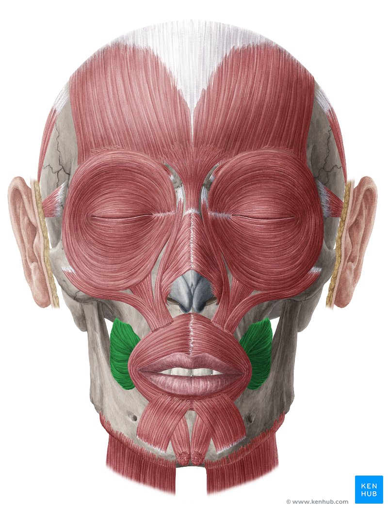
Buccinator
Location:
cheeks
interval between the maxilla and the mandible
Function:
holding food in place
compresses the cheeks
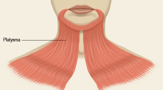
Platysma
Location:
neck
Function:
depress the lower jaw
pull down the corners of the mouth and lower lip
contribute to various facial expressions
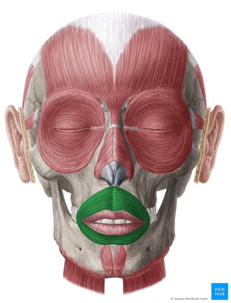
Orbicularis oris
Location:
around the mouth
lips
Function:
closes mouth
protrudes the lips
contributes to the form and shape of the lips
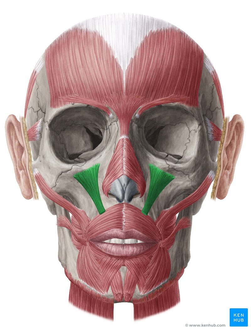
Levator labii superioris
Location:
begins at the infraorbital margin of the maxilla and extends toward the upper lip
originates on the maxilla and zygomatic bone
Function:
elevates and everts the upper lip
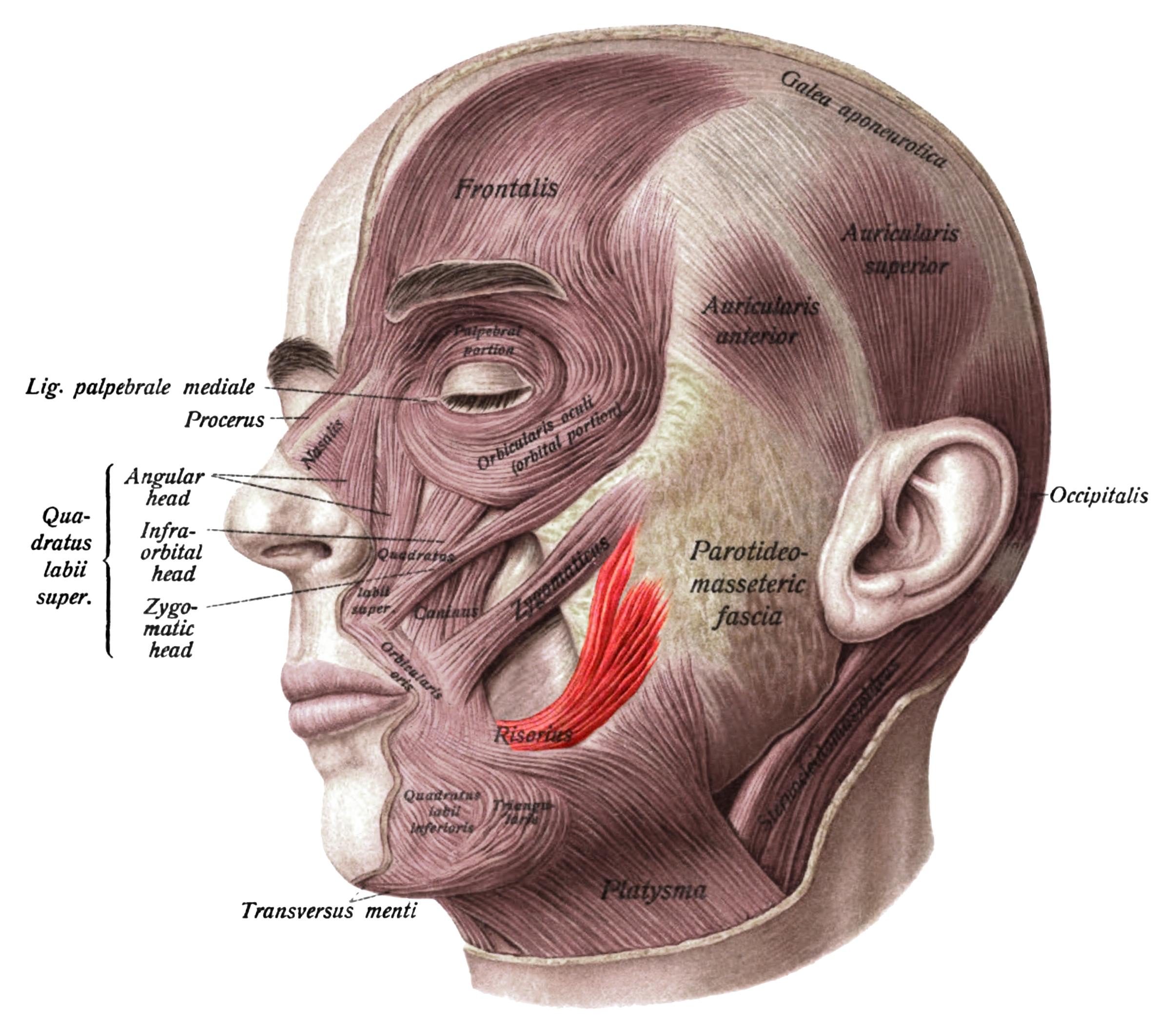
Risorius
Location:
either side of the lips
superficial layer of the facial muscles
Function:
pulls the corners of the mouth laterally
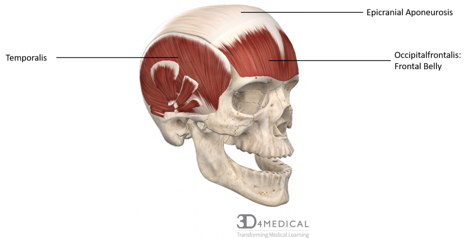
Epicranial aponeurosis
Location:
extends over the cranium
upper part of the skull
Function:
help support a muscle in your skull that controls your facial expressions
stabilizing layer that allows for movement of the skin over the skull and plays a role in facial expressions
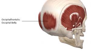
Occipital belly
Location:
back of the skull
Function:
retracts the scalp
move the eyebrows
wrinkle the forehead
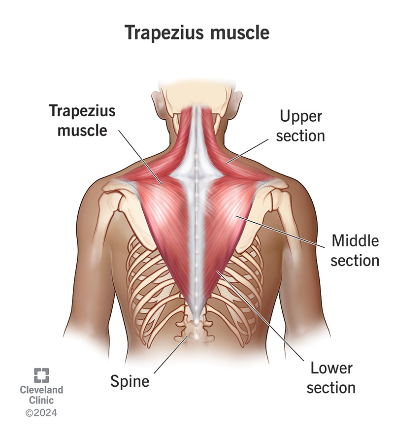
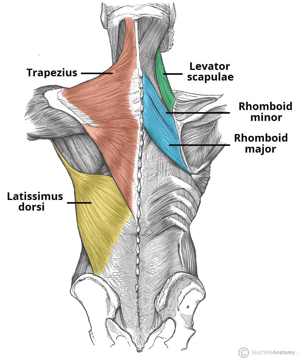
Trapezius
Location:
upper back
extending from the base of the skull to the mid-back
Function:
stabilize and move the scapula
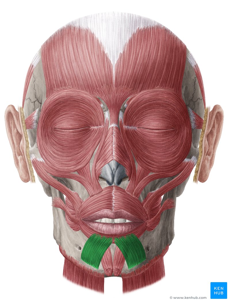
Depressor Labii inferioris
Location:
chin region of the face
Function:
allows you to pull your lower lip down or to the side
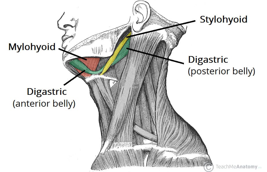
Digastric
Location:
neck
under the jaw
Function:
depress the mandible (lower jaw) for opening the mouth
elevates the hyoid bone
crucial for swallowing and speech
raise the tongue base
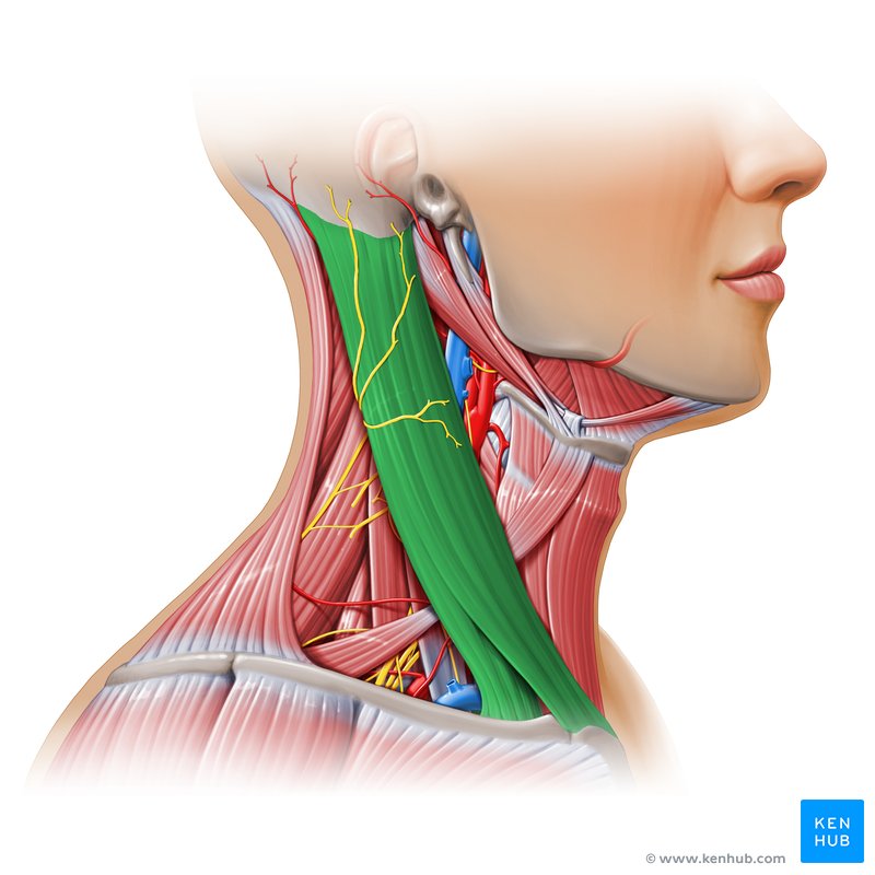
Sternocleidomastoid
Location:
the sides of the neck
down both sides of your neck until it ends at your sternum or clavicle
extends from the base of your skull
Function:
allows for head flexion and rotation
expanding the chest to aid in deep breathing
bend your neck and turn and tilt your head
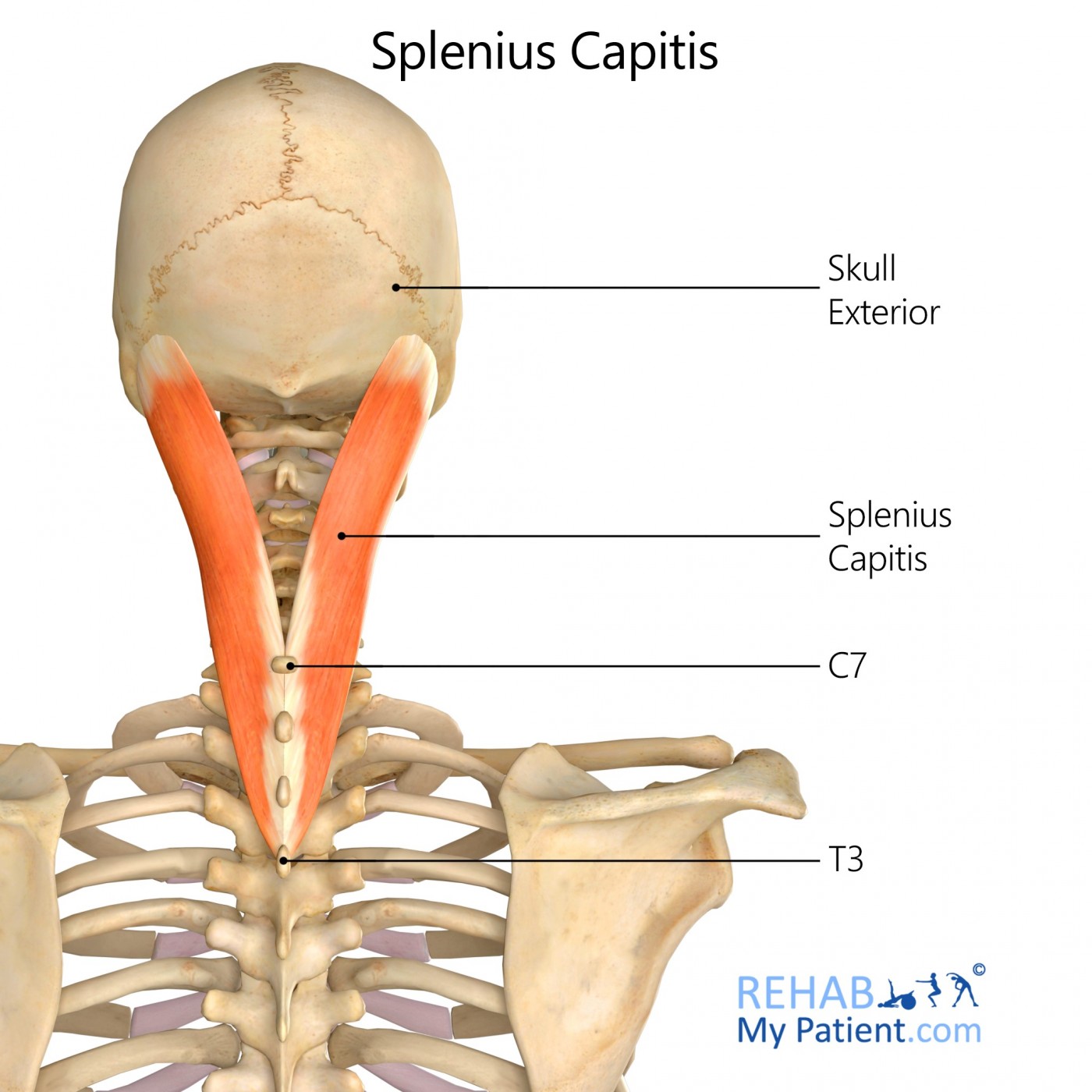
Splenius capitis
Location:
posterior and lateral parts of the neck
Function:
assists in supporting the head in the erect position
primarily to extend the head and neck
assists in lateral flexion and rotation
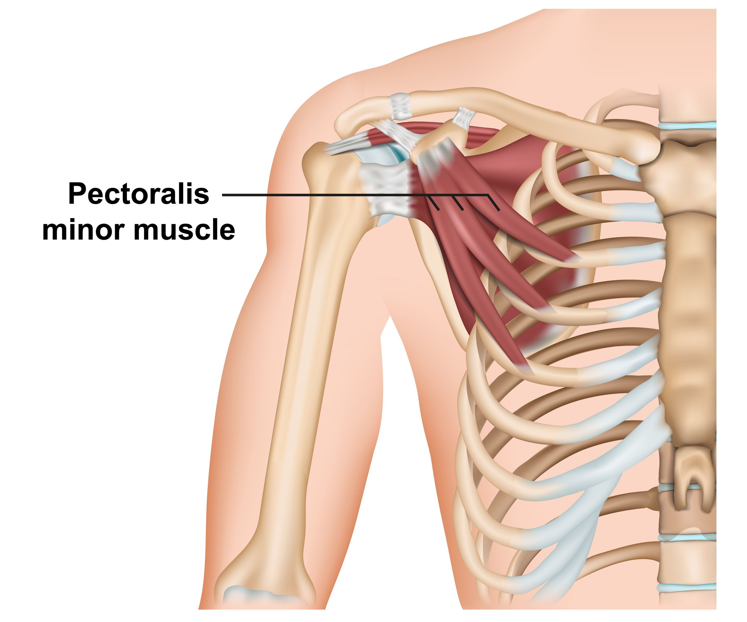
Pectoralis minor
Location:
upper part of the chest
beneath the pectoralis major
Function:
stabilization of the scapula
assist in rib elevation during deep inspiration
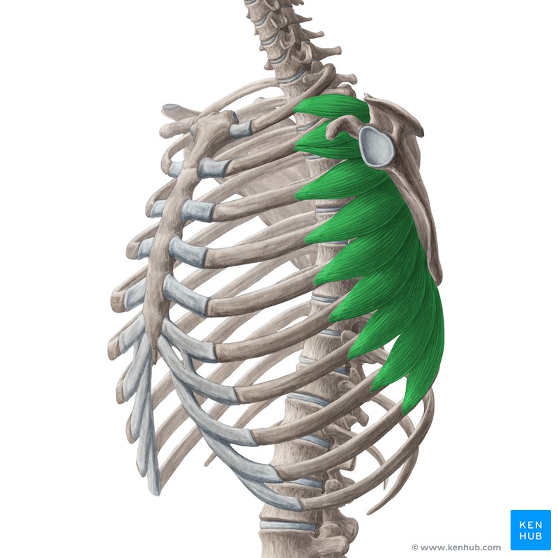
Serratus anterior
Location:
anterior chest wall
Function:
lift the ribs
assisting with respirations
scapula movement, including protraction and upward rotation
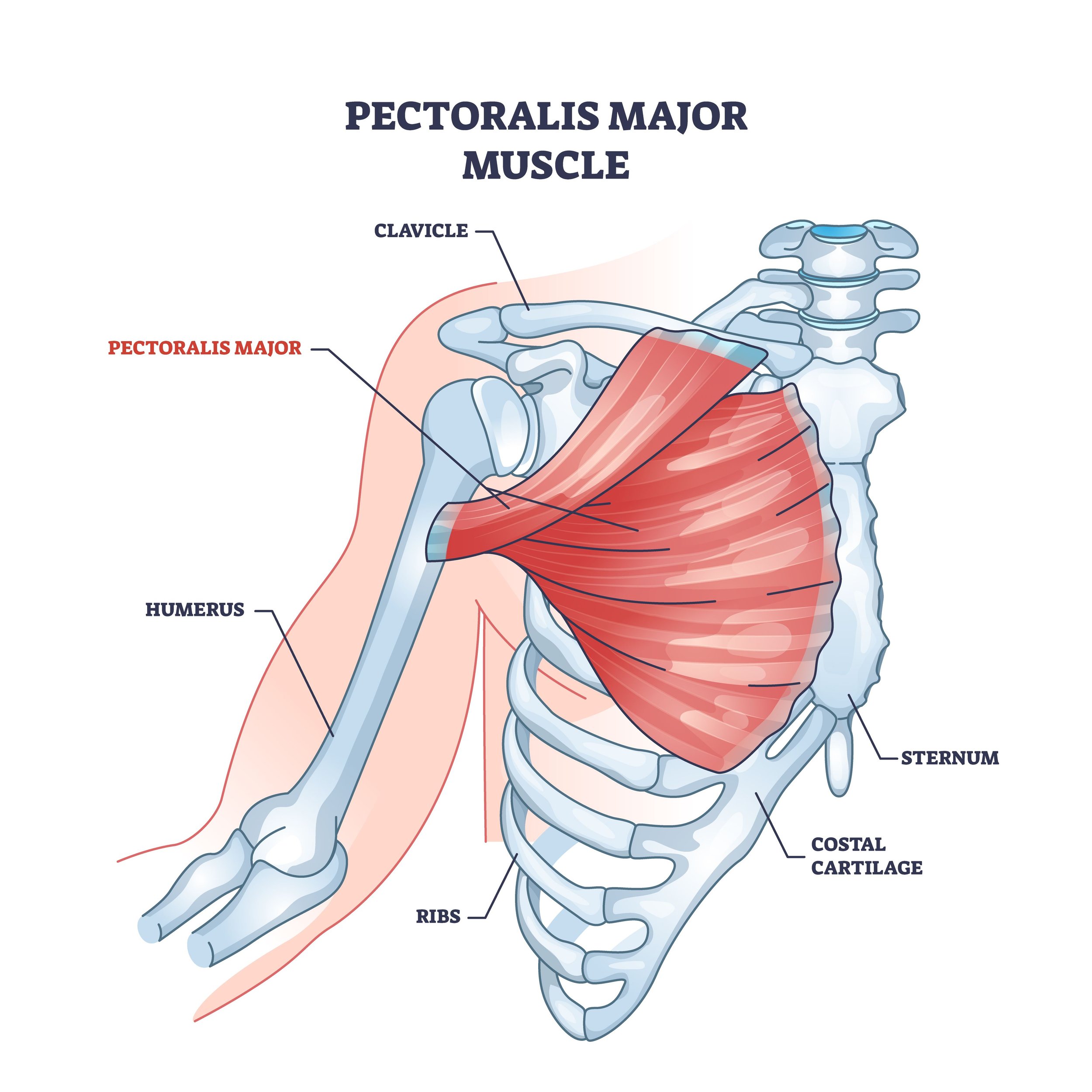
Pectoralis major
Location:
anterior chest wall
both sides of the sternum
Function:
adduction and internal rotation of the arm in the shoulder joint
internally rotating the humerus at the shoulder joint
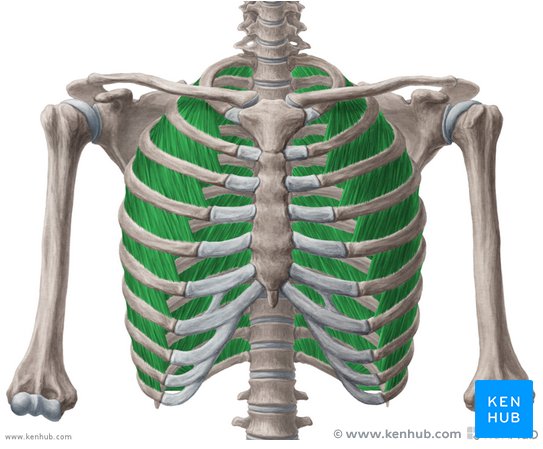
Intercostals
Location:
the spaces between the ribs, spanning the 11 intercostal spaces
rib cage
Function:
help form and move the chest wall
assisting in both inhalation and exhalation by elevating and depressing the rib cage
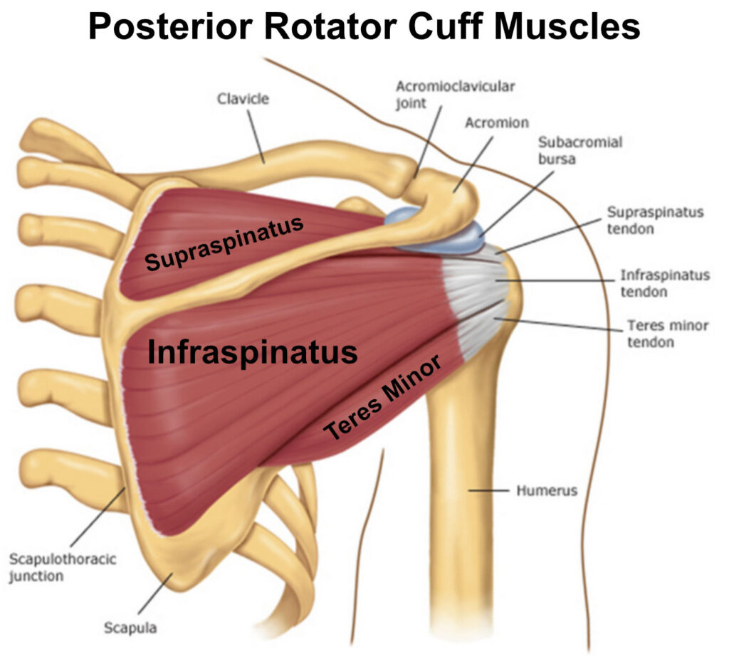
Infraspinatus
Location:
dorsal surface of the scapula
deep to trapezius
Function:
externally rotate the humerus
stabilize the shoulder joint
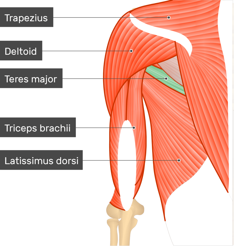
Teres major
Location:
along the lateral border of the scapula
Function:
extend, adduct, and internally rotate the humerus
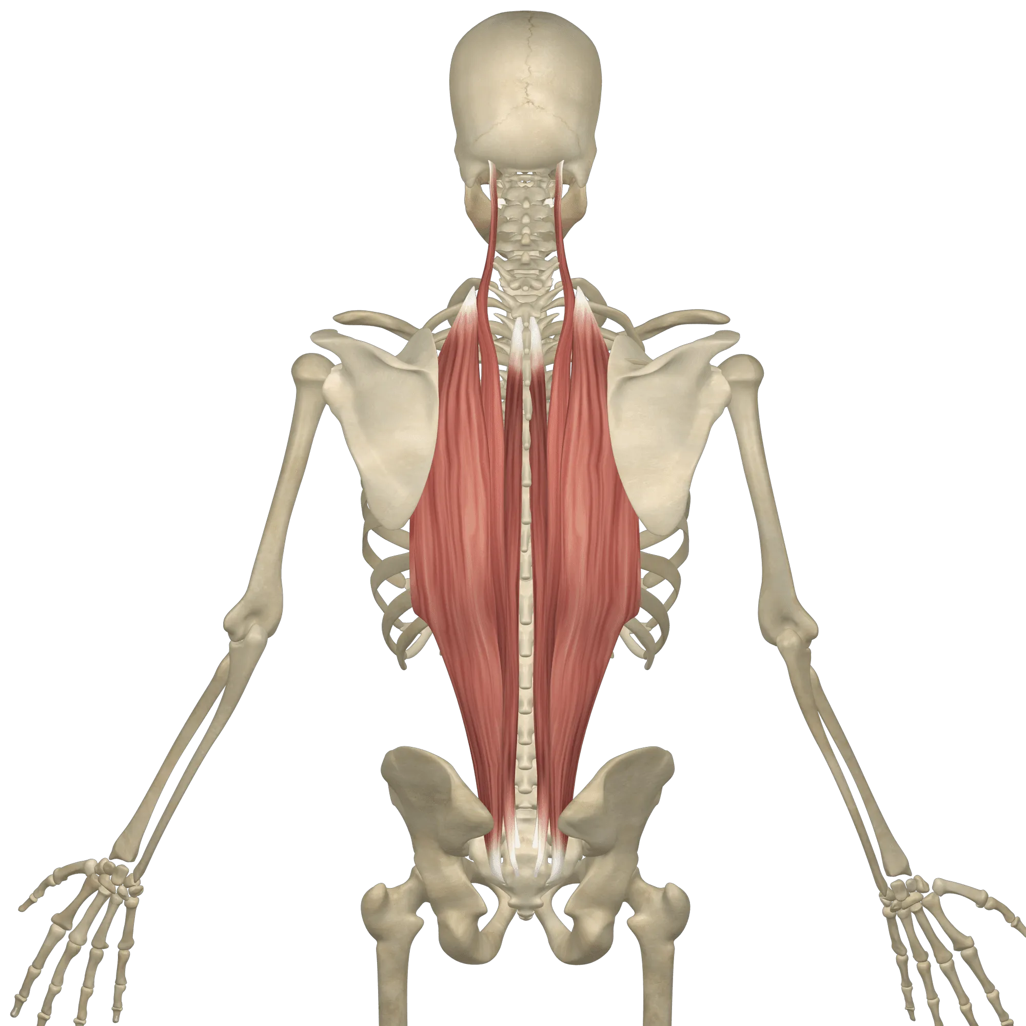
Erector spinae
Location:
either side of the vertebral column
between the base of the cranium superiorly and pelvis inferiorly
Function:
extend and laterally flex the spine
maintain correct posture and curvature of the spinal column
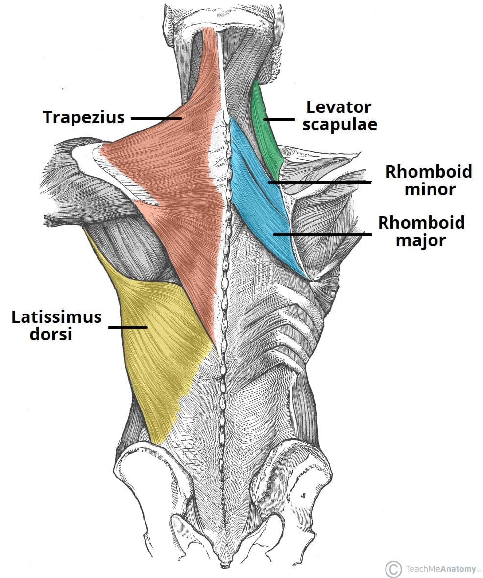
Rhomboid minor
Location:
deep to the trapezius
superior to the rhomboid major
Function:
retracts the scapula when trapezius is contracted
contributing to shoulder stability and proper movement
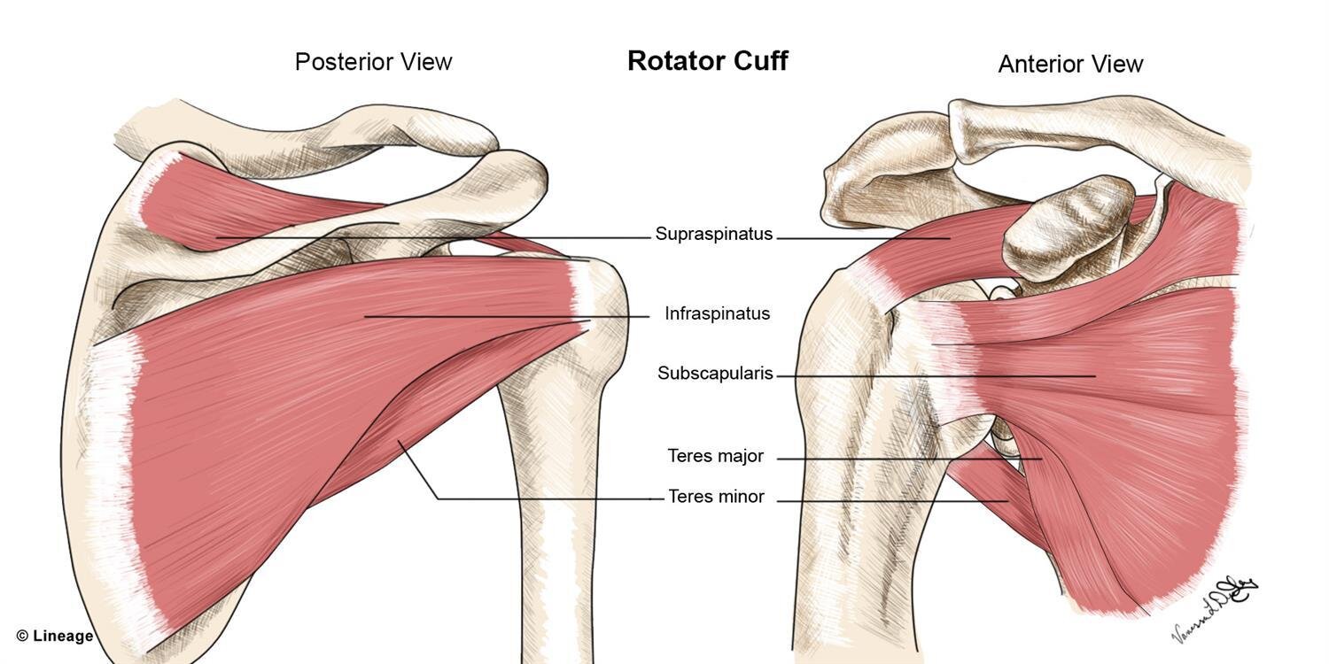
Supraspinatus
Location:
upper portion of the back
in the supraspinous fossa of the scapula
Function:
stabilization of the head of humerus in the joint
initiate and assist in arm abduction
Latissimus dorsi
Location:
superficially in the lower two-thirds of the trunk
extending from the shoulder blades to the lower spine
Function:
pull the inferior angle of the scapula in various directions
producing movements on the shoulder joint
extend, adduct, and internally rotate the arm at the shoulder joint
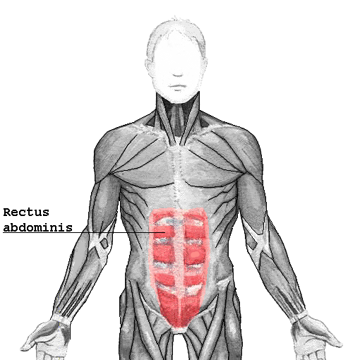
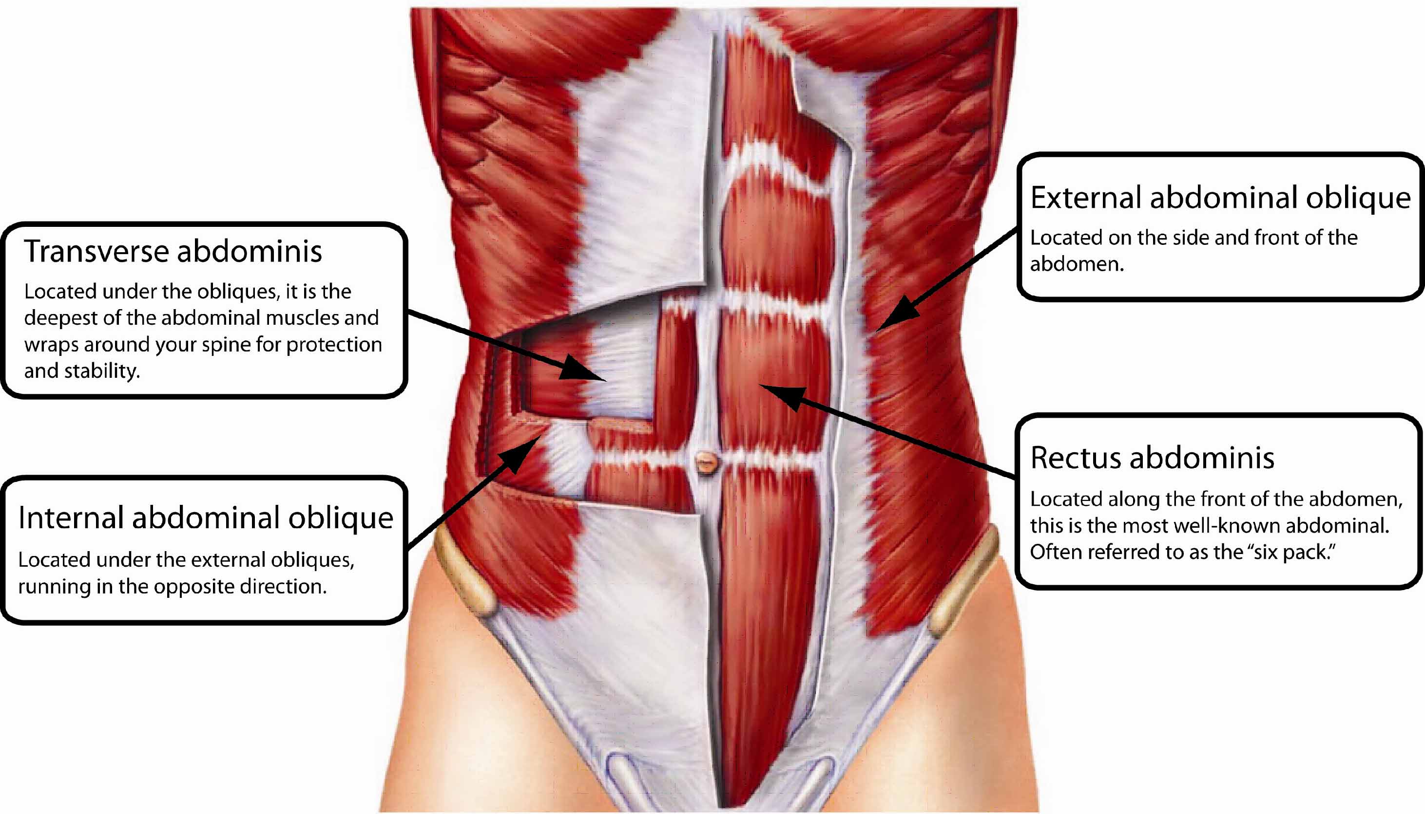
Rectus abdominis
Location:
the middle of your abdomen
midline of the anterior abdominal wall
running vertically from the ribs to the pelvis
Function:
flex the spine
stabilizing the pelvis
assisting in movements like bending and twisting the torso
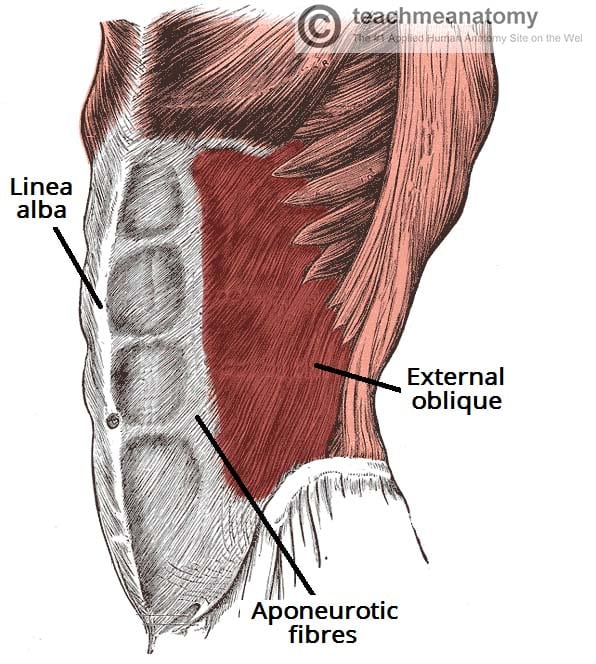
External oblique
Location:
the outer surface of the sides of the abdomen
each side of the rectus abdominis
Function:
movement of the trunk, spine, and core
rotate the trunk to the opposite side
assist in bending the trunk from side to side
stabilize the core
Transverse abdominis
Location:
in the abdomen
wrapping around the torso from front to back
spans from the lower ribs down to the pelvis
Function:
stabilizing the core
supporting the spine and pelvis
maintaining internal abdominal pressure
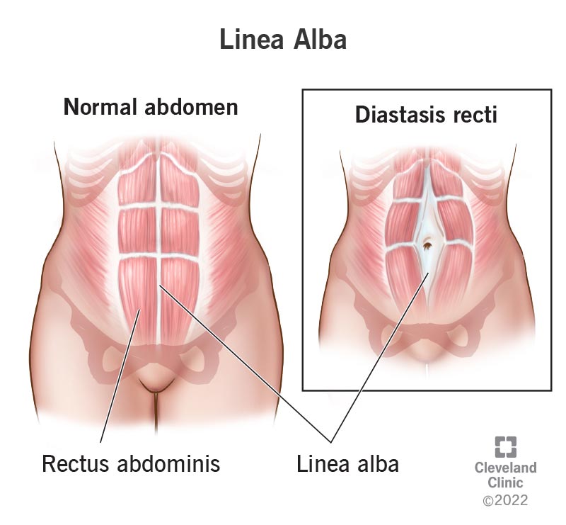
Linea Alba
Location:
front of the abdomen
Function:
keep the right and left abdominal muscles separated
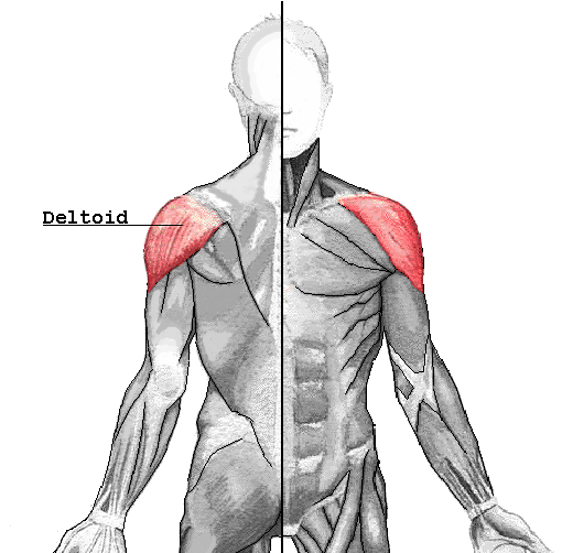
Deltoid
Location:
the upper arms and shoulders
Function:
stabilizing the shoulder joint
shoulder abduction
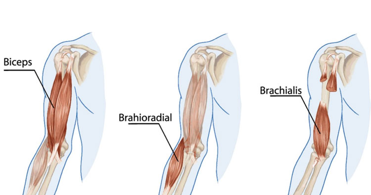
Brachialis
Location:
beneath the biceps brachii
lower anterior compartment of the upper arm
Function:
flex the forearm at the elbow joint
provides elbow flexion
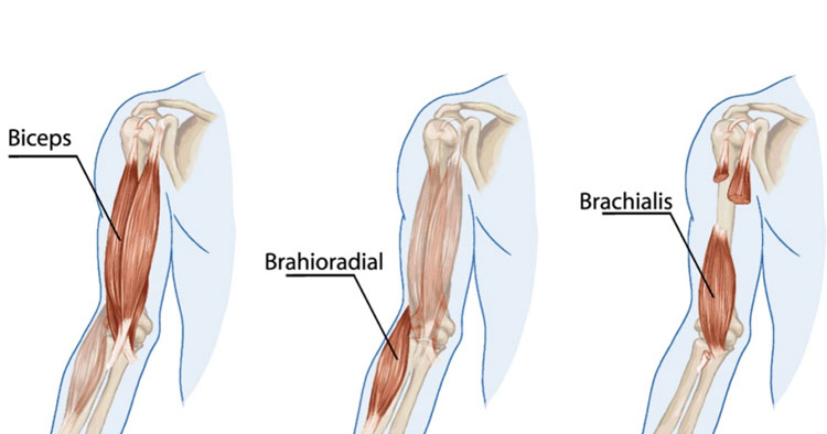
Biceps
Location:
front of the upper arm
between the shoulder and the elbow
Function:
flexion and supination of the forearm
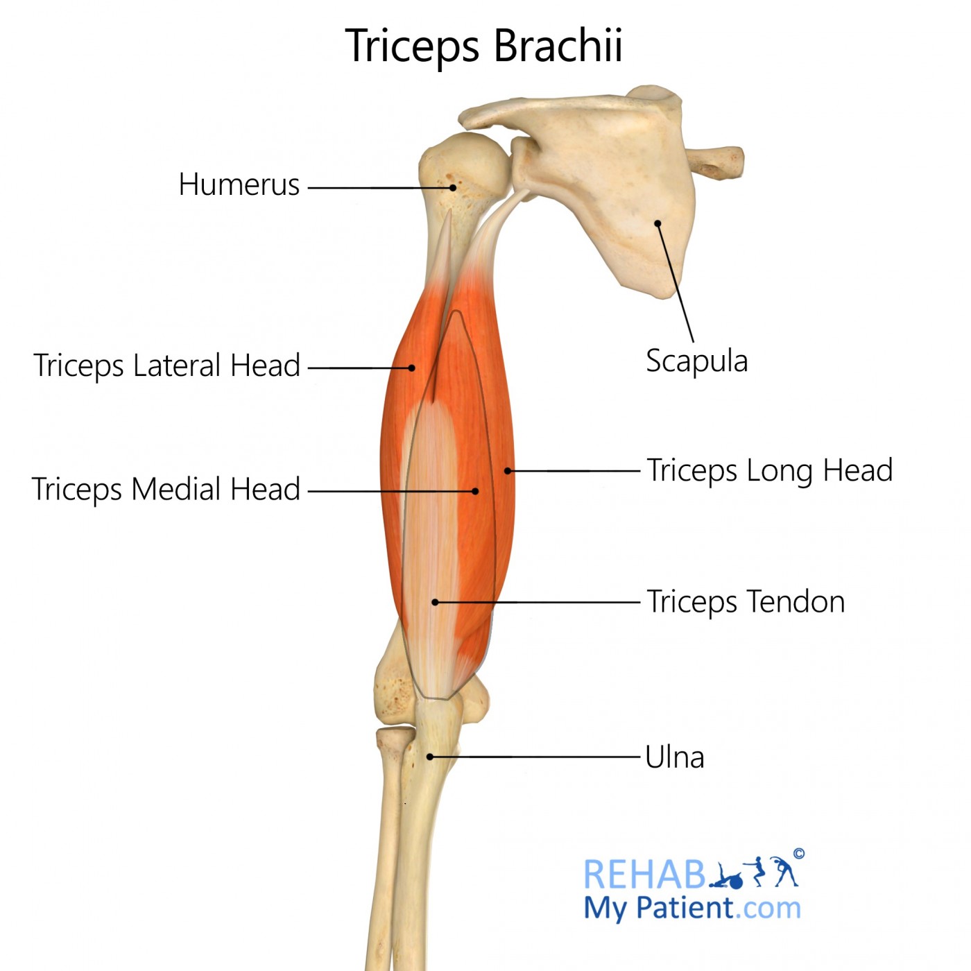
Triceps
Location:
dorsal part of the upper arm
between the shoulder and elbow joints
Function:
extend the forearm at the elbow joint
straightening the arm
helps stabilize the shoulder joint
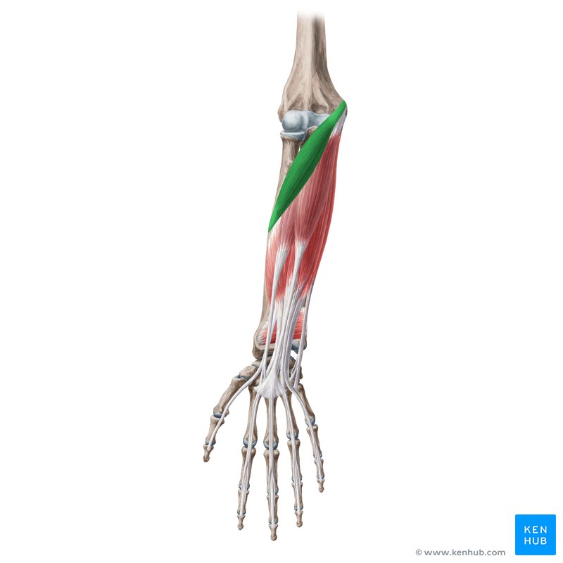
Pronator Teres
Location:
anterior aspect of the forearm
Function:
pronates the forearm
assists in flexion of the elbow joint
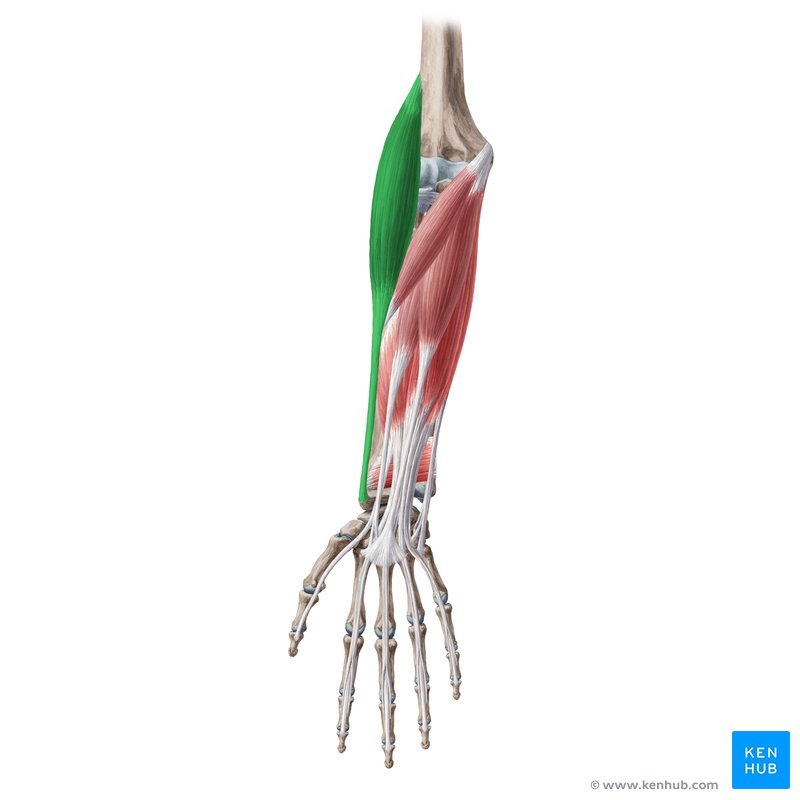
Brachioradialis
Location:
the radial side of the forearm
Function:
flexes the forearm at the elbow
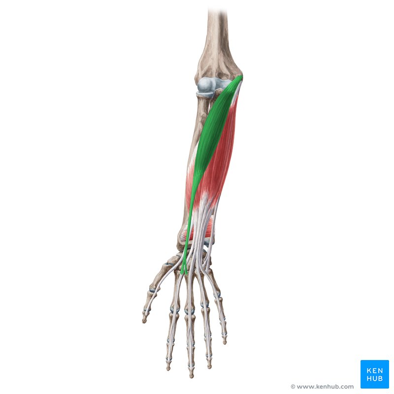
Flexor carpi radialis
Location:
anterior part of the forearm
Function:
flex and abduct the wrist
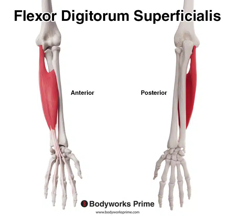
Flexor digitorum superficialis
Location:
anterior aspect of the forearm
anterior compartment of the forearm
Function:
flexes the middle phalanges of the four fingers
assists in flexing the metacarpophalangeal (MCP) joints and the wrist
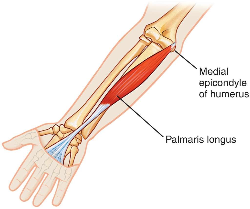
Palmaris longus
Location:
anterior forearm
between the flexor carpi radialis and flexor carpi ulnaris muscles
Function:
assists in wrist flexion
tightening the palmar aponeurosis
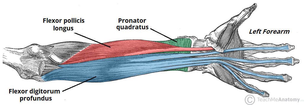
Flexor pollicis longus
Location:
Function:
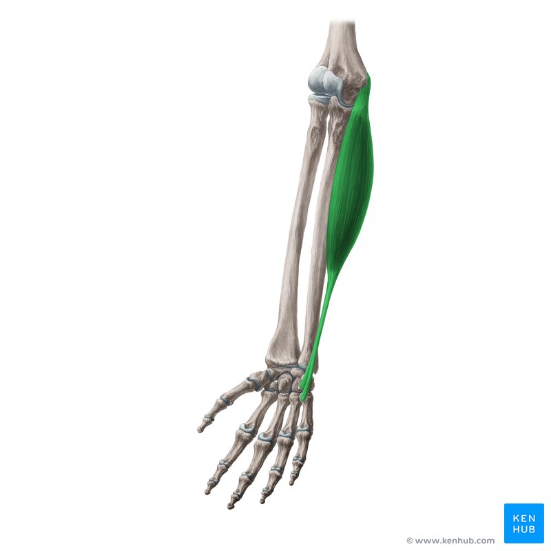
Flexor carpi ulnaris
Location:
within the deep compartment of the anterior forearm
originates on the anterior surface of the radius
Function:
flexion of the thumb at the interphalangeal joint
flex the thumb at the interphalangeal (IP) and metacarpophalangeal (MCP) joints
contributes to wrist flexion
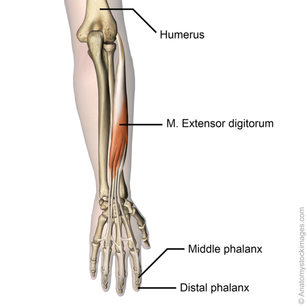
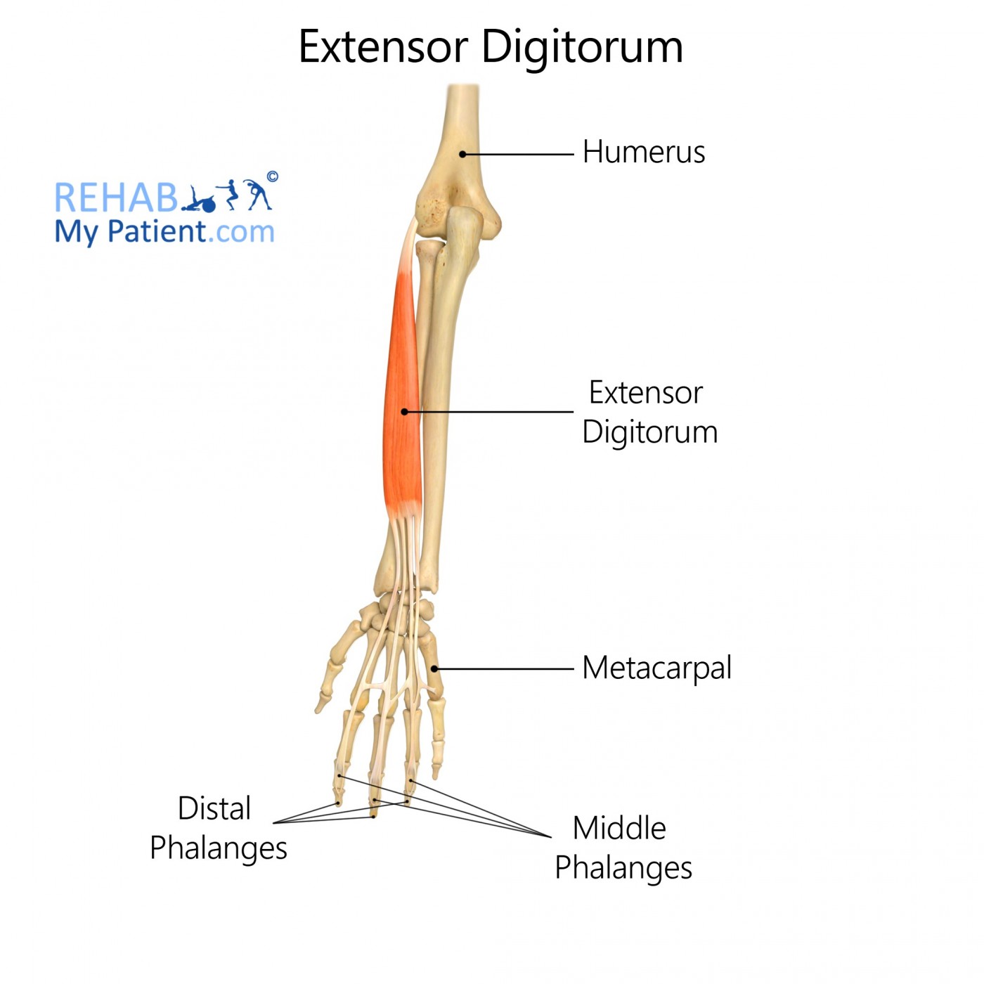
Extensor digitorum
Location:
posterior compartment of the forearm
within the superficial layer of muscles
Function:
extends the phalanges, then the wrist, and finally the elbow.
extend the four medial fingers
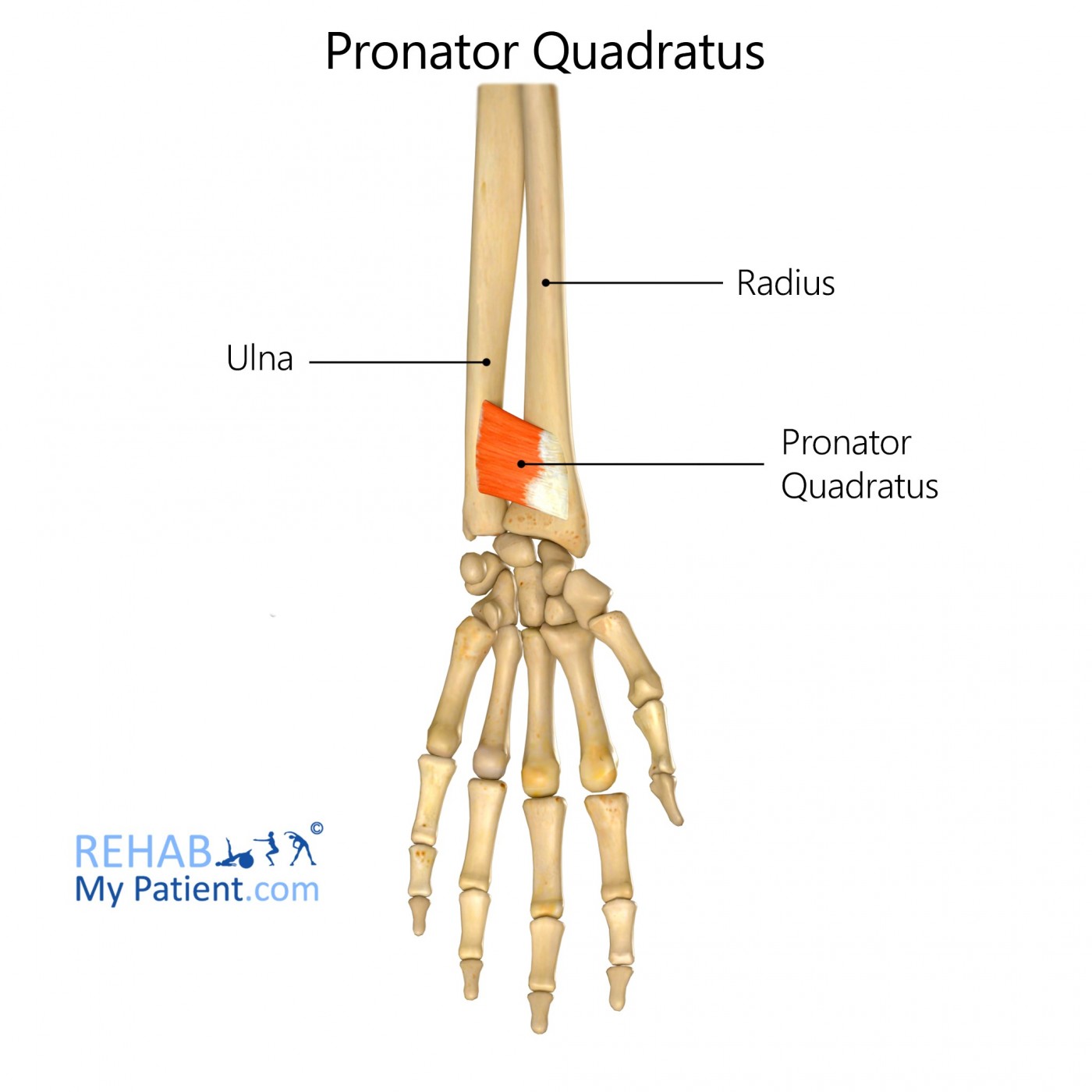
Pronator quadratus
Location:
within the anterior compartment of forearm
Function:
pulls the lateral side of the radius towards the ulna, thus pronating the hand
pronate the forearm
turning the palm downwards
stabilize the connection between the radius and ulna
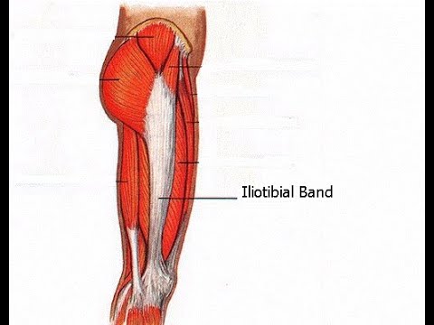
Iliotibial tract (IT Band)
Location:
along the lateral thigh
Function:
pelvic stabilisation and posture control
stabilizing the hip and knee during walking and running
contributes to lateral knee support
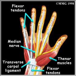
Thenar muscles of thumb
Location:
the base of the thumb on the palm (volar) side of the hand
Function:
produce the movements of the thumb
enabling fine motor control and precision grasping
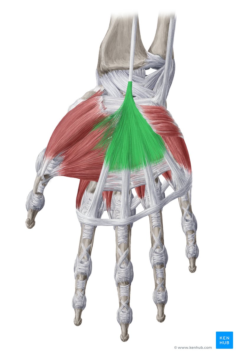
Palmar aponeurosis
Location:
the palm of the hand
Function:
improve grip
protect underlying tendons, vessels, and nerves, and provide a firm attachment to the skin
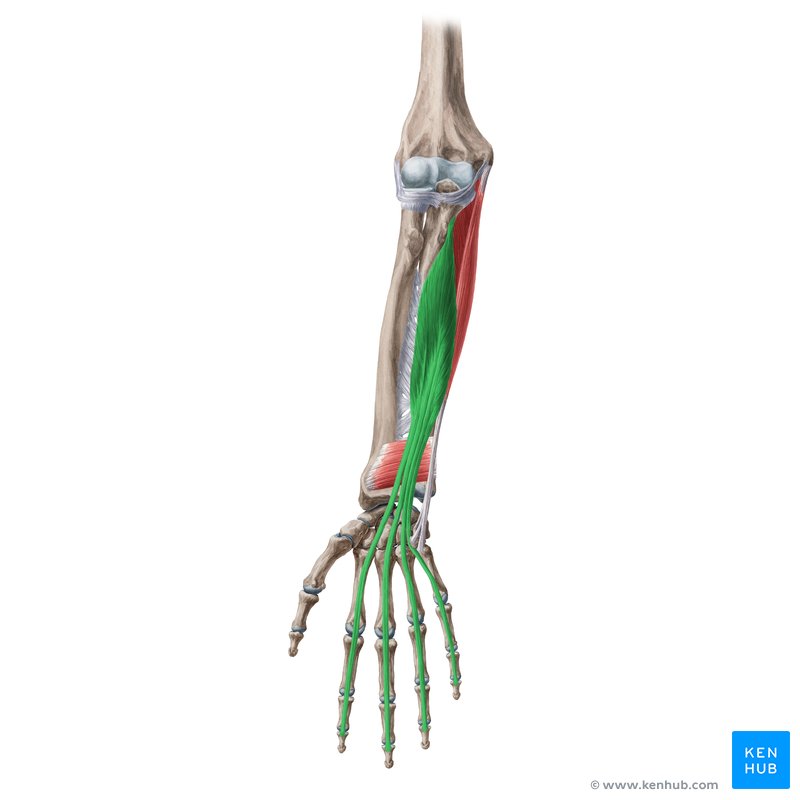
Flexor digitorum profundus
Location:
deep in the forearm flexor compartment
Function:
lexes the distal phalanges of the medial 4 digits at the DIP joints
assists in flexing the hand and wrist
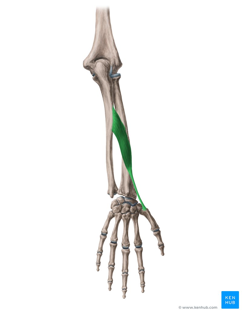
Abductor pollicis longus
Location:
below the supinator
posterior compartment of the forearm
Function:
abduct the thumb at the carpometacarpal joint
moving the thumb anteriorly
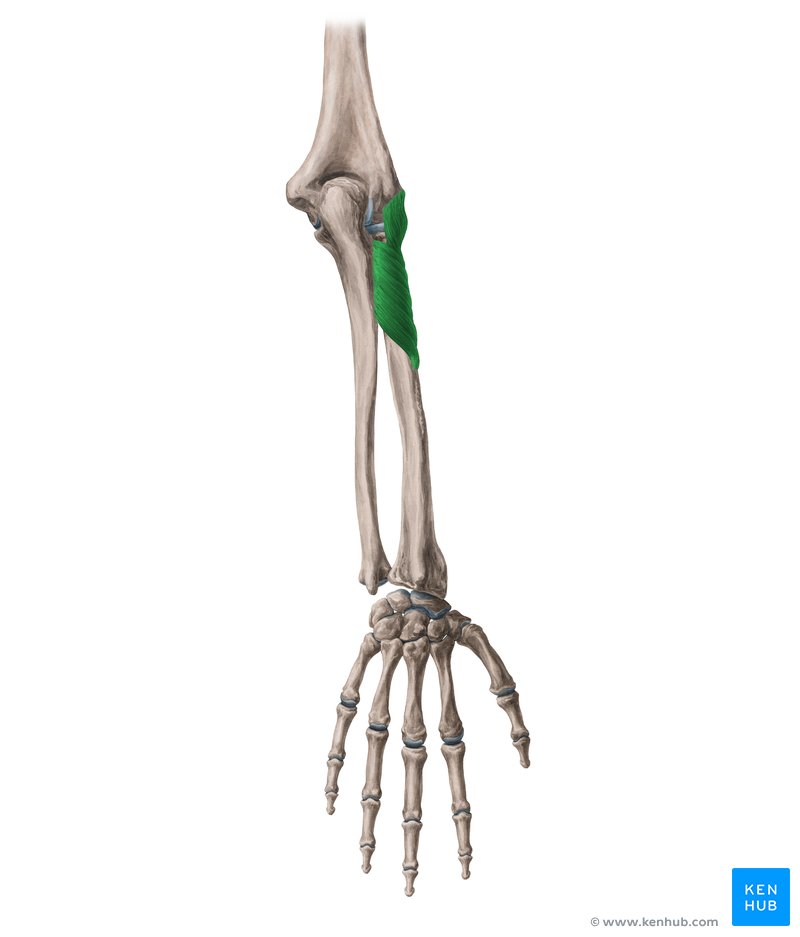
Supinator
Location:
superior and posterior compartment of the forearm
curving around the upper third of the radius
Function:
to supinate the forearm
rotating it so the palm faces upwards
assists with forearm flexion
Tendons of extensor digitorum
Location:
pass through the 4th extensor compartment at the wrist
Function:
extends medial four digits at the metacarpophalangeal joints and secondarily at the interphalangeal joints
extending the medial four digits (fingers 2-5) at both the metacarpophalangeal (MCP) and interphalangeal (IP) joints
contribute to wrist extension
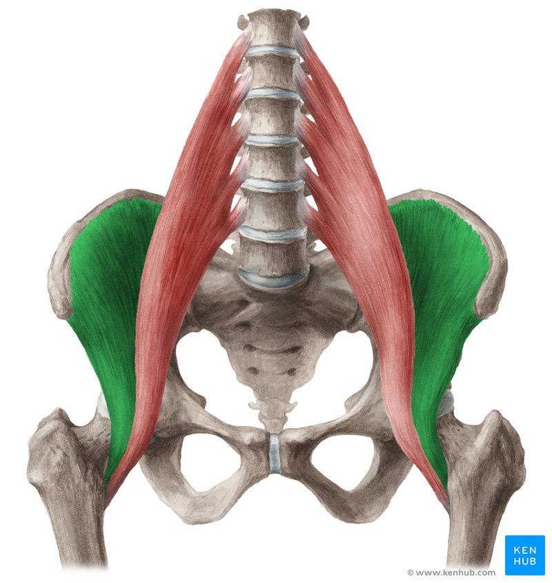
Illiacus
Location:
attached to the iliac fossa
Function:
flexes and externally rotates the femur
flex the thigh at the hip joint
contribute to hip stabilization and posture
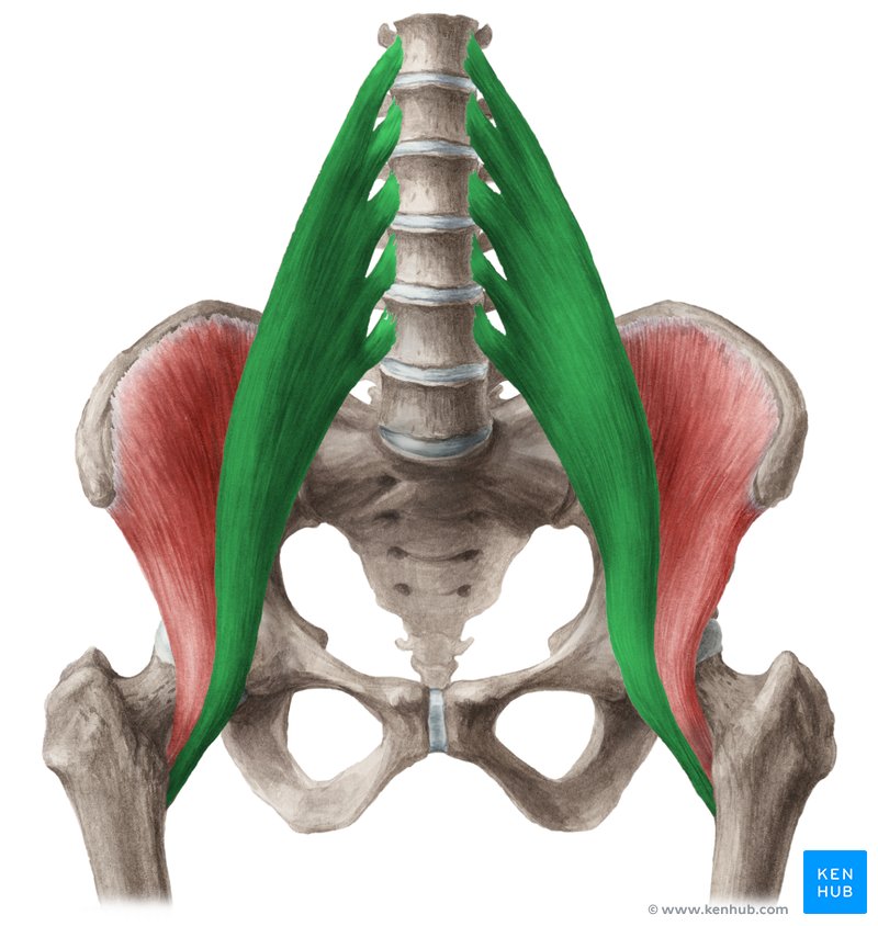
Psoas major
Location:
lateral to the lumbar vertebrae
Function:
flex the thigh at the hip
flex the trunk at the hip
laterally rotate the thigh
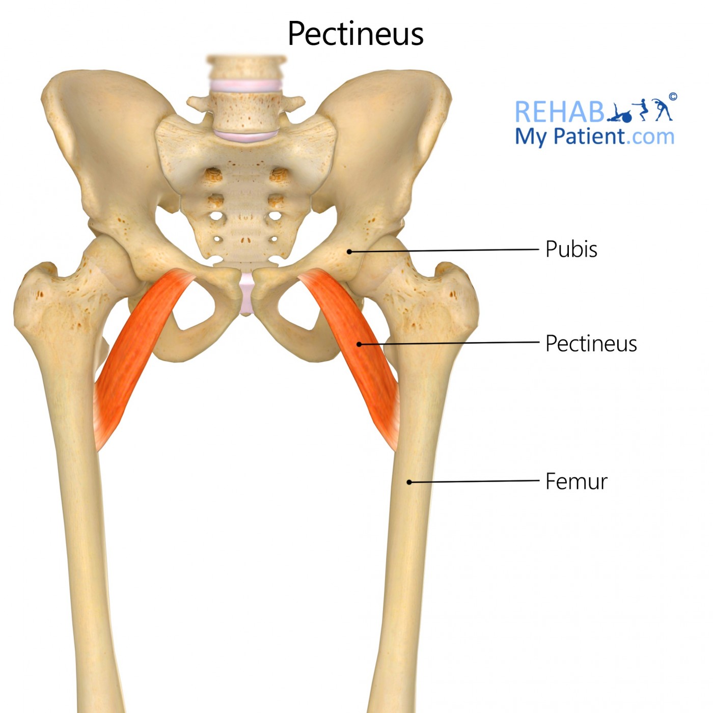
Pectineus
Location:
medial compartment of the thigh
anterior and medial part of the upper thigh
Function:
flex and adduct the hip joint
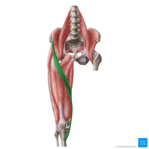
Sartorius
Location:
anterior compartment of the thigh
Function:
At the hip it flexes, weakly abducts, and rotates the thigh laterally
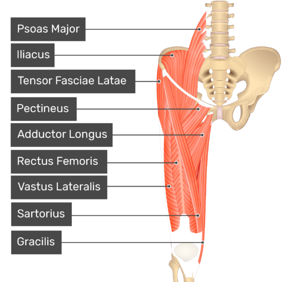
Adductor longus
Location:
medial aspect of the thigh
Function:
adduct the thigh at the hip joint
contributes to hip flexion and potentially internal rotation
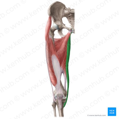
Gracilis
Location:
medial compartment of the thigh
Function:
adduct the thigh at the hip joint and flex
internally rotate the knee
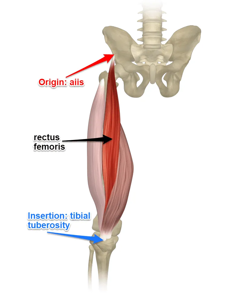
Rectus femoris
Location:
anterior thigh compartment's
Function:
extend the knee
assists iliopsoas in hip flexion
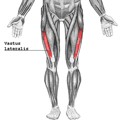
Vastus lateralis
Location:
lateral side of the thigh
outer anterior compartment of the thigh
Function:
extend the knee joint
promote movement as well as strength and stability
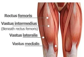
Vastus medialis
Location:
anterior compartment of the thigh
medial side of the thigh
Function:
involved in knee extension
extend the knee joint
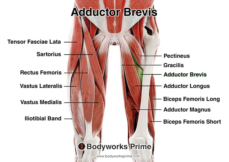
Adductor brevis
Location:
medial compartment of the thigh
Function:
adduction of the thigh
adduct (pull) the thigh towards the midline of the body
contributes to hip flexion and lateral rotation
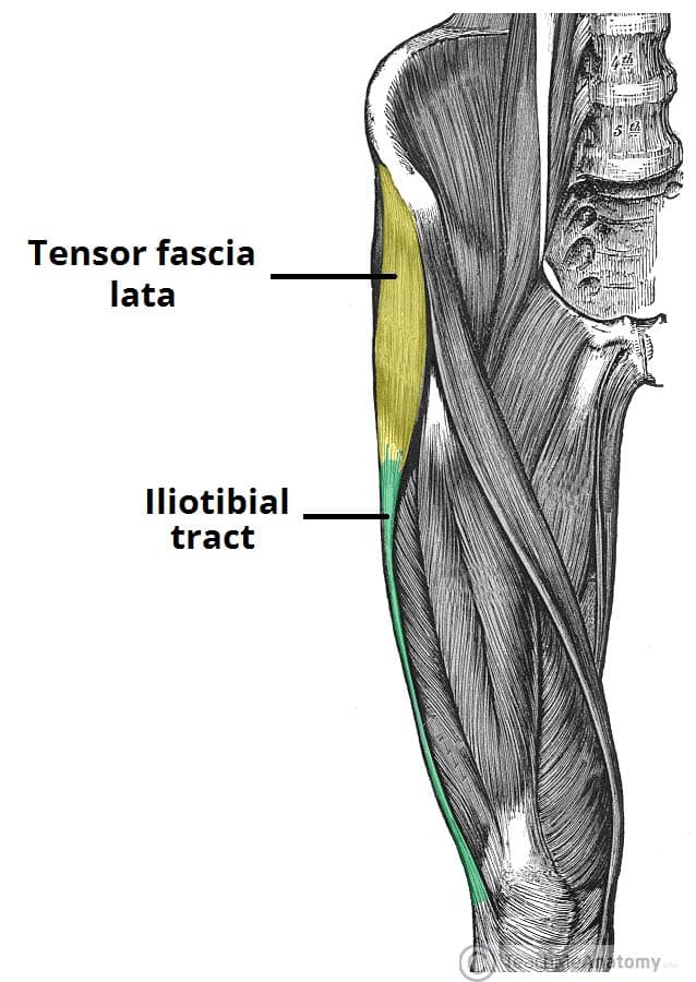
Tensor fascia lata
Location:
thigh
proximal anterolateral thigh
between the superficial and deep fibers of the iliotibial (IT) band
Function:
accessory knee flexor
stabilize the hip and knee joints
assist in hip flexion and abduction
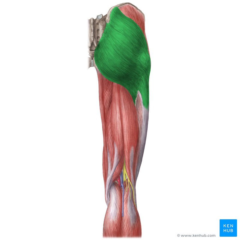
Gluteus maximus
Location:
buttock and hip area
posterior (back) of the pelvis
Function:
facilitating core stability and hip extension
stabilizing the pelvis
extend and externally rotate the thigh
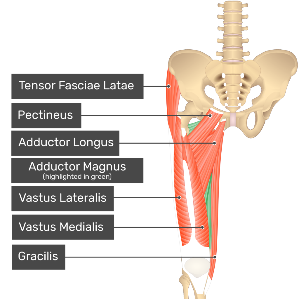
Adductor magnus
Location:
both in the posterior and medial fascial compartments of the thigh
medial compartment of the thigh
Function:
dynamic stabilizer of the pelvis and femur and a prime femoral adductor
contributes to hip extension and rotation
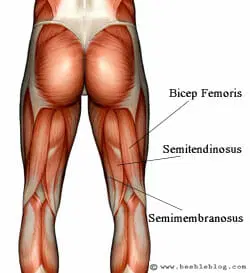
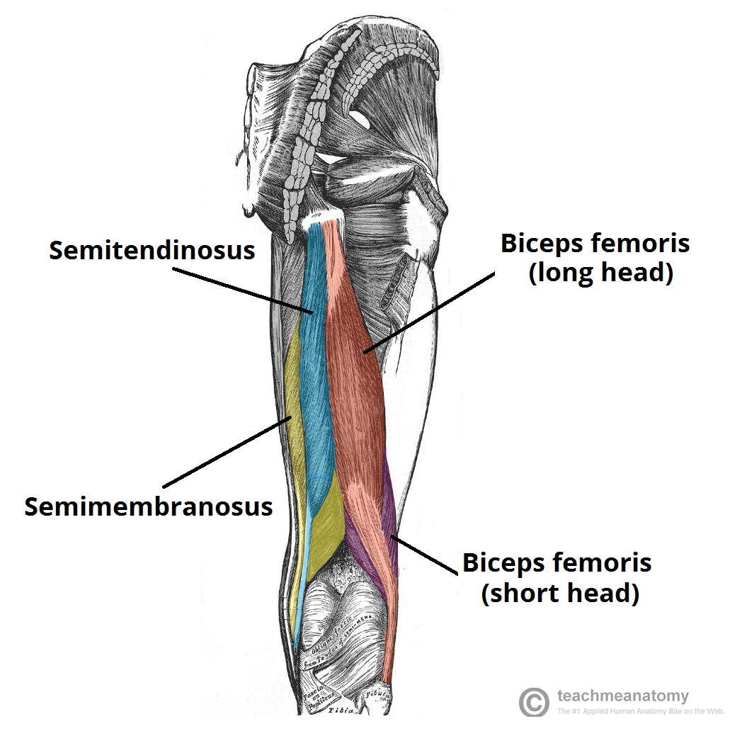
Biceps femoris
Location:
posterior compartment of the thigh
Function:
movement at both the hip joint and knee joint
flex the knee and extend the thigh at the hip
assisting in lateral rotation of the lower leg
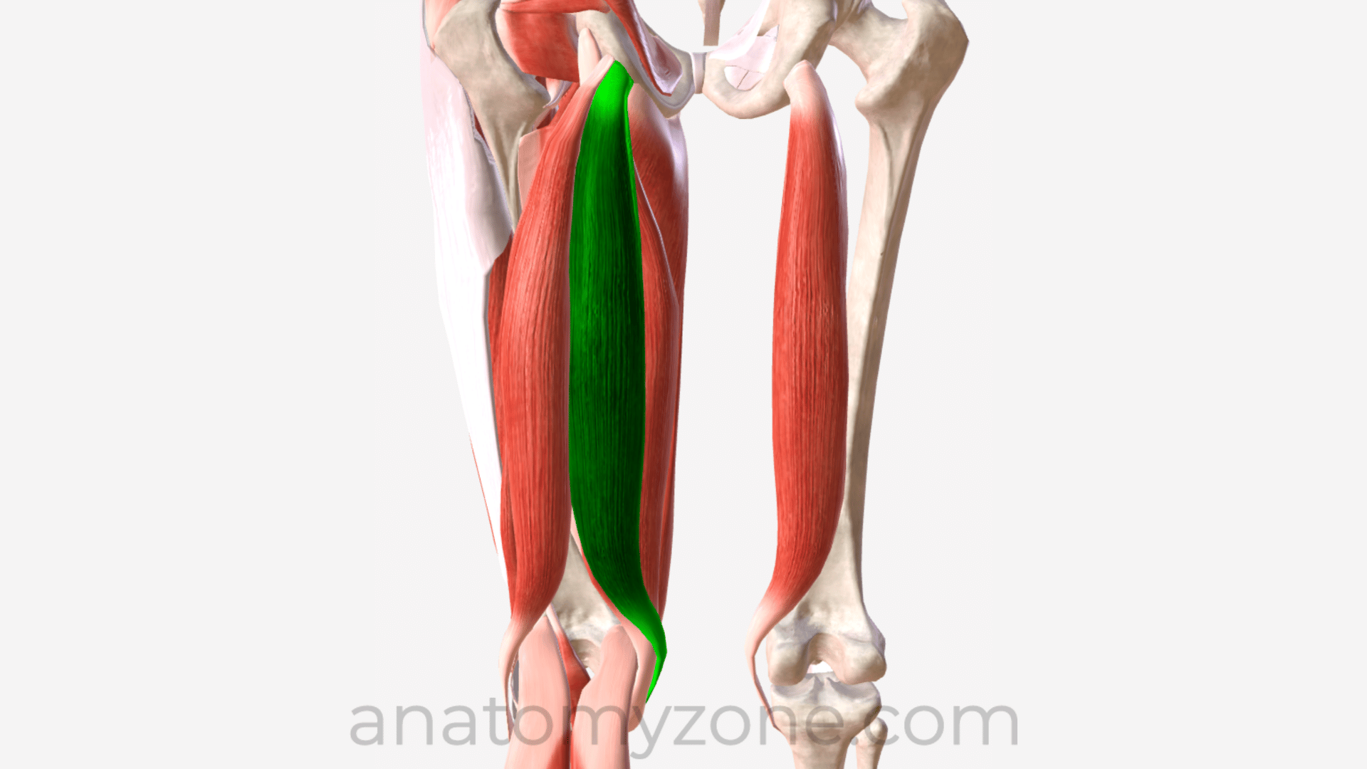
Semitendinosus
Location:
posterior and medial aspect of the thigh
Function:
extend at the hip and flex at the knee
flex the knee
extend the hip
Semimembranosus
Location:
the posteromedial side of the thigh deep to Semitendinosus
medial to the Biceps Femoris
Function:
flexion and internal rotation of the knee joint
knee flexion and medial rotation of the tibia
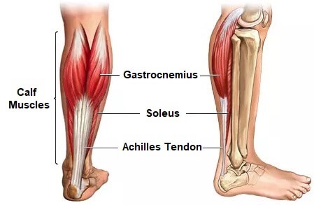
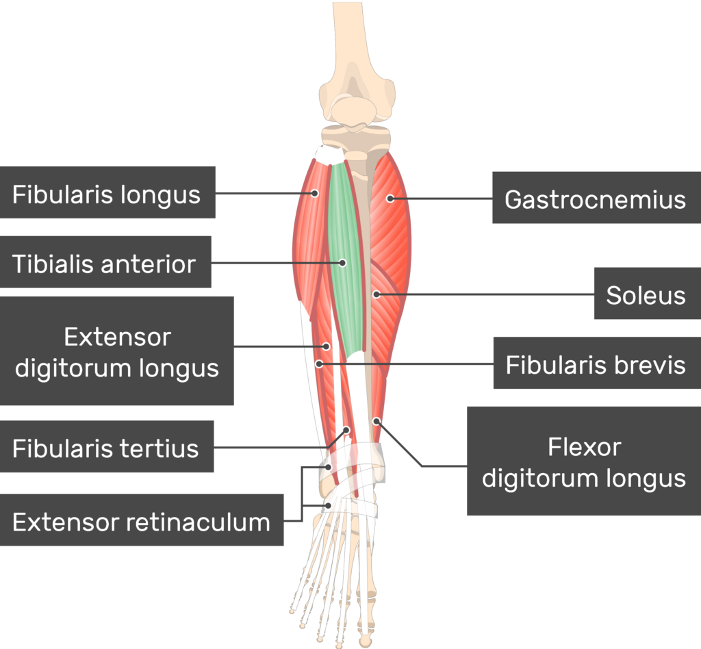
Gastrocnemius
Location:
under your skin at the back of your lower leg
posterior (back) part of the lower leg
Function:
walking and posture
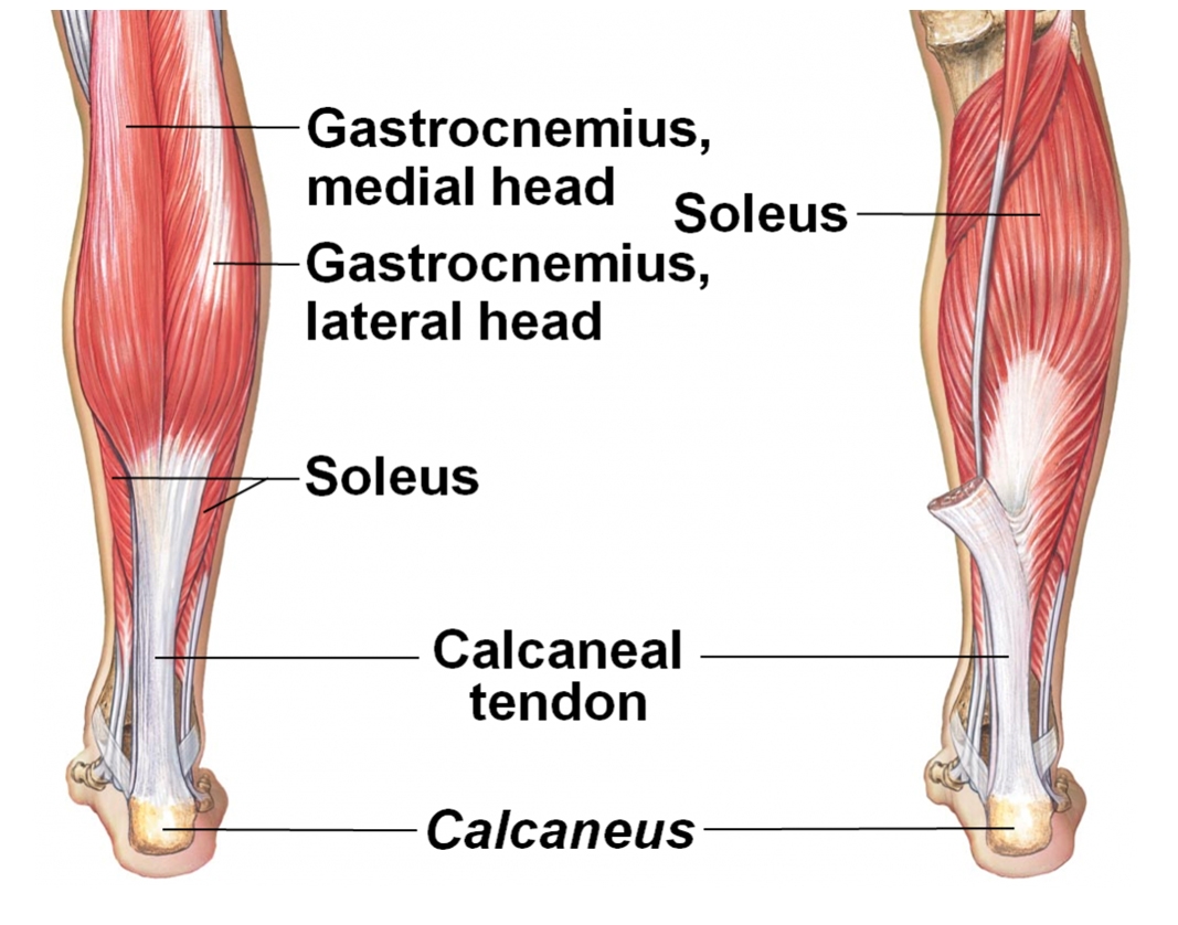
Soleus
Location:
posterior compartment of the lower leg
deep to the gastrocnemius muscle
Function:
primary plantar flexor of the ankle
crucial for walking, running, and maintaining upright posture
"skeletal muscle pump" to assist in venous blood return
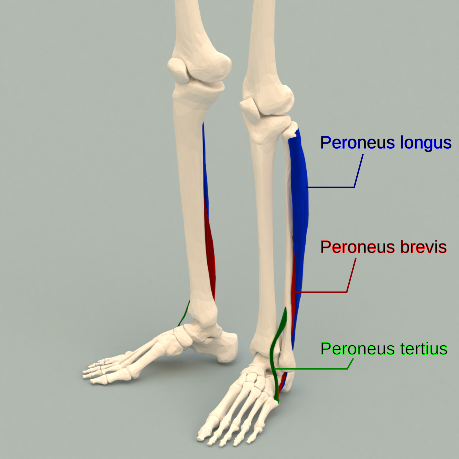
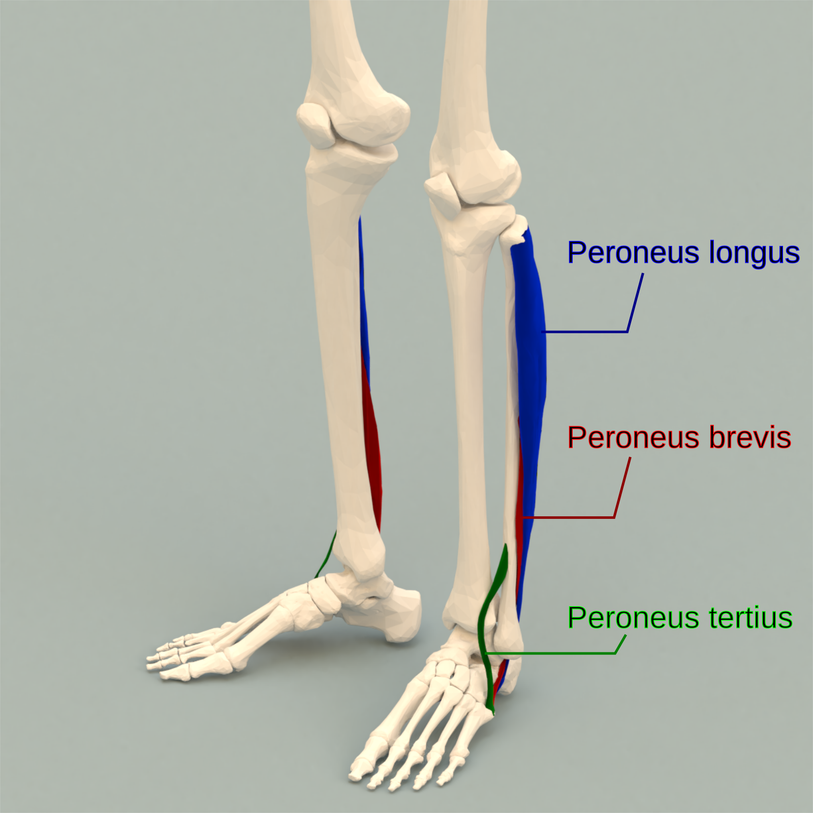
Fibularis longus
Location:
lateral compartment of the lower leg
near the fibula bone
Function:
to tilt the sole of the foot away from the midline of the body
to extend the foot downward away from the body
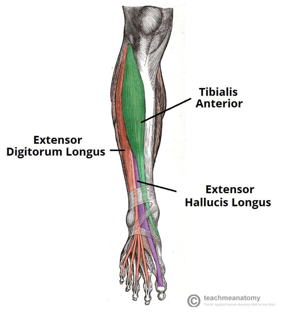
Tibialis anterior
Location:
lateral side of the tibia
anterior compartment of the lower leg
Function:
Inversion of the foot
Adduction of the foot
maintaining the medial arch of the foot
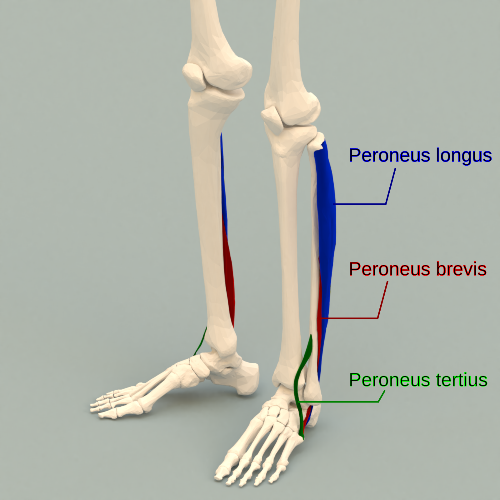
Fibularis brevis
Location:
the lateral part of the lower leg deep to the Fibularis Longus
Function:
responsible for 63% of the power needed to evert the foot as well as assists in plantar flexion
evert the foot and assists in plantar flexion
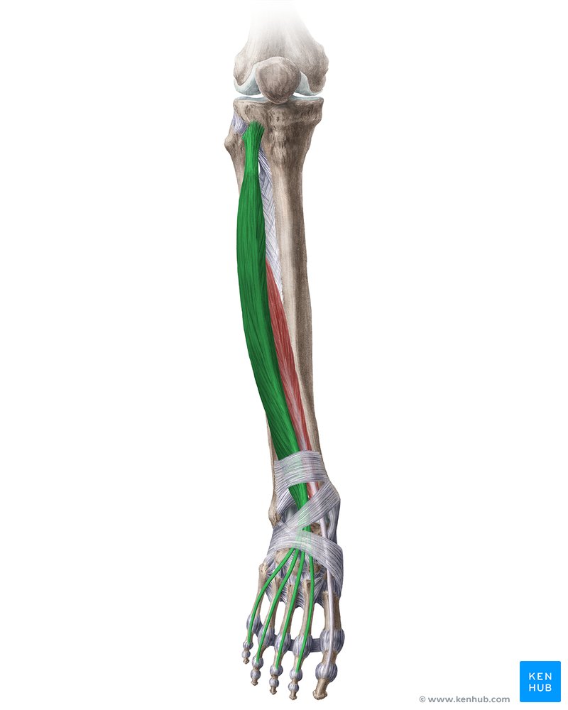
Extensor digitorum longus
Location:
lateral to the tibialis anterior
anterior compartment of the lower leg
Function:
allows you to extend your toes
dorsiflex your feet / ankle
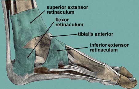
Superior and inferior extensor retinacula
Location:
distally in the leg just above the ankle joint
attaching to the anterior aspects of the fibula and tibia
proximally on the dorsal aspect of the ankle joint
Function:
stabilize the extensor tendons of the foot and ankle
Extensor hallicus longus
Location:
anterior compartment of the lower leg
deep to tibialis anterior and extensor digitorum longus
Function:
extension of the big toe (hallux)
assists in dorsiflexing the foot at the ankle joint
Fibularis tertius
Location:
on the lower lateral aspect of the leg
anterior compartment of the lower leg
Function:
tilt the sole of the foot away from the midline of the body (eversion)
to pull the foot upward toward the body (dorsiflexion)