Acquired Disorders of Bone & Cartilage
 Call Kai
 Learn
 Practice Test
 Spaced Repetition
 Match
 Flashcards
 Knowt Play
1/9

There's no tags or description
Looks like no tags are added yet.
10 Terms
Describe Osteoporosis
Dif to Osteopenia
Types
Epi
Bone Mass Throughout Life
Factors → Loss (5)
Morphology (3)
Osteoporosis
Osteopenia vs Osteoporosis
Osteopenia = dec. bone mass
1- 2.5 SD below mean
Osteoporosis = Severe Osteopenia
Increases fracture risk
> 2.5 SD below mean
Types:
Localized
Generalized
Primary/Secondary Forms
MC: senile and postmenopausal types
Epi:
one million Americans experience fractures related to osteoporosis each year
Bone Mass throughout Life
Young Adult = Peak bone Mass
Then, Decreases 0.7%/yr
Factors -> Loss:
Age-Related (senile osteoporosis)
Osteoblasts ->
reduced proliferative + biosynthetic capacity
Impaired response to growth factors
Reduced physical activity
Physical Activity -> mechanical forces -> stimulates bone remodeling
Athletes have increased bone density
Resistance training + weight-bearing endurance exercise help prevent osteoporosis
Genetic Factors:
Polymorphisms in:
RANK, RANKL
Osteoprotegerin (OPG)
Estrogen receptor
Vitamin D receptor
Calcium deficiency
Hormonal influences (postmenopausal osteoporosis)
Decreased Estrogen -> Increased RANKL + Decreased OPG -> Increased Osteoclast act.
STATS:
40% of postmenopausal women
Yearly reductions in bone mass may reach up to 2% of cortical bone and 9% of cancellous bone
Morphology
Hallmark = normal bone that is decreased in quantity
Thinning of cortical & cancellous bone
In postmenopausal osteoporosis
Vertebral Body cancellous bone particularly affected -> compression fractures


Osteoporotic vertebral body (right) shortened by compression fractures compared with a normal vertebral body (left).
Histologic section of normal cancellous bone. Note the interconnections between the trabeculae.
Histologic section of bone from a case of advanced osteoporosis. The cortical bone (black arrow) is markedly thinned, and there is extensive loss of cancellous bone with a couple of trabeculae appearing as “isolated islands” of bone (blue arrows).
Epi of Paget’s
Average age at Dx = 70 yrs
Striking geographic variation in prevalence
1% of the US population > age 40 is affected
Describe Paget’s Disease (Osteitis Deformans)
What is it
Factors:
Gene (2)
Env. (2)
Clinical Features (6)
Labs (2)
Morph of 3 phases?
What is it?
Disorder where there is increased bone mass/turnover but disordered + structurally unsound
Genetic factors
50% familial cases + 10% sporadic: SQSTM1 gene
Encodes p62 -> RANK signaling
-> NF-kB act. -> osteoclastogenesis
For Juvenile: LOF mutation in OPG
Environmental Factors
Viral infection
Intranuclear inclusions (Measles?) identified in osteoclasts & precursors
Bone marrow shown to contain paramyxovirus RNA
Paramyxovirus Infection -> inc. RANK expression
NOTE: NO live viruses have been isolated from bone
Clinical Features
Most = asymptomatic
Pain Localized
Polyostotic in 85%
Skeletal System
Lower-Extremities Long Bones Fracture
VC Compression Fracture
Leontiasis ossea
Enlargement of craniofacial skeleton
Platybasia
Invagination of the skull base -> Posterior Fossa Compression
Bone Sarcoma
(< 1%)
Labs:
Increased serum alkaline phosphatase
Normal serum calcium and phosphorus
Morphology
Osteolytic phase
Marked osteoclastic activity
Enlarged osteoclasts with inc. nuclei #
X-rays = Lytic lesions
Mixed phase
Inc. osteoclastic/blastic act.
Periosseous fibrovascular tissue
X-Rays = Enlarged Bones
Osteosclerotic (quiescent) phase
Thick trabeculae with “mosaic” pattern
Little cellular activity
Periosseous fibrovascular tissue recedes
X-rays = thickened distorted bones
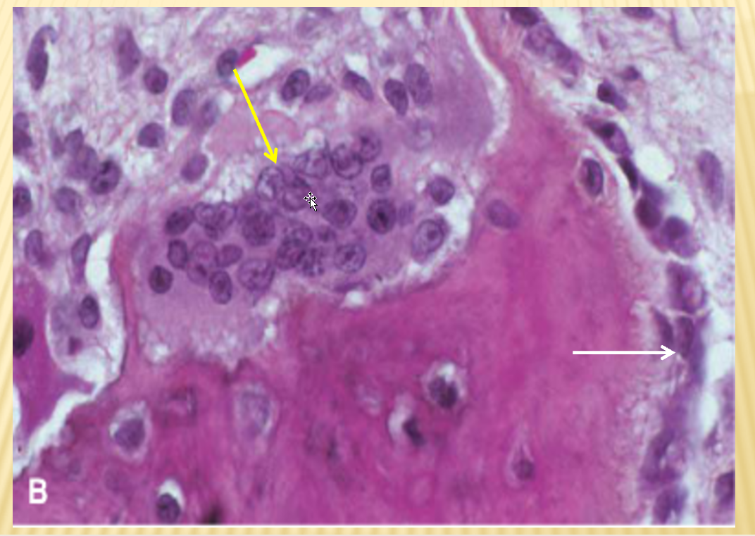
Histologic section of bone in the Mixed phase. The surface of the bone can be seen to be lined by osteoblasts (white arrow), as well as an osteoclast (yellow arrow). The osteoclast exhibits increased numbers of nuclei, well more than the usual 10-12 nuclei, which is typically seen in this disorder.

Left: Normal bone with cellular marrow.
Right: Histologic section of bone from patient with Paget disease in the Osteosclerotic (Quiescent) phase. The trabeculae are thickened and exhibit the so-called “mosaic” pattern, which is the histologic hallmark of Paget disease. This jigsaw puzzle-like appearance is produced by unusually prominent cement lines, which join haphazardly oriented units of lamellar bone. There is little or no detectable osteoclastic or osteoblastic activity, hence the term “quiescent” for this phase.
Describe Rickets
Dif. to Osteomalacia
Causes (4)
Clinical Features (3) (4)
Morphology (6)
Dif. to Osteomalacia:
Osteomalacia:
Adult Disorder
Inadequate Bone mineralization
Rickets:
Child Disorder
Inadequate Bone mineralization + cartilaginous growth plate
Causes:
Dietary deficiency of vitamin D
Disorders of vitamin D metabolism
Inadequate exposure to sunlight
Malabsorptive disorders
Clinical Features
Loss of Rigidity -> Skeletal deformities:
Frontal bossing
Leg Bowing
Rachitic rosary
Pectus carinatum (pigeon breast)
Shortened limbs
Frequent fractures
Morphology
Excess of Unmineralized bone matrix
Growth plate abnormalities:
Mineralization Zone inadequately mineralized
Osteoclasts do not resorb poorly mineralized cartilage
Growth plate cartilage continues growing
marked thickening of growth plate
Proliferating cartilage extend -> metaphyseal region
Only minimal amount of primary spongiosum is formed

Left: Frontal bossing
Right: Rachitic rosary

Left: Pectus carinatum (pigeon breast). Pectus carinatum is also known as “pigeon chest.”
Right: Toddler exhibiting “bowing” of the legs.