DENT 267: LO4 LS1&LS2- Plan Perio Therapy for Mucogingival Deformities
 Call Kai
 Learn
Learn Practice Test
 Spaced Repetition
 Match
Match Flashcards
 Knowt Play
1/29

There's no tags or description
Looks like no tags are added yet.
Name | Mastery | Learn | Test | Matching | Spaced | Call with Kai | Chat |
|---|
No analytics yet
Send a link to your students to track their progress
30 Terms
normal mucogingival conditions
-the absence of diseased state
-absence of gingivitis, periodontitis, and gingival recession
-perio health can be maintained despite lack of keratinized tissue as well as presence of frena, shallow vestibular depth
periodontal biotypes
-AKA gingival biotype, periodontal morphotype, phenotype
-describes the individual differences in gingival anatomy and morphology
3 categories of periodontal biotypes
-thin scalloped biotype
-thick flat biotype
-thick scalloped biotype
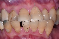
thin scalloped biotype
-slender triangular-shaped tooth crowns
-subtle cervical convexity (scalloping)
-interprox contacts close to incisal edge
-narrow zone of keratinized tissue
-clear, thin delicate gingiva
-thin alveolar bone
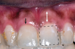
thick flat biotype
-square-shaped tooth crowns
-pronounced tissue scalloping
-large interprox contact areas more apical
-thick, fibrotic gingiva
-thick alveolar bone
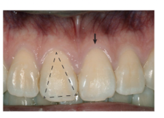
thick scalloped biotype
slender tooth crowns
-pronounced gingival scalloping
-thick fibrotic gingiva
-narrow zone of keratinized tissue
mucogingival deformities
-significant alteration of morphology, size and interrelationships b/w gingiva and alveolar mucosa that may involve underlying bone
-affects a large number of patients
what is the most common mucogingival deformity
-gingival recession
gingival recession
-most common mucogingival deformity
-apical displacement of gingival margin w/ respect of the CEJ
-associated w/ attachment loss with root exposure
risk factors for gingival recession
-thin periodontal biotype
-absence of attached gingiva
-reduced thickness of alveolar bone due to abnormal tooth position in the arch
how much keratinized tissue and attached gingiva are desirable to maintain health?
-2mm keratinized tissue
-1mm attached gingiva
Miller Classification System for recession of GM
-most common
-based on level of GM with respect to MGJ and underlying bone
-sometimes difficult to identify exact location of apical extent of recession defect w/ respect to MGJ
-difficult to differentiate b/w class I and II
-not clear in defining amount of interprox soft/hard tissue loss b/w class III and IV
-reliability has never been tested
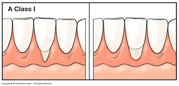
Miller Classification System for recession of GM: Class I
-marginal recession but does not extend to MGJ
-no loss of bone or soft tissue in the inter-dental area
-100% root coverage can be anticipated

Miller Classification System for recession of GM: Class II
-marginal recession that extends to or beyond MGJ
-no loss of bone or soft tissue in interdental area
-100% root coverage can be anticipated

Miller Classification System for recession of GM: Class III
-marginal recession that extends to or beyond MGJ
-bone and soft tissue loss in interdental area
-may have tooth malpositioning that prevents coverage of certain roots
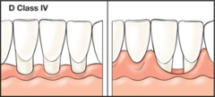
Miller Classification System for recession of GM: Class IV
-marginal recession that extends to or beyond MGJ
-bone and soft tissue loss very severe in interdental area
-may have tooth malpositioning so severe that prevents coverage of many roots
Cairo Classification System for Recession of GM
-new system proposed based on CAL measurements (more objective measurements to classify extent and severity than Miller System) at buccal and interprox sites
Cairo Classification System Recession Types (RT)
-RT1
-RT2
-RT3
Cairo Classification System Recession Types: RT1
-gingival recession w/ no loss of interprox attachment
-most likely associated w/ traumatic toothbrushing
Cairo Classification System Recession Types: RT2
-gingival recession w/ loss of interprox attachment
-most likely associated w/ periodontitis-associated horizontal bone loss
Cairo Classification System Recession Types: RT3
-same as RT2 (gingival recession w/ loss of interprox attachment) but with interproximal loss greater than buccal loss and infrabody defects
mucogingival deformities and conditions around teeth
-periodontal biotype
-gingival/soft tissue recession
-lack of keratinizd gingiva
-decreased vestibular depth
-aberrant frenum/muscle position
-gingival excess
-abnormal color

Stillman's cleft
-type of gingival recession
-narrow, triangular or apostrophe shaped
-exposes cementum
Stillman's cleft: clinical characteristics
-apical position
-inflamed or healthy
-localized or generalized
-more common w/ increase in age
-clients with good OH can have
causes of gum recession
-genetics
-brushing aggressively
-mouth injury or trauma
-perio disease
-braces and dental restos
-poor OH
-teeth grinding or clenching
-high frenal attachment
etiology of recession
-inflammation
-uneven gingival margins
-impact of cervical restos: sites with minimal or no attached gingiva associated w/ intrasulcular resorations are more prone to recession and inflammation
-over vigorously brushing
-excessive occlusal force
-tension of a frenum/high frenal attachment
-inadequate zone of attached gingiva
-habits
-ortho
-following perio surgery
-use of abrasive prophylaxis paste
-dentoalveolar ablation
-piercings
-root to bone angle and attrition
mucogingival involvement (MGI)
-when a perio pocket extends to or beyond the MGJ, the probe may pass through the pocket directly into the alvolar mucosa
minimally attached gingiva
-limited amount of attached gingiva, which is the portion of gum tissue firmly bound to the tooth and underlying bone surrounding the teeth
MAG vs MGI
-MAG:
prognosis of recession
-favourable if no MGI, if recession is not over extensive and if risk factors are corrected
-surgery may be needed: pedicle graft, free gingival graft