Hair, Skin, and Nails
 Call Kai
 Learn
Learn Practice Test
 Spaced Repetition
 Match
Match Flashcards
 Knowt Play
1/103

There's no tags or description
Looks like no tags are added yet.
Name | Mastery | Learn | Test | Matching | Spaced | Call with Kai | Chat |
|---|
No analytics yet
Send a link to your students to track their progress
104 Terms
derm/o
skin
sub
below
hypo
low, slow
hyper
fast, increased
adip/o
fat
lip/o
fat
cutane/o
skin
cyan/o
blue
erythem/o
red
hidr/o
sweat
ichthy/o
dry
kerat/o
hard tissue
melan/o
black
myc/o
fungus
squam/o
scale
therm/o
heat
xer/o
dry
pil/o
hair
trich/o
hair
onych/o
nail
cyte
cell
derma
skin
oma
tumor
phoresis
carrying, transmission
plasty
surgical repair
itis
inflammation of
Hair vellus
fine, short, soft hair “peach fuzz”
Hair terminal
thicker, longer hair
What are the 3 layers of the skin?
epidermis, dermis, and subcutaneous
Epidermis
outermost layer for protection
Dermis
contains blood vessels, nerves, glands, and hair follicles
Subcutaneous
contains fat and provides cushion, energy, and storage
What are the parts of the hair?
root, shaft, and follicle
Root
part of the hair below the skin
Shaft
part of the hair that extends above the skin
Follicle
structure of the skin where the hair grows
Eccrine glands
regulate body temperature through temperature
Apocrine glands
larger glands that are deeper in the skin AND activate at puberty to contribute to body odor
Sebaceous gland
produce sebum and helps keep the skin and hair lubricated and moisturized
Nails
epidermal cells converted to hard plates of keratin
Parts of the nail
free edge, nail body, and nail root
Skin physiology
Gives protection
Retains body fluids and electrolytes
Sensory perception
Body temperature and wound regulation
Absorption, excretion, and production
Skin Cancer Risk Factors
personal history of skin cancer, family history, older age, exposure to UV radiation, fair skin, blonde/red hair, blue or green eyes, moles
OLD CARTS
Onset, Location, Duration, Characteristics, Aggravating factors, Relieving factors, Timing, Severity
Beginning 5
2 patient identifiers, hand hygiene, provide privacy - cover patient with a drape, temperature and comfort educate, and gain permission
What are the abnormal skin colors?
Cyanosis - blue due to inadequate oxygenation
Pallor - unhealthy pale maybe from anemia or illness
Jaundice - yellowing of the skin and sclera

Pigmented Nevi
A mole about 10-40 scattered throughout the body
Freckles
small, flat, hyperpigmented macule
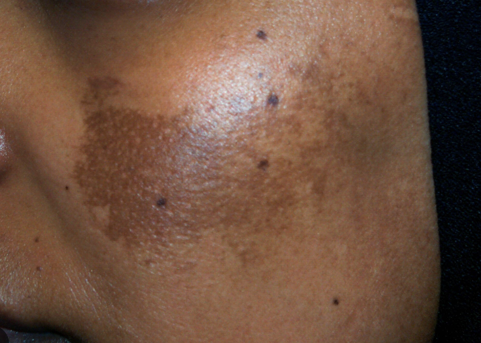
Patch
area of darker skin pigmentation

Striae
Silver or pink stretch marks
Which part of the hand do you use to check temperature when palpating the skin?
dorsal (non palm side)
What is the expected angle of the nail base?
160 degrees
Beau’s Lines - abnormal nail finding
grooves or transverse depressions running across the nails, can be from trauma
Koilonychia or Spoon Nails - abnormal nail finding
deep depression, nails become concave
Clubbing - abnormal nail finding
increased curvature, >180 degrees @ nails base (chronic respiratory or cardiovascular disease)
What do you assess when looking at a lession?
location/distribution, color, shape, pattern, edges, depth, size, other things
ABCDE signs of melanoma
Asymmetry, Border, Color, Diameter, Evolving
Primary lesions
develop on previously unaltered skin

Primary lesion - macule
FRECKLES, flat circumscribed area, change in color of skin <1cm big
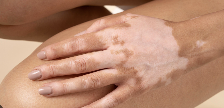
Primary lesion - patch
non-palpable irregular shaped macule >1cm

Primary lesion - papule
elevated firm & <1cm

Primary lesion - plaque
elevated, firm, and rough lesion >1cm

Primary lesion - nodule
elevated firm lesion deeper in dermis (1-2cm)

Primary lesion - tumor
elevated solid lesion deep in dermis (>2cm)

Primary lesion - wheal
elevated irregular shaped area in cutaneous edema

Primary lesion - urticaria
HIVES wheals that form an extensive reaction
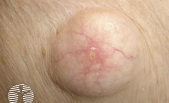
Primary lesion - cyst
elevated encapsuled lesion, filled with liquid/semi-solid material
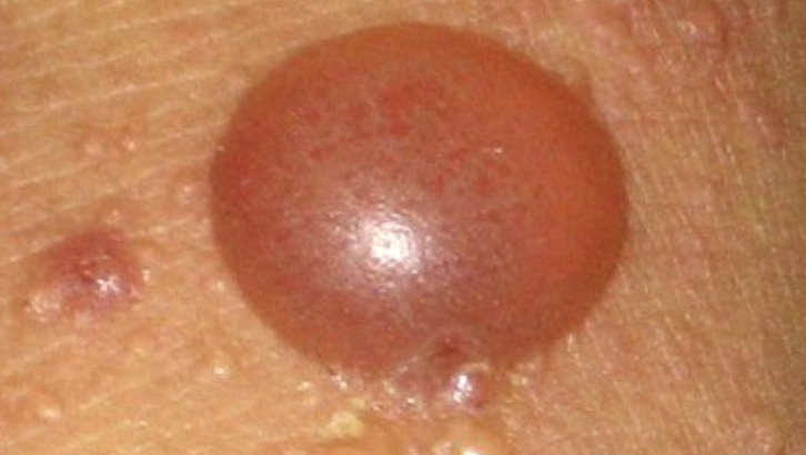
Primary lesion - bulla
vesicle >1cm

Primary lesion - pustule
elevated lesion filled with prulent fluid
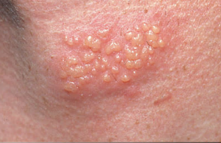
Primary lesion - vesicle
elevated not in dermis filled with serous fluid
Secondary lesions
develop after a primary lesion OR as a result of changes to the original lesion

Secondary lesion - scar
fibrous tissue that replaces normal skin after an injury

Secondary lesion - keloid
elevated irregular scar that continues to grow

Secondary lesion - Striae
Stretch marks, thinning of epidermis from stretching

Secondary lesion - excoriation
loss of epidermis

Secondary lesion - scale
heaped up keratin, flaky skin

Secondary lesion - lichenification
rough, thickened epidermis

Secondary lesion - crust
dried drainage or blood
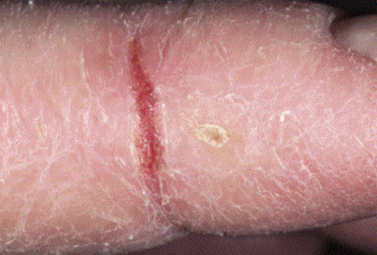
Secondary lesion - fissure
crack or break from the epidermis to dermis

Secondary lesions - erosion
loss of part of the epidermis

Secondary lesion - ulcer
loss of epidermis + dermis and concave
Pressure Ulcer Stage 1
redness and skin intact
Pressure Ulcer Stage 2
partial skin loss and dermis can be open
Pressure Ulcer Stage 3
full skin loss extending into subcutaneous tissue
Pressure Ulcer Stage 4
full tissue loss with exposed structures like bone, tendon, or muscle
Unstageable pressure injury
thickness skin loss and do NOT know depth bc covered
Deep tissue pressure injury
damage to underlying tissue from pressure, not open, and don’t know how deep
The tool that is used to help assess a patient’s risk of developing a pressure ulcer.
Braden Scale
Vascular lesions
changes related to blood vessels or bleeding under the skin

Vascular lesion - ecchymosis
bruising

Vascular lesion - Petechiae
tiny, flat, reddish-purple spots <0.5 mm
small amounts of bleeding/hemorrhaging into skin

Vascular lesion - purpura
reddish-purple discoloration fo the skin >5cm (infection or bleeding disorder)

Vascular lesion - angioma
benign tumor consisting of small blood vessels

Vascular lesion - telangiectasia
dialation of small blood vessels
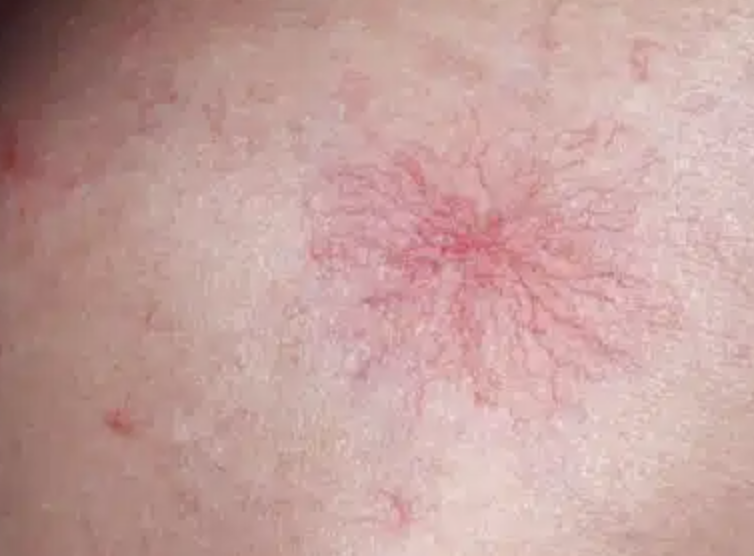
Vascular lesion - vascular spider
small, central red areas
What are some of the differences with cyanosis, ecchymosis, erythema, jaundice, and pallor with darker skin colors?
cyanosis = ashen-grey color
ecchymosis = deeper blueish-black tone
erythema = deeper brown/purple
jaundice = yellowish-green color
pallor = skin tone lighter than normal
What are some developmental considerations with infants and children?
jaundice due to increased bilirubin, nails thin and fragile, erythema toxicum (newborn rash), diaper rash, birthmarks, stock bites (red mark @ base of skull)
What are some developmental considerations with adolescents?
acne due to increase in sebaceous gland activity
What are some developmental considerations with aging skin?
dry skin, less elasticity, loss in subcutaneous tissue, facial hair in women, balding, increased capillary fragility, solar lentigines