A&P Slideshow Chapter 12
 Call Kai
 Learn
 Practice Test
 Spaced Repetition
 Match
 Flashcards
 Knowt Play
1/253

There's no tags or description
Looks like no tags are added yet.
254 Terms
What is cephalization?
evolutionary development of rostral (anterior) portion of CNS
resulted in increased number of neurons
highest level reached in human brain
Embryologically, the brain and spinal cord begin as a _____.
neural tube
Neural tube’s anterior end expands, and constrictions form 3 _____.
primary brain vesicles
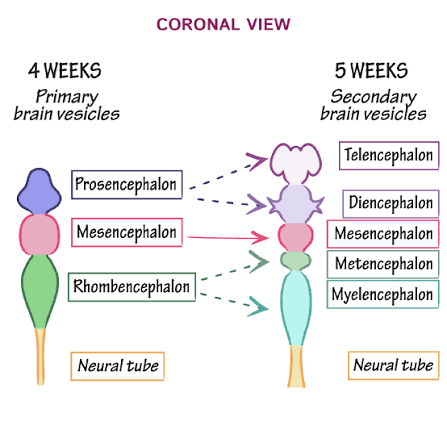
What are the 3 primary brain vesicles that constrictions form when the neural tube’s anterior end expands?
prosencephalon, or forebrain
mesencephalon, or midbrain
rhombencephalon, or hindbrain

When the neural tube’s anterior end expends and constrictions form 3 primary brain vesicles, what does the posterior end become?
spinal cord
Primary vesicles give rise to 5 _____.
secondary brain vesicles
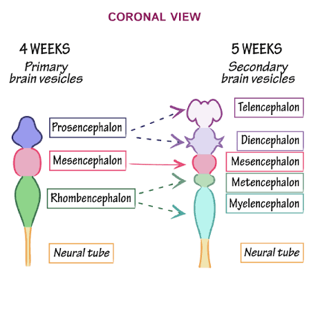
Primary vesicles give rise to what five secondary brain vesicles?
forebrain becomes telencephalon and diencephalon
midbrain remains undivided
hindbrain becomes mesencephalon and myelencephalon

When primary vesicles give rise to 5 secondary brain vesicles, the forebrain _____.
becomes telencephalon and diencephalon

When primary vesicles give rise to 5 secondary brain vesicles, the midbrain _____.
remains undivided
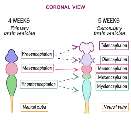
When primary vesicles give rise to 5 secondary brain vesicles, the hindbrain _____.
becomes mesencephalon and myelencephalon

Telencephalon gives rise to _____.
2 cerebral hemispheres
together make up the cerebrum

What do the 2 cerebral hemispheres that the telencephalon gives rise to make up?
together make up the cerebrum

Diencephalon becomes the _____.
epithalamus, thalamus, hypothalamus, and retina

Mesencephalon becomes _____.
remains the midbrain
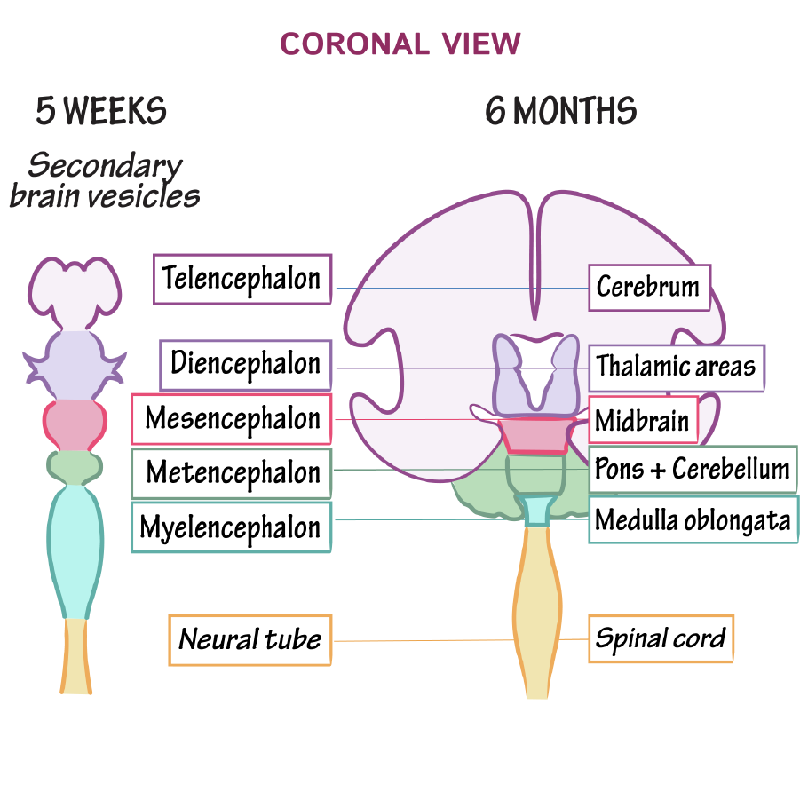
Mesencephalon becomes _____.
the pons and cerebellum

Myelencephalon becomes _____.
medulla oblongata

Central cavity of neural tube becomes _____.
ventricles

The brain grows _____ than surrounding membranous skull. (faster/slower)
faster, so it must fold to occupy available space
What must the brain do because it grows faster than the surrounding membranous skull?
it must fold to occupy available space
Forebrain moves toward _____.
brain stem (midbrain, pons, medulla oblongata)
What do the cerebral hemispheres do during brain development?
double back and envelop diencephalon and midbrain while creasing and folding to increase surface area
What is the forebrain also called?
prosencephalon
What is the prosencephalon also called?
forebrain
What is the midbrain also called?
mesencephalon
What is the mesencephalon also called?
midbrain
What is the hindbrain also called?
rhombencephalon
What is the rhombencephalon also called?
hindbrain
Adult brains have 4 regions:
cerebral hemispheres
diencephalon
brain stem, consisting of:
midbrain
pons
medulla oblongata
cerebellum

What does the brain stem consist of?
midbrain
pons
medulla oblongata

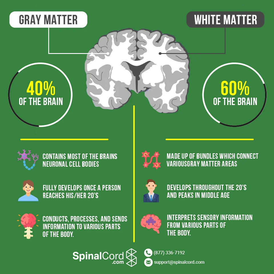
What is gray matter?
short, nonmyelinated neurons and cell bodies
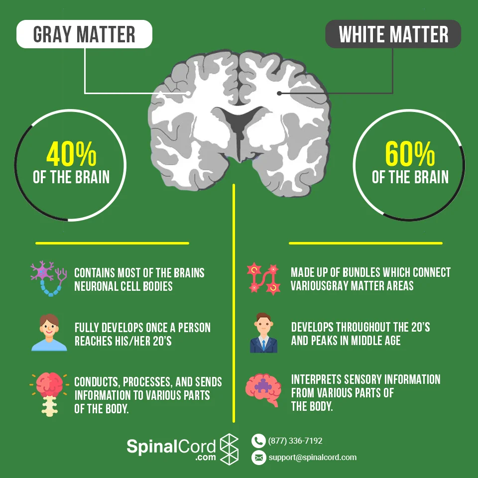
What is white matter?
myelinated and nonmyelinated axons
What is the basic pattern found in the CNS?
central cavity surrounded by gray matter, with white matter external to gray matter
What are some features and changes of the basic pattern found in the CNS?
spinal cord exhibits this basic pattern; however, pattern changes with ascent into brain stem
brain stem has additional gray matter nuclei scattered within white matter
cerebral hemispheres (cerebrum) and cerebellum contain outer layer of gray matter called the cortex
cerebrum and cerebellum also have scattered areas of gray matter nucleus amid white matter
Does the spinal cord exhibit the basic pattern found in the CNS?
exhibits this basic pattern; however, pattern changes with ascent into brain stem
Does the brain stem exhibit the basic pattern found in the CNS?
brain stem has additional gray matter nuclei scattered within white matter
Cerebral hemispheres (cerebrum) and cerebellum contain outer layer of gray matter called _____.
the cortex
Do the cerebrum and cerebellum exhibit the basic pattern found in the CNS?
Cerebral hemispehres (cerebrum) and cerebellum contain outer layer of gray matter called the cortex
cerebrum and cerebellum also have scattered areas of gray matter nuclei amid white matter
What are ventricles?
fluid-filled chambers that are continuous to one another and to central canal of spinal cord
filled with cerebrospinal fluid (CSF)
lined by ependymal cells (neuroglial cells)
What are ventricles lined by?
ependymal cells (neuroglial cells)
What are the lateral ventricles?
(paired) are large, C-shaped chambers located deep in each hemisphere
pair is separated by membranous septum pellucidum
What are paired lateral ventricles separated by?
membranous septum pellucidum

Each lateral ventricle is connected to the _____ via interventricular foramen.
third ventricle

Each lateral ventricle is connected to the third ventricle via _____.
interventricular foramen

Third ventricle lies in _____.
diencephalon
Third ventricle is connected to the _____ via cerebral aqueduct.
fourth ventricle

Third ventricle is connected to the fourth ventricle via _____.
cerebral aqueduct

Where does the fourth ventricle lie?
lies in hindbrain
3 openings connect the fourth ventricle to subarachnoid space that surrounds brain:
paired lateral apertures in side walls
median aperture in roof
_____ form superior part of brain.
cerebral hemispheres

Cerebral hemispheres form _____ part of brain.
superior

Cerebral hemispheres account for ___% of brain mass.
83%
What are the surface markings of the cerebral hemispheres?
gyri: ridges
sulci: shallow grooves
fissures: deep grooves
longitudinal fissure
separates 2 hemispheres
transverse cerebral fissure
separates cerebrum and cerebellum
What is the longitudinal fissure?
separates 2 hemispheres

What is the transverse cerebral fissure?
separates cerebrum and cerebellum

Several sulci divide each hemisphere into 5 lobes:
frontal lobe
parietal lobe
temporal lobe
occipital lobe
insula

Where is the insular lobe located?
buried under portions of temporal, parietal, and frontal lobes
What are the major sulci that divide lobes?
central sulcus separates pre central gyrus of frontal lobe and post central gyrus of parietal lobe
parieto-occipitaq sulcus separates occipital and parietal lobes
lateral sulcus outlines temporal lobes
What is the central sulcus?
separates pre central gyrus of frontal lobe and post central gyrus of parietal lobe

What is the parieto-occipital sulcus?
separates occipital and parietal lobes
What is the lateral sulcus?
outlines temporal lobes

Each hemisphere has 3 basic regions:
cerebral cortex of gray matter superficially
internal white matter
basal nuclei deep within white matter
_____ is “executive suite” of brain.
cerebral cortex
What is the site of conscious mind?
awareness, sensory perception, voluntary motor initiation, communication, memory storage, understanding
What part of the brain holds the site of conscious mind?
cerebral cortex
What does the cerebral cortex consist of?
thin (2-4 mm) superficial layer of gray matter
composed of neuron cell bodies, dendrites, glial cells, and blood vessels, but no axons
The cerebral cortex consists of ___& of total brain mass.
40%
Functional imaging (PET and MRI) of brain show specific motor and sensory functions are located in discrete cortical areas called _____.
domains
What are 4 general considerations of the cerebral cortex?
contains 3 types of functional areas:
motor areas
sensory areas
association areas
each hemisphere is concerned with contralateral (opposite) side of body
lateralization (specialization) of cortical function can occur in only one hemisphere
conscious behavior involves entire cortex in one way or another
What are the 3 types of functional areas in the cerebral cortex?
motor areas: control voluntary movement
sensory areas: conscious awareness of sensation
association areas: integrate diverse information
What do motor areas of the cerebral cortex do?
control voluntary movement
What do sensory areas of the cerebral cortex do?
conscious awareness of sensation
What do association areas of the cerebral cortex do?
integrate diverse information
Each hemisphere of the cerebral cortex is concerned with _____ side of body.
contralateral (opposite)
_____ of cortical function can occur in only one hemisphere.
lateralization (specialization)
What are the motor areas of the cerebral cortex?
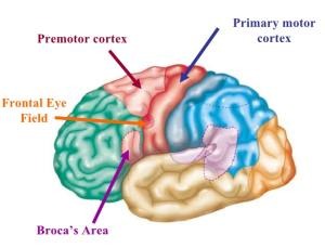
located in frontal lobe, motor areas act to control voluntary movement
primary (somatic) motor cortex in pre central gyrus
premotor cortex anterior to pre central gyrus
Broca’s area anterior to inferior premotor area
frontal eye field within and anterior to premotor cortex; superior to Broca’s area

What is the primary (somatic) motor cortex?
in the motor area of the cerebral cortex
located in pre central gyrus of frontal lobe
pyramidal cells: large neurons that allow conscious control of precise, skilled, skeletal muscle movements
pyramidal (corticospinal) tracts: formed from long axons that project down spinal cord
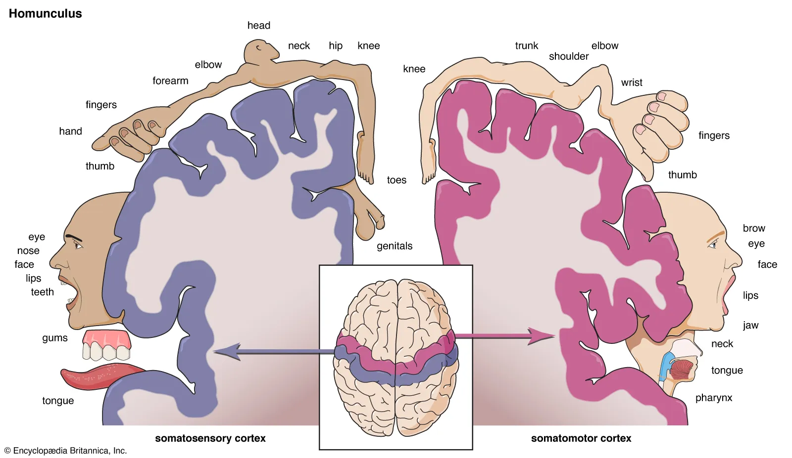
somatotopy: all muscles of body can be mapped to area on primary motor cortex
motor homunculus: upside-down caricatures represent contralateral motor innervation of body regions

What are pyramidal cells?
large neurons that allow conscious control of precise, skilled, skeletal muscle movements

What are pyramidal (corticospinal) tracts?
formed from long axons that project down spinal cord

What is somatotopy?
all muscles of body can be mapped to area on primary motor cortex
(W) the point-for-point correspondence between an area of the body and a specific point within the central nervous system, such as the brain or spinal cord
What is the motor homunculus?
upside-down caricatures represent contralateral motor innervation of body regions
(W) a distorted, topographical organized map of the human body laid across the primary motor cortex
What is the premotor cortex?
helps plan movements
staging area for skilled motor activities
controls learned, repetitious, or patterned motor skills
coordinates simultaneous or sequential actions
controls voluntary actions that depend on sensory feedback

What is Broca’s area?
present in one hemisphere (usually the left)
motor speech area that directs muscles of speech production
active in planning speech and voluntary motor activities
What is the fontal eye field?
controls voluntary eye movements

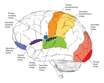
What are the sensory areas of the cerebral cortex?
areas of cortex concerned with conscious awareness of sensation
occur in parietal, insular, temporal, and occipital lobes
8 main areas include: primary somatosensory cortex, somatosensory association cortex, visual areas, auditory areas, vestibular cortex, olfactory cortex, gustatory cortex, and visceral sensory area

What are the 8 main areas of the sensory areas?
primary somatosensory cortex
somatosensory association cortex
visual areas
auditory areas
vestibular cortex
olfactory cortex
gustatory cortex
visceral sensory areas
What is the primary somatosensory cortex?
located in postcentral gyro of parietal lobe
receives general sensory information from skin
receives general sensory information from skin and proprioceptors of skeletal muscle, joints, and tendons
capable of spatial discrimination: identification of body region being stimulated
somatosensory homunculus: upside-down caricatures represent contralateral sensory input from body regions
What is spatial discrimination?
identification of body region being stimulated
(W) the ability to perceive, detect, and distinguish between 2 or more spatial stimuli or locations
What is somatosensory homunculus?
upside-down caricatures represent contralateral sensory input from body regions
(W) a distorted neurological map of the human body laid across the primary somatosensory cortex)

What is the somatosensory association cortex?
posterior to primary somatosensory cortex
integrates sensory input from primary somatosensory cortex for understanding of object
determines size, texture, and relationship of parts of objects being felt

What are the visual areas of the sensory areas of the cerebral cortex?
primary visual (striate) cortex
visual association area
What is the primary visual (striate) cortex?
located on extreme posterior tip of occipital lobe
receives visual information from retinas

What is the visual association area?
surrounds primary visual cortex
uses past visual experiences to interpret visual stimuli (color, form, or movement)
ex: ability to recognize faces
complex processing involves entire posterior half of cerebral hemispheres

What are the auditory areas of the cerebral cortex?
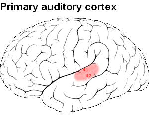
primary auditory cortex
auditory association area
What is the primary auditory cortex?
superior margin of temporal lobes
interprets information from inner ear as pitch, loudness, and location
What is the auditory association area?
sensory area of cerebral cortex
located posterior to primary auditory cortex
stores memories of sounds and permits perception of sound stimulus
What is the vestibular cortex?
sensory area
posterior part of insulation and adjacent parietal cortex
responsible for conscious awareness of balance (position of head in space)

What is the olfactory cortex?
primary olfactory (smell) cortex
medial aspect of temporal lobes (in piriform lobes)
part of primitive rhinencephalon, along with olfactory bulbs and tracts
remainder of rhinencephalon in humans becomes part of limbic system
involved in conscious awareness of odors

What is the gustatory cortex?
in insula just deep to temporal lobe
involved in perception of taste
What is the visceral sensory area?
posterior to gustatory cortex
conscious perception of visceral sensations, such as upset stomach or full bladder

What is the visceral sensory area?
posterior to gustatory cortex
conscious perception of visceral sensations, such as upset stomach or full bladder