Exam 2
 Call Kai
 Learn
 Practice Test
 Spaced Repetition
 Match
 Flashcards
 Knowt Play
1/256

There's no tags or description
Looks like no tags are added yet.
257 Terms
Obligate Intracellular Parasites
Viruses lack the ability to reproduce on their own and depend entirely on host cells to provide building materials and cellular machinery while supplying only the genetic blueprint
Microscopic Size of Viruses
Typical virus particles range between 10 and 100 nm in size .
Giant virus exceptions exist
CroV (730 kb genome)
Megavirus chilensis (1.2 Mb genome encoding 1,200 proteins)
Mimivirus (400 nm in diameter with a 1.2 Mb genome)
Structure of viruses: Viruses are
Intracellular obligate parasites
Typically between 10 and 100 nm
Genomes of DNA or RNA are typically between a few thousand to 200,000 nucleotides in length
Walter Reed
showed in 1901 that a human disease, yellow fever, was caused by a virus transmitted by mosquitoes
There are exceptions to the small size of viruses
CroV virus of marine single-celled organisms has a 730k bp genome
Megavirus chilensis (a virus of amoebas) has a genome over 1.2 megabase pairs, encodes 1,200 proteins
Mimivirus (a dsDNA virus of amoebas) can be 400 nm in diameter, with a 1.2 megabase pair genome coding for 979 proteins
Structure of viruses
Single or double-stranded DNA or RNA
Protein shell (capsid) around genome composed of many capsomere proteins
Capsid and genome together = nucleocapsid
Possible envelope (plasma membrane around capsid)
Virus Genomic Diversity
Viral genomes consist of single-stranded (ss) or double-stranded (ds) DNA or RNA, with sizes spanning from a few thousand to over 1.2 million base pairs
Capsid
A protective protein shell assembled from individual capsomere proteins that encloses the viral genome
Nucleocapsid
The combined complex of the capsid and the genome
Envelope
An optional outer lipid membrane surrounding the nucleocapsid. Viruses with a membrane are enveloped (e.g., Influenza virus, HIV, Variola virus), whereas those lacking a membrane are naked or non-enveloped (e.g., Poliovirus, Tobacco mosaic virus)
Replication Cycle Steps of a virus
A virus must adhere to a target host cell, penetrate and uncoat its genome, express viral genes and replicate nucleic acids, assemble new viral particles, and exit the host cell
Viral capsids generally exhibit ….
symmetrical structures or form irregular, complex shapes
Helical Shape of viruses
Identical protein subunits assemble in a spiral around the nucleic acid to form a cylindrical tube.
Example: Tobacco mosaic virus (TMV)
Icosahedral Shape of a Virus
A regular geometric polyhedron featuring 20 triangular faces (capsid faces)
Displays distinct 2-fold, 3-fold, and 5-fold axes of symmetry
Examples: Poliovirus and Adenovirus
Head-Tail / Biphage Structure:
T4 bacteriophage combines an icosahedral head containing the genome with a helical tail assembly and tail fibers for attachment

Complex Enveloped & Fusiform Shapes in viruses
Poxviruses (such as Variola virus) exhibit complex, brick-like enveloped structures, while other viral families display fusiform (spindle-like) geometries
Structure of viruses: Viral envelopes
If a plasma membrane surrounds the nucleocapsid, the virus is “enveloped.”
If there is no plasma membrane, the virus is “naked.”
Replication cycle Step-by-Step
Stick to a host cell (adhere)
Get into the cell (penetrate) and release its genome (uncoat)
Express its genes to make proteins (synthesis)
Replicate its genome (synthesis)
Put everything together (assembly) and get the new virus particles out (exit)

Replication cycle: Entry
Entry is arguably the most important part in the viral
replication cycle.
Mechanisms for entry vary depending on the host cell.
Animal viruses don’t have to contend with a cell wall structure.
Plant, fungal, and bacterial viruses do
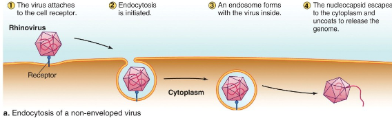
Endocytosis of non-enveloped viruses
Membrane fusion of an enveloped virus

How the flu enters

Replication cycle: Entry into plant cells
Often depends on some damage to the plant tissues to open a spot in the cell wall
• Insects feeding on plants
• Wind damage
• Hail/rain damage
• Fire damage
• Human-induced damage
Replication cycle: Entry into bacteria

How did viruses originate?
The evolutionary history of viruses isn’t very clear, but three hypotheses have emerged:
o Coevolution
o Regressive
o Progressive
Coevolution Hypothesis
Viruses evolved along with their host cells.
• This could explain the origin of many RNA viruses.
• There is little supporting evidence as of yet
Regressive hypothesis
Viruses are cells that lost some of the replicative and metabolic traits over time.
• Examples of this phenomenon exist in the biological world.
• This doesn’t adequately explain origins of RNA viruses
Progressive hypothesis
Existing genetic elements gradually gained the ability to move from cell to cell.
Evidence in the form of transposons (for DNA viruses) and retrotransposons (for retroviruses)

What are the specific traits that make viruses much tricker to work with than bacteria when trying to grow and quantify them?
they are very small
they only replicate within appropriate host cells
Bacteriophages (viruses that infect bacteria) generally follow one of two primary replication pathways depending on their viral type:
Lytic Cycle (Virulent Phages):
Lysogenic Cycle (Temperate Phages):
Lytic Cycle (Virulent Phages):
Virulent phages (such as T2 and T4) infect host bacteria, rapidly replicate their nucleic acids and capsids, and ultimately lyse (burst open) the host bacterial cell to release mature phage particles
Lysogenic Cycle (Temperate Phages):
Temperate phages (such as Lambda phage) can choose between a lytic outcome or lysogeny.
During the lysogenic phase, the phage genome circularizes and integrates into the host bacterial chromosome as a prophage (or is maintained as a plasmid)
The prophage DNA is silently replicated along with the host chromosome during cell division.
If environmental or host cell conditions change (regulated by molecular switches like the cI and cro repressor proteins), the prophage excises from the host DNA and initiates the lytic cycle
Plaques
are clear, discrete zones of cell lysis or cell death that form within a continuous lawn of susceptible host cells (or on plant leaves) following viral infection
In laboratory quantification, discrete plaques are counted as…
Plaque-Forming Units (PFUs) to determine the infectious viral titer of a sample
Animal viruses progress through a structured multi-step replication cycle:
Adherence (Attachment)
Penetration (Entry)
Uncoating
Gene Expression (Synthesis)
Genome Replication (Synthesis)
Assembly and Egress (Exit)
Adherence (Attachment) - Animal Virus Life Cycle
Viral attachment proteins specifically recognize and bind to host cell surface receptors (such as CD4 for HIV or sialic acid for Influenza virus), establishing the virus's host range
Penetration (Entry) - Animal Virus Life Cycle
The virus crosses the host plasma membrane.
Enveloped viruses enter via receptor-mediated endocytosis or direct membrane fusion, whereas non-enveloped viruses enter via endocytosis
Uncoating - Animal virus life cycle
The protein capsid breaks down or undergoes conformational changes, releasing the viral genome into the cytoplasm or nucleus
Gene Expression (Synthesis) - animal virus life cycle
The viral nucleic acid is transcribed into mRNA and translated by host ribosomes to synthesize structural and enzymatic viral proteins
Genome Replication (Synthesis) - animal virus life cycle
The virus copies its DNA or RNA genome using host or virus-encoded polymerases according to its genomic structure
Assembly and Egress (Exit) - animal virus life cycle
Assembly: Viral capsids self-assemble around new genomic copies (or package nucleic acids into pre-formed capsids).
Egress: Enveloped viruses exit by budding out of the cell membrane (acquiring a lipid envelope), while naked viruses typically exit by causing host cell lysis
Because viruses are obligate intracellular parasites, specialized methodologies are required to grow, isolate, and quantify them:
Cultivation
Purification
Quantification (Determining Viral Titer)
Cultivation
Bacteriophages are cultured on lawns of bacterial host cells.
Animal viruses are grown in sterile tissue cultures using specific cell lines (such as HeLa cells)
Purification
Initial filtration removes large host cells and debris.
Centrifugation (differential or sucrose density gradient centrifugation) isolates intact virus particles based on size and density
Quantification (Determining Viral Titer):
Plaque Assay
Direct Count
Hemagglutination Assay
Endpoint Assays
Nucleic Acid Quantification
Plaque Assay
Counts Plaque-Forming Units (PFU) on host lawns to measure infectious virus concentration
Direct Count
Visualizes and counts virus particles directly using an electron microscope (cannot distinguish infectious from non-infectious virions)
Hemagglutination Assay
Exploits the ability of certain viruses to bind red blood cells into a gel mat (rapid and simple, but non-quantitative for exact viral count)
Endpoint Assays
Calculates the TCID50 (tissue culture infectious dose causing effect in 50% of cultured cells) or LD50 (lethal dose killing 50% of test animals)
Nucleic Acid Quantification
Uses PCR or RT-PCR (Reverse Transcriptase PCR) to amplify and sequence viral DNA or RNA
What are Cytopathic Effects (CPE)?
Cytopathic Effects (CPE) refer to visually observable structural changes, damage, or cell death occurring in virally infected cultured host cells.
Common examples include cell rounding, cell fusion/clumping, and detachment from culture vessel surfaces.
CPE serves as a key indicator of viral infection in tissue cultures and is used to quantify viral infectivity in endpoint assays like TCID50
Lytic:
Phages will replicate in and burst open (lyse) the host cells
Lysogenic:
Phages can integrate their genome into the host cell genome (becoming a prophage) and be replicated each time the host cell replicates
Cultivation, Purification, and Quantification of Viruses:

Viral purification typically begins with
simple filtration to remove large cells and cellular debris
Viral purification typically begins with simple filtration to remove large cells and cellular debris. Following filtration, viruses are purified and concentrated using two main types of centrifugation techniques:
Differential Centrifugation
Gradient Centrifugation (Density Gradient Centrifugation)
Differential Centrifugation
Differential centrifugation separates components in a cell suspension step-by-step using progressively higher centrifugal speeds:
Low-Speed Centrifugation: The initial mixture of host cells and viruses is centrifuged at low speed to pellet intact host cells and large cell fragments. The liquid supernatant containing the viruses is transferred to a new tube.
Medium-Speed Centrifugation: The supernatant is centrifuged at medium speed to pellet cellular nuclei and other large organelles. The viral supernatant is again collected.
High-Speed Ultracentrifugation: The remaining supernatant is spun at high speed, causing the virus particles to settle into a concentrated viral pellet at the bottom of the tube

Gradient Centrifugation (Density Gradient Centrifugation)
Gradient centrifugation separates viral particles and cellular components according to their differing physical densities:
Gradient Preparation: A centrifuge tube is filled with layers of decreasing concentrations of a solute (such as sucrose, ranging from 20% to 70%) to create a continuous density gradient.
Layering & Separation: The viral suspension is layered on top of the sucrose gradient and centrifuged.
Band Formation: As the sample travels through the gradient during centrifugation, each component settles at the specific depth corresponding to its buoyant density, producing distinct bands (e.g., separating the band of intact virus particles from bands of residual cell debris)

How do you quantify viruses?
Direct Count
Plaque Assay
Hemagglutination Assay
Endpoint Assays
Nucleic Acid Analysis (PCR & RT-PCR)
Direct Count
Mechanism: A known volume of a viral sample is directly visualized using an electron microscope, and the visible viral particles are counted and scaled up to determine the total concentration.
Limitations: Requires specialized, expensive equipment and cannot distinguish between infectious and non-infectious (viable vs. non-viable) virions
Hemagglutination assay
Mechanism: Capitalizes on the property of certain viruses to bind to surface molecules on red blood cells (RBCs), causing the blood cells to clump together into a gel mat.
Pros & Cons: It is rapid, low-cost, and does not require a microscope, but it does not provide an exact particle count, cannot differentiate viable from non-viable viruses, and only works for viruses capable of hemagglutination

Plaque Assay
Mechanism: Serial dilutions of a viral sample are mixed with or added onto a monolayer or lawn of susceptible host cells. As viruses infect and lyse the host cells, clear zones of cell destruction called plaques emerge.
Output: Counting discrete plaques allows scientists to calculate the titer in Plaque-Forming Units (PFU) per unit volume

Endpoint Assays
Tissue Culture Infectious Dose 50 (TCID50): Quantifies the concentration or dilution of virus required to induce observable cytopathic effects (CPE) in 50% of inoculated tissue culture cell samples.
Lethal Dose 50 (LD50): Measures the dose of virus required to kill 50% of a test animal host population

Nucleic Acid Analysis (PCR & RT-PCR)
Mechanism: Uses Polymerase Chain Reaction (PCR) to detect and quantify DNA viral genomes, or Reverse-Transcriptase PCR (RT-PCR) for RNA viruses.
RT-PCR Process: The enzyme reverse transcriptase converts isolated viral RNA into a complementary DNA (cDNA) strand. Taq polymerase then exponentially amplifies the cDNA sequence, allowing precise identification and quantification of viral nucleic acids
Virus names:
Historically, quite varied!
• Simple letter/number combinations (T4 phage)
• Organism(s) they infect (tobacco mosaic virus)
• Location of discovery (Ebola River, Zaire)
• Appearance (coronavirus, “crown”)
• Disease caused (hepatitis viruses)
Virus classification uses what?
The ICTV classification scheme
The ICTV classification scheme
ICTV = International Committee on Taxonomy of Viruses
Classify viruses based on Order, Family, Subfamily, Genus, and Species
ICTV Classification Scheme
The ICTV (International Committee on Taxonomy of Viruses) provides a standardized taxonomic framework for classifying viruses.
Taxonomic Ranks: Rather than relying solely on informal names, the ICTV classifies viruses across structured hierarchical ranks: Order, Family, Subfamily, Genus, and Species.
Naming Conventions: Viral family names categorized under the ICTV scheme systematically utilize the "-viridae" suffix (e.g., Coronaviridae, Picornaviridae, Herpesviridae, Flaviviridae)
The Baltimore classification system
Developed by Nobel laureate David Baltimore
Based around mRNA production methods
Separates viruses into seven classes
Baltimore Classification System
Developed by Nobel laureate David Baltimore, this system organizes viruses based on their genome structure and methods of mRNA production. It divides all viruses into seven distinct classes consisting of 3 DNA virus groups and 4 RNA virus groups
DNA Virus Groups
Class I (dsDNA): Double-stranded DNA viruses (e.g., Adenovirus, Herpesvirus, Poxvirus).
Class II (ssDNA): Single-stranded DNA viruses (e.g., Parvovirus).
Class VII (dsDNA using Reverse Transcriptase): Double-stranded DNA viruses that replicate through an RNA intermediate using reverse transcriptase (e.g., Hepadnavirus)
RNA Virus Groups
Class III (dsRNA): Double-stranded RNA viruses (e.g., Reovirus).
Class IV (ssRNA (+)): Single-stranded positive-sense RNA viruses whose genomes can be directly translated into viral proteins or serve directly as mRNA (e.g., Picornavirus, Coronavirus, Flavivirus).
Class V (ssRNA (−)): Single-stranded negative-sense RNA viruses that must transcribe a complementary positive-sense strand to function as mRNA (e.g., Rhabdovirus, Paramyxovirus, Orthomyxovirus).
Class VI (ssRNA (+) using Reverse Transcriptase): Single-stranded positive-sense RNA retroviruses that use reverse transcriptase to convert their RNA genome into DNA for cellular integration (e.g., Retroviridae / HIV).
What is PCR?
Polymerase Chain Reaction (PCR) is a nucleic acid analysis technique used to amplify DNA sequences, identify viruses by their genome sequence, and study viral evolutionary patterns
RT-PCR (Reverse Transcription PCR)
is an adaptation of the standard PCR process that converts RNA into complementary DNA (cDNA) so that it can be amplified and analyzed. Because RNA is less stable than DNA, converting viral or cellular RNA into cDNA allows researchers to study RNA molecules, identify viral genomes by sequence, and analyze patterns of viral evolution.
The method relies on reverse transcriptase (RT), an enzyme discovered in viruses during the 1970s by David Baltimore and Howard Temin
How RT-PCR Works (Step-by-Step)
RNA Isolation: Total RNA is extracted from the sample of interest.
First-Strand Synthesis: Primers, reverse transcriptase (RT), and nucleotides are added. Reverse transcriptase uses the primer to synthesize a single-stranded cDNA copy from the RNA template.
Heat Denaturation: The sample is heated to separate the RNA-cDNA strands and inactivate the RT enzyme.
Second-Strand Synthesis: Specific primers, Taq polymerase, and nucleotides are introduced. Taq polymerase synthesizes a complementary second DNA strand, forming double-stranded DNA.
DNA Amplification: The reaction undergoes standard PCR cycles to repeatedly amplify the resulting DNA template into millions of copies for sequence identification

What are some good properties of viruses?
Gene Therapy Vectors: Because viruses naturally excel at entering host cells and delivering genetic material, modified viral vectors (such as adeno-associated virus / AAV) are used in gene therapy to deliver functional copies of genes to replace damaged or mutated versions.
Oncolytic Cancer Therapies: Certain specialized viruses, known as oncolytic viruses, selectively infect, replicate within, and destroy cancer cells.
Molecular Vectors for Cloning: Bacteriophages and phage-derived vectors (such as lambda phage vectors and cosmids) serve as molecular transport vehicles to package and insert large foreign DNA fragments (spanning 20 to 45 kb) into recipient host cells for genetic engineering.
Therapeutic Transduction: Bacteriophages can transfer genetic material into host bacteria through transduction. Researchers harness this mechanism to engineer beneficial bacteria—such as using a phage/cosmid system to transduce Lactobacillus gasseri so that it secretes CCL5, a chemokine with anti-HIV activity.
Essential Molecular Tools: Viruses are the source of key enzymes used in laboratory research. Most notably, reverse transcriptase (discovered in retroviruses) converts unstable RNA into stable cDNA, enabling RT-PCR testing, gene expression analysis, and viral genome sequencing.
Regulation of Other Viruses (Virophages): Sub-viral entities called virophages infect helper viruses and impair their replication, playing an important role in the lateral transfer of genetic material between organisms.
What is an oncovirus?
An oncovirus is a cancer-causing virus studied within virology and medicine
oncolytic viruses
specialized viruses that instead target and destroy cancer cells
What are virions?
A virion is a complete, individual virus particle. It represents the extracellular phase of the viral life cycle, serving as the vehicle that carries the viral genome from an infected host cell to a new target cell
Structural Components of a Virion
Viral Genome: The genetic material inside the virion, consisting of single-stranded or double-stranded DNA or RNA.
Capsid: A protective protein shell that encloses the genome, composed of repeating protein subunits called capsomeres.
Nucleocapsid: The combined structure formed by the nucleic acid genome and its protein capsid.
Envelope (if present):
Enveloped Virions: Possess an outer lipid membrane derived from the host plasma membrane during cell exit (budding). These membranes usually contain viral attachment proteins or spikes (e.g., gp120/gp41 in HIV or hemagglutinin in Influenza).
Naked (Non-Enveloped) Virions: Lack a lipid membrane and consist solely of the nucleocapsid
Function of Virion in the Replication Cycle
The primary role of the virion is to adhere to specific target host cell surface receptors, penetrate the host cell membrane, and uncoat its nucleic acid genome so viral gene expression and replication can take place. New virions then assemble within the host cell and are released (via cell lysis or budding) to infect adjacent cells or new hosts
What are viroids?
Viroids are sub-viral, infectious entities that are even simpler in structure than viruses
Viroid possess several distinct characteristics:
Genome Size: Extremely small, containing fewer than 400 nucleotides.
Structure & Stability: Possess a high degree of internal complementarity, which provides structural folding and makes them resistant to ribonucleases.
Host Range: To date, viroids have only been observed to infect and cause disease in plants.
What are prions?
Proteinaceous Infectious Particles: Prions are unique infectious agents composed entirely of protein, containing no nucleic acid (neither DNA nor RNA) and no genes.
Agents of Neurodegenerative Disease: They are responsible for a group of fatal neurodegenerative conditions known as transmissible spongiform encephalopathies (TSEs), which include mad cow disease and scrapie
How are Prions Transmitted?
Conformational Infection: Unlike traditional pathogens that transmit via genetic replication, prions transmit disease when an abnormal, misfolded protein enters a host environment and physically alters the structure of the host's existing normal proteins
Why Do Prions Cause Disease?
Prion pathology is driven by a cascade of protein misfolding and aggregation:
Direct Contact: An abnormal prion protein (designated PrP^Sc} as seen in scrapie) makes contact with a normal cellular protein (PrP^C).
Conformational Change: The abnormal PrP^Sc forces the normal PrP^ protein to undergo a structural alteration, converting it into a misfolded prion.
Amplification Cascade: A small number of PrP^Sc proteins progressively convert large quantities of host PrP^C proteins into the disease-associated form over time.
Fibril Accumulation: These misfolded prions aggregate to form fibrils, accumulating in nervous tissue and leading to severe neurodegenerative damage and host disease.
What are examples of prion diseases?
Mad Cow Disease (Bovine Spongiform Encephalopathy): A prominent example of a transmissible spongiform encephalopathy in cattle.
Scrapie: A prion disease affecting sheep and goats (from which the abnormal prion protein designation, PrP^Sc, takes its name)
Chronic Wasting Disease (deer and elk)
Kuru (cannibalism)
Creutzfield-jakob disease (it randomly spawns/sporadic, there is also a genetic one)
mink-wasting disease (mink)
What are satellite viruses?
Satellite viruses are sub-viral infectious agents defined by two key characteristics:
Dependence on a Helper Virus: Satellite viruses cannot replicate on their own; they require co-infection with a specific helper virus to provide essential functions for replication.
Encoding Their Own Capsid: Unlike satellite RNAs, satellite viruses contain genes that code for their own capsid protein.
Example: Hepatitis D virus (HDV) is a well-known satellite virus. It depends on Hepatitis B virus (HBV) as its helper virus to supply envelope proteins for its structure
Satellite Viruses vs. Satellite RNAs
Satellite Viruses: Require a helper virus to replicate, but carry their own capsid protein gene.
Satellite RNAs: Also require a helper virus for replication, but rely on the helper virus to provide the capsid protein
What are satellite RNAs?
Satellite RNAs are sub-viral infectious molecules characterized by the following features:
Requirement for a Helper Virus: Like satellite viruses, satellite RNAs cannot replicate on their own and require co-infection with a specific helper virus to carry out replication.
Dependence on Helper Capsid Proteins: Unlike satellite viruses (which carry their own capsid protein gene), satellite RNAs rely entirely on the helper virus to provide the capsid protein needed for packaging
West Nile virus in the news: what happened in 2012?
In the summer of 2021 there was the largest outbreak of West Nile virus seen since 2002. This was the largest manifestation of the disease and caused hundreds of deaths.
How did West Nile virus get to the United States?
It was first found in a febrile woman in the West Nile, and the first virus in the US that was detected was in NY in 1999. Then the illness started to spread to other states.
How did West Nile virus spread across the country so quickly?
So this virus needs to be spread by a host cell. So, it spread by traveling from mosquito to bird to mosquito to bird. So a mosquito will bite a bird, which will then amplify the virus, then another mosquito will bite it and get the more concentrated virus, and then bite a human giving them the virus.
How do people get infected with West Nile virus?
By getting bitten by a mosquito infected with West Nile virus. So, after someone gets bitten the infected saliva gets into their blood stream, where the virus grows and affects the immune system.
Why do some people get West Nile fever or neuroinvasive disease? Did they become infected with a “bad” virus?
It’s not known why some people get the fever and others are unaffected. Some things they believe might affect this is how much of the virus is delivered through the saliva or genetics. Some risk factors are age and if you have transplant tissue. Some other possible factors are gender and immunosuppression, and some possible things could be underlying health conditions.
Why was 2012 so bad?
For a WNV outbreak to happen three things must occur, a critical mass of infected mosquitoes, susceptible birds, and humans unprotected from biting mosquitos. This disease tends to happen during summer months, because that’s when there is an abundance of mosquitos. Another factor that affects if the mosquitos can flourish is the balance between drought and rainfall. In 2012, the US had above average temperatures and below average precipitation, making the perfect storm for mosquitos to grow.
If we know what conditions cause West Nile virus outbreaks, can we predict when a new outbreak will emerge and what its severity might be?
We can attempt to predict how it will happen, however there are a variety of factors that intersect to make this “perfect storm”, which makes it hard for us to tell. Also these outbreaks are driven by the weather condition, which is very hard to predict months in advance.
Can the severity of WNND change from year to year?
There are a few known mutations of WNND, which affect the activity of the disease. However, WNV cannot spread directly between people except under special circumstances, so any treatments that we use will not increase the chance that a resistant or more virulent strain will emerge. So, any strain that is increasingly harmful in humans will also have to be able to replicate in both mosquitoes and birds. So, it’s not impossible, however, it's just very difficult.
Can West Nile virus outbreaks be prevented?
The only way that we can “prevent” an outbreak is by watching local mosquito populations and implementing effective mosquito control when infections are detected. There are also some sprays that are to kill off the mosquitos. Unfortunately there is no treatment or vaccine for the disease.
Influenza virus (1918 flu NOVA Special)
Nucleic Acid: Single-stranded, negative-sense RNA.
Segmented Genome: Yes.
Number of Segments: 8 unique segments of viral RNA.
Number of Genes: Encodes 10 to 14 proteins depending on the specific strain, utilizing overlapping reading frames and alternative splicing on the 8 segments.
Envelope: Yes, it is a lipid membrane derived from the host cell during budding, studded with viral glycoproteins.