VM1 anatomy E1
 Call Kai
 Learn
Learn Practice Test
 Spaced Repetition
 Match
Match Flashcards
 Knowt Play
1/88

There's no tags or description
Looks like no tags are added yet.
Name | Mastery | Learn | Test | Matching | Spaced | Call with Kai | Chat |
|---|
No analytics yet
Send a link to your students to track their progress
89 Terms
dorsal
toward the back
ventral
toward belly
rostral
can also be used as “cranial” toward nose
caudal
toward tail
median plane
plane that divides animal into equal left and right halves
ipsilateral
referring to structures on the same side of the body
contralateral
referring to structures on opposite sides of body
medial
direction toward median plane
lateral
direction away from median plane or midline of body
sagital
plane that divides body into unequal left and right portions
dorsal plane
divides body into dorsal and ventral halves
transverse plane
divides body into cranial and caudal sections
adduction
limb moves toward median plane
abduction
limb moves away from median plane
circumduction
movement of a limb in a circular fashion, combining flexion, extension, adduction, and abduction
muscle, epithelial, connective, nervous
four tissue types
epithelium
covers, lines, secretes, absorbs
connective tissue
structural support tissue
muscle tisssue
tissue that contracts to produce movement
nervous tissue
tissue that integrates sensory information with motor outcomes
light microscopy (LM)
photons interacting with tissue components to create magnified image
electron microscopy (EM)
electrons interacting with tissue components to create a magnified image ; higher resolution than LM
transmission electron microscopy (TEM)
shows internal structure of a cell ; electrons are transmitted through slices of tissue
scanning electron microscopy (SEM)
shows external (surface) of cell ; electrons bounce off tissue creating a 3D image
basophilic stain
a dye that binds to acidic structures but is attracted to basic components of cells ; appear blue or purple
acidophilic
stain that binds to basic structures but is attracted to acidic components ; appear pink, red, or orange
periodic acid schiff (PAS)
stains carbohydrates magenta or purple
silver
stains reticular fibers black
trichrome
stains collagen fibers blue or green
orcein
stains elastic fibers reddish brown
nuclear envelope (nucleus, DNA), and cytoplasm (cytoskeleton, matrix)
what two basic parts of the cell does the plasma membrane surround
unit membrane
selective barrier around cells and some organelles ; appears trilaminar under TEM
E-face
outer layer of plasma membrane that faces extracellular environment
p-face
inner layer of plasma membrane that faces cytoplasm
diffusion
gradient dependent; passive
membrane transport
requires membrane transport proteins ; may be active or passive
vesicular transport
transportation of material into or out of cell by membrane bound vesicles ; active transport
exocytosis
release of material out of cell via vesicular transport ; material is packaged in secretory vesicles by golgi and transported to plasma membrane
endocytosis
uptake of material; via vesicular transport
regulated secretion
method of exocytosis where vesicles congregate near plasma membrane, have secretory granules, condense secretions, and have a signaled release into extracellular space
constitutive secretion
method of exocytosis where there is continuous secretion, no accumulation of secretory vesicles, no signaling required
pinocytosis
type of endocytosis that is a generalized type of absorption ; water and small dissolved solutes ; forms caveolae to fuse with lysosomes for processing ; aids in membrane trafficking ; endothelial cells lining capillaries
phagocytosis
type of endocytosis involved in engulfing larger particles
pseudopodia surround the material
phagosomes are formed
lysosomes degrade material
nucleus
nuclear pores on the nuclear envelope contain chromatin (DNA) whereas nucleus contains rRNA
euchromatin
uncoiled, active DNA that is more electron lucent
heterochromatin
coiled, inactive DNA that is more electron dense
polyribososmes
free ribosomes ; create cytoplasmic proteins
ER-bound ribosomes
create membrane and secretory proteins
rough endoplasmic reticulum
ribosomes stuck to outside of it ; interconnected, flattened membranous sacs for protein synthesis
golgi apparatus
interconnected flattened sacs where proteins from rER are processed, sorted, and packaged
cis face
face of golgi that faces rER
trans face
face of golgi that faces plasma membrane
lysosomes
digestive compartments ; spherical and membrane-enclosed ; varying electron densities ; contain enzymes that degrade lysosomal contents
autophagosomes
endocytotic vesicles
phagosomes - in specialized cells
lysosomes fuse with…
mitochondria
chemical energy conversion ; site of ATP synthesis ; 2 unit membranes (outer - smooth, and inner - folded)
cristae
extend into the matrix as broad flattened folds of inner membrane
vesicular mitochondria
steroid hormone producing cells
smooth endoplasmic reticulum
no ribosomes attached, interconnected membrane-enclosed tubules ; abundant in cells that detoxify, and secrete steroid hormones
actin
thin filament proteins that help anchor cells to each other ; form structural core of microvilli
intermediate filaments
cell-to-cell adhesion ; high tensile strength
microtubules
largest cytoskeletal component ; provide rigid structure to cell shape
epithelium
type of tissue that covers, lines, secretes, and absorbs ; avascular ; exhibit polarity
connective tissue
type of tissue used for structural support
muscle tissue
tissue type that contracts to produce movement
nervous tissue
tissue type that integrates sensory information with motor outcomes
epithelial cells
cells that are closely packed together and attached basally to basement membrane ; face lumen (outside world)
domains of epithelial cells
apical, lateral, basal (borders basement membrane ; related to connective tissue)
apical domain
domain of epithelial cell that faces lumen ; define function of epithelial cell ; contain microvilli for absorption and cilia for movement
microvili
absorption factor of apical domain of epithelial cell ; extend lumen to increase surface area
cilia
motion factor of apical domain of epithelial cell ; contain microtubules ; locked into cell by basal body
microtubules
contain 9 peripheral doublets and 2 central singlets ; basal bodies are developed from the 9+2 arrangement
lateral domain
domain of epithelial cell that forms a selective barrier ; adherence ; cell-to-cell communication ; surfaces show infoldings prominent in cells engaged in fluid transport to increase lateral surface area
junctional complex - lateral domain
zonula occludens
zonula adherens
macula adherents (desmosomes)
zonula occludens
most apical part of junctional complex that forms protective barrier between epithelial cells ; separates luminal space from intercellular space ; freeze fracture
free fracture
technique used to study membrane structures by separating bilayers to reveal protein and lipid distribution ; exposes location where two cells connect
zona adherence
part of junctional complex that aids in cell-to-cell adhesion ; cells do not contact each other ; supported by actin ; spanned by integral membrane proteins

macula adherens
desmosomes ; cell-to-cell adhesion ; contain attachment plaques and linking proteins

gap junctions
not part of junctional complex ; communicating junctions ; cells approach each other but have a gap ; direct passage of signaling molecules from one cell to another ; found in tissues that accomplish coordinated functions (cardiac cells, neurons)
connexon
creates channel through membrane ; when one from cell A lines up with one from cell B it forms a gap junction
substances in extracellular space cannot enter this ; substances in this cannot leak into intracellular space
basal domain
the surface of an epithelial cell that is attached to the underlying basement membrane; involved in adhesion and communication with the extracellular matrix
basement membrane
basal lamina (thin black line) + reticular lamina (large space)
hemidesmosome
½ of a desmosome
focal adhesions
associated with actin filaments important for mechanoreception
squamus

cubodial
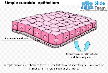
columnar

stratified epithelium
a type of epithelial tissue consisting of multiple layers of cells
non-keratinized
epithelium that remains moist and does not have a layer of keratin, often found in areas like the oral cavity and esophagus. ; alive cells
keratinized
a type of epithelial tissue characterized by a layer of cells that are filled with keratin, providing protection and moisture resistance ; dead ; toughest - makes up most of skin