BIOC
 Call Kai
 Learn
 Practice Test
 Spaced Repetition
 Match
 Flashcards
 Knowt Play
1/63

There's no tags or description
Looks like no tags are added yet.
64 Terms
Seven AAs to remember charge
Asp, Gly, Cys, Tyr
Pro: 0
Dep: -1
Arg, His, Lys
Pro: +1
Dep: 0
3 macromolecules
Proteins: myoglobin stores O2 in muscle tissue
Polysaccharides
Nucleic acids
Polypeptide vs Oligopeptide
P: many amino acids, usually a complete protein
O: few amino acids, usually a fragment
Polarity Defined as
Different electronegativities (tendency to hold bonding electrons)
Hydrogen bond acceptors vs donors
Acceptors: electronegative atom with available lone pair of electrons
Donor: hydrogen atom covalently bonded to other electronegative atom
Electronegative elements
O>N>S>C=H (C about H)
Henderson-Hasselbalch equation (to find % dep and pro)
pH = pka + log(Dep/Pro)
Dep = X
Pro = 1 - X
Hydrogen bond donor & acceptors (aa)
Donors: Arg, Lys, Trp
Acceptors: Asp, Glu
Both: Ser, Thr, Asn, Gln, Tyr, His
Column chromatography
buffer on top then separation occurs
Partition Chromatography
Mobile phase: silica gel (polar)
Stationary phase: liquid solvent/buffer
NON polar amino acids move faster
Thin Layer Chromatography
Thin sheet of silica
Sheet: polar
Buffer/liquid: non-polar
Elution volume
Volume of buffer needed to move through column
Affinity chromatography
Tagged proteins bind to ligand
Immobilized metal affinity chromatography
His binds to Ni2+ or Co2+
Resin contains Ni2+
Imidazole elutes bound protein
One step purification
Gel filtration (molecular exclusion chromatography)
Gel filled with pores
Large molecules pass through first
Electrophoresis
Movement of charged particles in an electric field
Polyacrylamide gel: sodium dodecyl sulfate (SDS) surrounds protein with negative charge
Isoelectric focusing: separate by isoelectric point (when net charge=0). Then increasing pH deprotonates aa and they move to + electrode
Two dimensional gel: includes both methods
Mass Spectrometry
To identify proteins
Larger particles move more slowly
Specific Activity equation
Specific Activity = Enzyme Activity/Total Protein
Step by step purification of a protein (3)
Enzyme activity decreases
Total protein decreases
Specific activity increases
(specific activity = measurement of purity)
1 Dalton to g/mol
1:1 ratio
1 Da = 1g/mol
Log (molar mass) is the _ axis and elution volume is the _ axis
Log (mm): Y
EV: X
y = mx + b
Protein structure hierarchy
Primary: linear
secondary: a-helical or b-sheet
Tertiary: myoglobin
Quaternary: Hemoglobin
Protease function
catalyzing hydrolysis (H2O is a poor nucelophile)
What does a nucleophile do
attack/break bonds
Sanger sequencing
N-terminal tagging: tags first aa
but then first aa is lost
Perh Edman sequencing (2 steps)
Coupling: Weak base
Cyclization: anhydrous acid (except Gly could cyclize early)
Trypsin binds & cuts:
Arg or Lys (unless pro follows: to right)
Cuts right side
Chymotrypsin targets:
Phe, Tyr, Trp
CnBr (Cyanogen Bromide) attacks:
S of Met
Met → Hse (homoserine: serine w extra Ch2)
a-helix and Å/turn
3.6 aa per turn of helix
5.4 Å per turn
So 5.4/3.6 = 1.5 Å (distance between amino acids, or amino acid every 1.5 Å)
Parallel vs antiparallel B-sheet
Parallel: angled bonds:
Strands run in same direction
Antiparallel: parallel bonds → better alignment → stronger
Strands run in opposite directions
B-sheet aas and breaker aas
B-sheet: VIT C WYF
Breakers: GPS ND
Collagen: triple helical shape (what aas)
G-P-X
Stabilizing interactions in protein structure (4)
Ion pairs (+/-)
H-bonds
Disulfide bonds (between cys)
Van der Waals (close interactions)
3 things an atom with a lone pair of electrons can do
Accept hydrogen bonds
Act as a base (capture H+)
Nucleophile: making a bond by sharing e-
In a-helix: h bonding occurs:
at every 5th aa
So atom n H bonds with atom n + 4
Denaturation of protein can be done by:
heat, solvents, detergents (SDS)
a vs b keratin
a: a helix
b: fibroin → antiparallel b sheet
What drives protein folding
hydrophobic effect
hydrophobic (non-polar) aas stay inside
hydrophilic (polar) aas like outside
5 tertiary structures
a-helix bundle: np sides face each other
B-sheet: antiparallel Bs
parallel B-sheet: B-a-B-a alternating structure
B-strands same direction, np side of sheet faces np side of a-helix
a-B-barrel (parallel): a-B sheet when helicases on outside
a-B sandwich (parallel): np B-sheet between 2 a-helix
Disulfide bond
SH of Cys reacts with O2 (oxidation) → condensation rxn
R-SH & SH-R + O2 → R-S-S-R + H20
Urea & 2-mercaptoethanol
Denatures protein
Urea: weakens hydrophobic effect: unfolds (denatures) protein
2-mercaptoethanol: reducing agent: restores disulfide bond to Cys
After removing both, protein regains native state (renaturation)
Elastase binds ____ and ____ best
Gly, Ala
Arrhenius Equation (rate)
Rate = pze^-(Ea/RT)
P probability factor (orientation increases)
Z collision frequency (proximity increases)
R constant
T temperature
Ea activation energy

4 types of catalysis
Nucleophilic: provides better nucleophile
e.g. Cys-S, His-H or Asp/Glu COO- (removing H+)
Electrophile:
Using electron seeking group (electrophile) to remove e- from substrate
General Base: Removes H+
General Acid: Donates H+
Prefixes of centi, milli, micro, none (g), kilo
Centi: -2
Milli: -3
Micro: -6
none: 0
kilo: 3
2 Absorbance equations
Io = intensity of light
I = Measured intensity (transmitted light)
E: extinction coefficient (L/mol/cm)
c = sample concentration (mol/L
L = sample thickness (cm)

4 Enzyme catalysis equations
Rate of reaction: change in substrate (product) over time
(mol/L min)
Enzyme activity: moles of substrate over time
rate x volume (mol/min)
Specific activity: enzyme units/mg total protein
Enzyme activity/total protein (mol/min mg)
Molar activity: activity per mole of enzyme
Specific activity x enzyme molar mass (1/min)
Rate equations for vo, vmax k1 & k-1
vo (initial rate) = k2(ES)
vmax: k2(E)t
k1 (forward) = k1(E)(S)
k-1 (reverse) = k2(ES) + k-1(ES)
equation at steady rate & km
(k-1 +k2)(ES) = k1(E)(S)
let km = (k-1 + k2)/k1
so km(ES) = (E)(S)
let (E) = (E)t - (ES)
then leads to (ES) = (E)t(S)/(km + (S))
let vo=k2(ES)
then leads to vo=k2(E)t(S)/(km + (S))
let vmax=k2(E)t
then leads to vo= Vmax(S)/ (km + (S))
Higher Vmax means
faster reaction rate, better catalysis
k2 is the true __________ and is the ___________________
constant
Turnover number
km is the concentration of (S) at which rate vo is equal to _____ of the Vmax
50%
If (S) = km, then vo/Vmax = 0.5
Km definition
[S] at ½ Vmax
Double reciprocal plot equation
1/Vo = (Km/Vmax)(1/(S)) + 1/Vmax
y = mx + b
slope: km/Vmax
y-int: 1/Vmax
x-int: -1/km
Competitive vs Non-competitive equilibrium
Competitive: inhibitor can only bind to (E)
km increased
Non-competitive: inhibitor can bind to (E) or (ES)
Vmax decreased

5: very polar aas
4 polar aas
5 mod polar aas
6 non polar aas
Very: Asp, Glu, Arg, His, Lys
Polar: Ser, Thr, Asn, Gln
Mod: Gly, Cys, Pro, Trp, Tyr
Non: Ala, Val, Leu, Ile, Phe, Met
Competitive vs Non-competitive reciprocal graphs

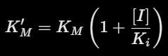
K’m equation
Which amino acid’s N-terminal is unavailable to H-bond
Proline
Most mobile in thin layer chromatography
non polar: elute first and move furthest
SDS gel electrophoresis to saparate amino acids by
SIZE
dithiothreitol/performic acid/Urea/2-mercetethanol
dithiothreitol: reducing agent, disulfide bonds → cys
performic acid: oxidizing agent: disulfide bonds → cys → cysteic acid
Urea: weakens hydrophobic effect
2-mercaptethanol: reducing agent, disulfide bonds → cys
Benefit of measuring initial rate vo at t=0
[P]=0 so reverse reaction can be ignored