Corneal disease
 Call Kai
 Learn
 Practice Test
 Spaced Repetition
 Match
 Flashcards
 Knowt Play
1/55

There's no tags or description
Looks like no tags are added yet.
56 Terms
What are the three layers of the eye?
Outer fibrous
Sclera and cornea
Middle vascular
Inner nerve
Where do sclera and cornea meet?
At limbus
Ahat are the three layers of the sclera?
Episcleral
Scleral stroma
Lamina fusca
Explain the blood supply of the sclera
Short posterior ciliary aa +nn -> ON choroid
Long posterior ciliary aa + nn -> ON CB
Anterior ciliary aa + vortex veins > CB
What are the features of the corneal anatomy?
Avascular, no pigments and low cell density
Main refractive surface of eye
4 layers
Epihtelium
Stroma
Descemet's
Endothelium
What are the features of normal corneal healing?
Good regenerative ability
'Slide and devide' healing
Limbal source of stem cells
Entire epithelium can regenratie within a week
What attached the corneal epithelium to the basement membrane?
Hemidesmosome
What are the features of the stroma layer of the cornea?
90% of corneal thickness
Provides rigidity and transparency
Lamellar sheets, equidistant and parallel
What are the features of stromal healing?
Avascular healing-> Superficial/ acute/ sterile
Neutrophils causing keratocyte death
Fibroblasts secreting collagen
Vascularised healing-> Deep/ Chronic/ Infected
Blood vessel invasion
Fibrovascular granulation
What are the differences between avascular healing and vascularised healing outcomes?
Avascular: scar, weeks to months, facet
Vascularised: denser scar, ghost blood vessels, months to year
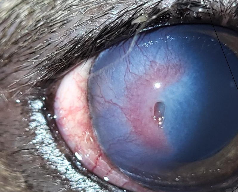
What are the features of descemet's membrane?
Very thin and elastic
Thicker as gets older
What signs can you get with descemet's membrane damage?
Can break- Haab's striae
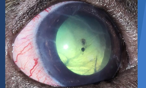
Exposed in deep ulcer-> descemetocoele
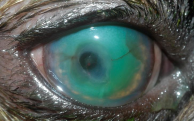
What are the features of the endothelium of the cornea?
Single cell thick
Dehydrates stroma
Limited capacity to regene
Decompensation-> oedema
What is the consequence of disurption to the cornea?
Opacification
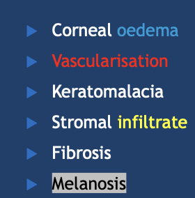
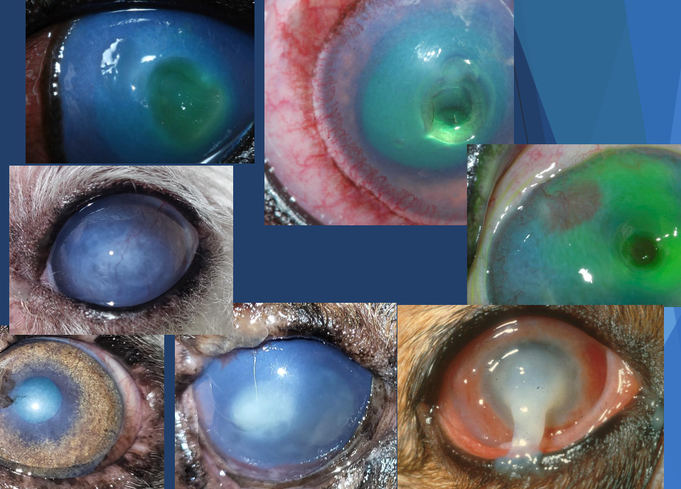
What can the different colours tell you about disruption to the cornea?
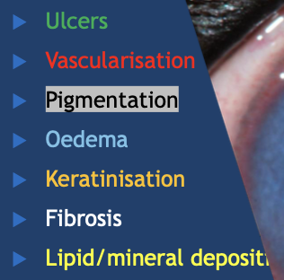
What is reflex uveitis? What causes it?
All corneal ulcers cause reflex uveitis
Mediated by axonal reflex within CN V- Painful!!
How do you treat reflex uveitis?
Systemic NSAIDs
Cyclopegic- paralyses ciliary muscle so stops spasming
What is a corneal ulcer?
A hole in the epithelium
Exposure of the stroma or Descemet’s membrane
A full thickness defect in the epithelium
NB- Clinical signs not diagnosis
How is a corneal ulcer diagnosed?
History
CS- ocular pain
Dx tests
STT, IOP, Fluorescin
(Further- bacteriology, AB sens, cytology)
What is a corneal facet?
Corneal defect (dent)
Fluorescein negative
Epithelialised deep ulcer
Non-painful
How do you diagnosed corneal perforation?
Seidel test
Check anterior chamber depth- shallow
Reduced IOP-care
What is a siedal test?
Test for corneal perforation and aqueous leakage
Apply stain, don’t rinse
Cobalt blue filter/Wood’s lamp
See clear aqueous running through green dye
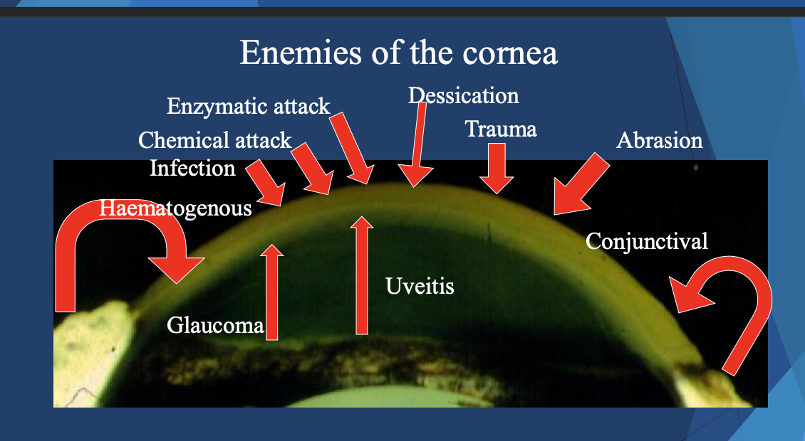
List some of the causes of ulcers?
Trauma
FB
Dry eye-q or q
Conformation:lagophthalmos/ mp fissure/ entropion/ facial folds
Neurological: CN V (neurotrophic) or VII (neuroparalytic) keratitis
XS irritation: lashes(ectopic cilia/distichiasis)/ masses/ blepharitis
Infections: primary FHV-1, M.bovis or 2ry infection
Miscellaneous
Why are brachys predisposed to corneal ulcer?
Over exposed corneas
Reduced corneal sensation
Reduced blinking (3-5/min)
Poor tear film quality
Irritation from medial entropion
What are the different types of ulcers?
Simple vs complicated
Superficial vs deep
How are ulcers treated?
IDENTIFY AND REMOVE CAUSE
PREVENT SELF TRAUMA
Antibiotics
Cycloplegic
Analgesia
Anti-collagenase
Antivirals
Lubricants
Surgery
What are your antibiotic choices for eye ulcers?
Chloramphenicol (fridge, ointment corneal penetration)
Not against pseudomonas
Fusidic acid (staphs only)
Chlortetracycline
Chlortetracycline
Oflaxacin
Resistance- limit use C&S
NB- C&S first is best
What are your options for cyclopegics for treatment of corneal ulcers?
Atropine (+mydriatic, better cyclocpegic)
Longer duration
Tropicamide (+mydriatic, not a good cyclopegic)
What analgesics can you use for corneal ulcers?
Systemic NSAID
Topical atropine to effect
Systemic opioids
Bandage lens
E-collar
NEVERE- topical anaethetics, topical steroids or NSAIDs as -> melting
What is keratomalacia?
Melting ulcers/ acute stromal collagenolysis
Can develop as complication of any ulcer type
Exogenous and endogenous protease activity
Rapid collagenolysis and risk of rupture
Can also be caused by chemicals- flush eye in this case
NB- urgent aggressive medical therapy, stitch eye
What are the risk factors of melting ulcers?
Brachycephalics
Anaesthesia
Other ocular disease: KCS, ulcers, corneal surgery
Trauma-mechanical or chemical
Steroids
Systemic disease(skin/endocrine)
How can you treat keratomalacia?
Anti collagenase treatment
Tetracycline
EDTA
N-acetylcysteine
Serum (pateint's own)
Stromease
N-acetylcyeine
AB
What is SCCEDs? What causes them?
Spontaneous chronic corneal epithelial defect
Boxer ulcers/ indolent ulcers
Non healing- painful and persistent
Hemidesmosome adhesion complex problem
How is SCCED treated?
Sedation
Topical LA
Debride loose epithelium with cotton bud
Flush eye
Topical AB
Bandage lens
Temporary lateral partial tarsorrhaphy
Ecollar
Remove collar and sutures 2 weeks
What are debriding, and grid keratotomy indicated for?
SCCEDs ONLY
NOT in normaly corneal ulceration
NEVER in cat
Summary of corneal ulcers and treatment
Superifical- superficial (just ulcer) or SCCED (appears beyond edge of ulcer)
Deep ulcer: walls and floor stain, desmetocoele, walls stain not floor (come up as black hole)
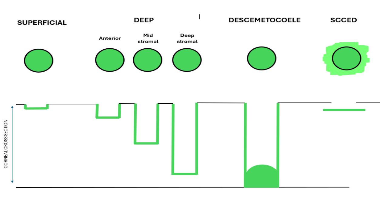
Medical- superificial and anterior deep
Surgical stromal (>50% depth) and deep stromal and descemetocoele
SCCED- only one do Grid keratotomy
List the corneal diseases other than ulcers
Congenital
ASD-PPM-leukoma
Microcornea/megalocornea
Dermoid
Acquired
Immune mediated
Neoplasia
What breeds are predisp to dermoids?
Shih Tzus
German Shepherds
Basset Hounds
Bulldogs
Labradors
Rare in cats
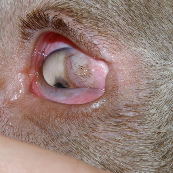
How are dermoids treated?
Surgical resection: lid margin, conjunctivectomy, keratectomy, ocular lubrication
What are the risks with corneal lacerations/ FB?
AC collapse
Iris prolapse
Endophthalmitis
Secondary glaucoma
Phacoclastic uveitis
NB- refer urgently
List the acquired corneal diseases
Corneal sequestrum
Corneal lipidosis:
Corneal arcus
Lipid keratopathy
Crystaline stromal dystrophy
Calcium degeneration
Endothelial dystrophy/degeneration
Pigmentary keratopathy
Trauma: lacerations/FB
What is calcium degeneration/ calcium keratopathy?
1° or 2° in older patients
Old animals
Ca infiltrate looks matte, chalky
Treat underlying cause
What are the features of corneal lipidosis?
CRYSTALLINE STROMAL DYSTROPHY
Young adult dogs
Slowly progresses
Superficial stromal lipid deposition in central/paracentral cornea-sparkly when illuminated-ground glass appearance
Not painful
FL-ve
No tx- Resection leads to recurrence
What are the features of lipid keratopathy?
2° to other disease
Inflammation
Always vascularised
Unilateral
Location based on prior disease
+/- calcification/ degen
What are the features of corneal arcus?
Rare
Due to systemic hyperlipoproteinaemia
Bilateral lipid infiltration of perilimbal cornea
Arc shape-follows limbal curvature
Grey-ish colour
Lucid interval of Vogt
+/-vascularisation later
Investigate and Tx: endocrine/liver/pancreatic disease
What animals are predisposed to endothelial degeneration?
Boston terrier- young animals
Mostly old animals (senile endothelial degeneration)
What are the features of endothelial degeneration?
Progressive slowly-> corneal oedema
Bilateral but not symmetrical
Not painful but bullae erupt ulceratoin
Visible but as progresses-> vision compromised
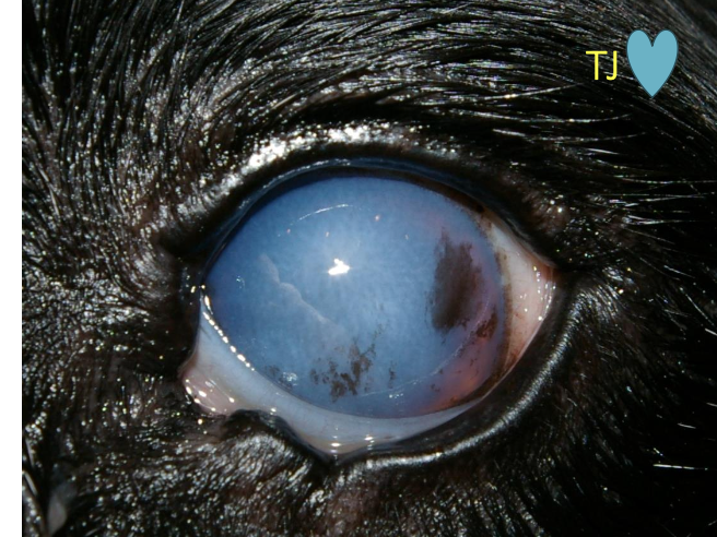
How is endothelial degeneration treated?
Surgery: Keratectomy + Gunderson Flap (refer)
What causes pigmentary keratopathy?
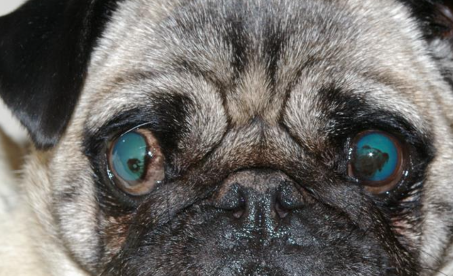
Response to chronic corneal surface irritation-any animal
(response to entropion/ trichiasis /CSK /KCS /lagophthalmos)
From limbus-pigmented limbus more likley affected
How is pigmentary keratopathy treated?
Remove irritation
Topical ciclosporine
Cryotherapy/keratectomy
Medial canthoplasty
What are the immune mediated diseases of the cornea?
Pannus
KCS
Eosinohophilic keratitis
What is pannus?
Chronic superficial keratitis (CSK)
Common in GSD/similar, Greyhounds
Lymphoplasmacytic cell anterior stromal infiltration from lateral/ventrolateral limbus-can involve entire cornea
Immune mediated/idiopathic
UV light (summer)
+/-plasmoma
Not painful
Vision compromised-especially if extensive
Scarring &pigment deposition
How is pannus diagnosed?
CS
Cytology
How is pannus treated?
Topical corticosteroid QID
initially(taper)
Topical Optimmune BID
Lifelong maintenance therapy
What are the features of epithelial inclusion cysts?
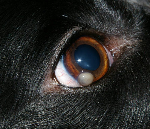
Non painful
Round, smooth corneal surface cysts
Size varies
Epithelium in stroma
After corneal surgery/trauma
Tx: Keratectomy
What are the neoplasias of the cornea (RARE)?
SCC
Haemangiosarc
Papilloma
Limbal melanoma