RADSCI 4615 - exam 1 added
 Call Kai
 Learn
 Practice Test
 Spaced Repetition
 Match
 Flashcards
 Knowt Play
1/211

There's no tags or description
Looks like no tags are added yet.
212 Terms
pathology
the study of the causes & effects of disease
disease
- the pattern of the body's response to some form of injury that causes a deviation from normal conditions
- causes = hereditary, trauma, infectious organisms, vascular / metabolic processes
signs
measurable / objective manifestations
symptoms
subjective manifestations (feelings that the patient describes)
asymptomatic
without subjective OR objective manifestations
diagnosis
identification of disease process
etiology
study of the cause of the disease process
idiopathic
underlying cause is unknown
prognosis
probable patient outcome
syndrome
linked combination of signs & symptoms
iatrogenic
disease caused by physician / treatment
nosocomial infection
- contracted from the acute case facility / hospital
- most common = UTI
iatrogenic vs nosocomial
all iatrogenic infections are nosocomial, but not all nosocomial infections are iatrogenic
community-acquired infection
contracted in a public setting outside of the acute care facility / hospital
inflammation
the initial response of body tissues to local injury (-itis)
5 clinical signs of inflammation
1. rubor (redness)
2. calor (heat)
3. tumor (swelling)
4. dolor (pain)
5. loss of function
events that occur in an inflammatory response
1. alterations (increased) blood flow & vascular permeability
2. migration of circulating white blood cells to the interstitium of the injured tissue
3. phagocytosis & enzymatic digestion of dead cells / tissue elements
4. repair of injury by regeneration of normal parenchymal cells / proliferation of granulation tissue
permeable membrane
allows fluids / cells to pass from 1 tissue to another tissue (or location)
granulation tissue
fibrous scar tissue replaces destroyed tissue
pyogenic bacteria
thick, yellow fluid called pus (dead white blood cells)
abcess
- large packet of pus
- localized, encapsulated collection of fluid
bacteremia
- potential involvement of other organs & tissues in the body by organisms invading the blood vessels
- leads to sepsis (bacteria deposited in organs --> shuts organs down)
edema
accumulation of abnormal amounts of fluid in the intercellular tissue spaces / body cavities ("tumor")
anasarca
- common in peds
- Generalized edema occurs with pronounced swelling of subcutaneous tissues throughout the body
elephantiasis
localized lymphatic obstruction resulting in localized edema (usually lower extremety)
ischemia
- interference with the blood supply to an organ / part of an organ (reduced blood supply, not obstruction)
- deprives cells & tissues of oxygen / nutrients
- may lead to infarction
causes of ischemia
- narrowing of an artery (atherosclerosis)
- thrombotic occlusion (blot clot that stays in place)
- embolic occlusion ('blood clot' that moves)
infarction
- localized area of ischemic necrosis within a tissue / organ
- produced by occlusion or arterial supply or venous drainage
hemorrhage
- rupture of a blood vessel that allows blood to escape within surrounding tissues / cavities
- bleeding into tissues = hematoma
hematoma
accumulation of blood trapped within body tissues
alterations of cell growth
- changes in the # of cells, size of cells, cell differentiation, cell arrangement
- may develop in response to physiologic stimuli
atrophy
- reduction in the size / # of cells in an organ or tissue
- corresponding decrease in function

hypertrophy
increase in the size of cells in an organ / tissue in response to a demand for increased function

hyperplasia
increase in the # of cells in an organ / tissue

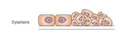
dysplasia
loss of uniformity of individual cells & their architectural orientation
metaplasia
changing of 1 type of cells to another (ex: Barrett's esophagus)

neoplasia
- abnormal proliferation of cells that are no longer controlled by the factors that govern the growth of normal cells
- malignant or benign

oncology
study of neoplasms (tumors)
benign
growth which closely resembles it's cells of origin in structure & function
malignant
neoplastic growth that invades & destroys adjacent structure
metastasize
malignant tumors that travel to distant sites
carcinoma
malignant tumor of epithelial cell origin
anaplastic
undifferentiated cell growth (without form)
sarcoma
highly malignant tumor originating from connective tissue
lymphatic spread
carcinomas metastasizing through lympathic system
hematogenous spread
malignant tumors that have invaded the circulatory system & travel as neoplastic emboli through the blood stream
grading
assessment of aggressiveness / degree of malignancy (lower grade = less malignant)
staging
- extensiveness of tumor at the primary site
- presence or absence of metastases to lymph nodes & distant organs
epidemiology
study of determinants of disease events in a given population
morbidity
rate that an illness / abnormality occurs
mortality
the # of deaths by disease per population
hereditary disease
- result from an abnormality in DNA
- pass from 1 generation to the next through the genetic info contained in the nucleus of each cell
autosomes
44 chromosomes OTHER THAN X and Y
dominant genes
always produce an effect
recessive genes
manifest themselves when a person is homogenous for that trait
mutations
alterations in the DNA structures that may becomes permanent hereditary changes
immunity
production of antibodies (immunoglobulins) that bind to antigens (foreign substances) to make them harmless
active immunity
- introduce pathogen --> body knows how to make antibodies against it
- vaccines / toxoids
passive immunity
preformed antibodies from a donor injected into a patient
types of immune reactions
1. anaphylactic
2. cytotoxic reaction
3. delayed
anaphylactic reaction
- hypersensitivity reaction resulting in histamine release
- ex: urticaria (hives), bronchospasm, laryngeal edema, vascular collapse (shock)
cytotoxic reaction
- cell destruction due to antigen inside / attached to cell
- ex: blood transfusion (blood types don't match)
delayed immune reaction
- manifestation after previously sensitized
- ex: poison ivy, contrast reaction
infectious disease protection precautions
- standard precautions from CDC
- use appropriate protective personnel equipment (PPE)
- utilize transmission-based precautions
respiratory system
- function = intake oxygen & remove CO2 from the blood
- alveoli = smallest functional unit of the respiratory system (where actual gas exchange occurs)
congenital / hereditary respiratory diseases
- cystic fibrosis
- hyaline membrane disease
cystic fibrosis
- location = bronchi
- caused by defective gene of chromosome 7
- characterized by the secretion of excessive viscous mucus (mucus is too thick to be expelled)
- traps bacteria & block air passage --> cysts, infection, abscesses
- also affects pancreas & digestive system (90% of mortality is a result of respiratory involvement)
- coarse reticular pattern (scar tissue formations around cysts)

hyaline membrane disease
- AKA infant respiratory distress syndrome (IRDS)
- location = alveoli
- one of the most common causes of respiratory distress in newborns (premature)
- caused by a lack of surfactant in immature lungs ( < 34 weeks)
- surfactant decreases surface tension --> alveoli can open back up (don't get stuck together)
- air bronchogram --> bronchus visible on CXR (air vs dense lung tissue)

inflammatory upper respiratory disorders
- croup
- epiglottitis
croup
- viral infection in young children --> produces inflammatory obstructive swelling (localized to the subglottic portion of the trachea)
- barking cough
- pencil sign (pinched trachea)
- treatment = steam OR corticosteroids

epiglottitis
- caused by haemophilus influenzae in children
- causes thickening of epiglottic tissue & the surrounding pharyngeal structures
- Hib vaccine = incidence decreased
- CXR = "thumb sign"

inflammatory lower respiratory disorders
- pneumonia
- anthrax
- lung abscess
- tuberculosis
- pulmonary mycosis
- respiratory syncytial virus (RSV)
pneumonia
- inflammation of the lung
- 3 radiographic patterns = alveolar pneumonia, bronchopneumonia, interstitial pneumonia

alveolar pneumonia
air bronchogram

bronchopneumonia
- location = mainstem bronchus
- central consolidations / densities (no peripheral issues)
- not patchy

interstitial pneumonia
patchy densities

lung abscess
- necrotic area of pulmonary parenchyma containing puslike material
- most common cause = aspiration
- treatment = antibiotics OR surgical intervention (drains)
- patients present with fever & foul smelling septum
- CXR shows air fluid levels
- CT shows lung cavities & air fluid levels

tuberculosis
- caused by mycobacterium tuberculosis (rod shaped bacterium with protective waxy coat)
- spreads by droplets in the air (less common = ingestion of infected cows milk)
- primary --> latent stage --> secondary (secondary occurs when immune system is weakened)
- treatment = double drug therapy (first part breaks down waxy coating & second part kills bacteria)
- "cheese lung"
- untreated = massive hemoptysis (death)
- hilar retraction
- air-fluid levels & abscesses (caseous lesions)

primary tuberculosis
hilar lymph node enlargement

tuberculoma
walled off scar tissue area where TB is (latent stage)

miliary tuberculosis

pulmonary mysosis
- fungal infection of the lung
- most common fungal infections = histoplasmosis (dry air) & coccidioidomycosis (ohio-river valley)
- usually asymptomatic (no treatment needed)
- CXR = fungal balls throughout the lungs

respiratory syncytial virus (RSV)
- most common cause of bronchiolitis (inflammation of bronchioles)
- attacks the lower respiratory tract = necrosis of the respiratory epithelium of the bronchi & bronchioles
- bronchi obstruction occurs due to necrotic material & edema from infection
- results in interstitial pneumonia
- almost all children contract RSV by age 2
- streaky ("camo pattern")

diffuse lung disease
- chronic obstructive pulmonary disease (COPD)
- sarcoidosis
- pneumoconiosis
chronic obstructive pulmonary disease (COPD)
- chronic obstruction of the airways leads to an ineffective exchange of respiratory gases (makes breathing difficult)
- types = chronic bronchitis, emphysema, asthma
chronic bronchitis
- 90% caused by smoking
- inflammation & excessive mucus production
- thickening of bronchial walls
- leads to emphysema
- tram lines = inflamed bronchus walls

emphysema
- inability of full expiration (air gets trapped each time you breathe)
- damage cilia --> can't cough up pathogens
- bronchial narrowing & loss of elasticity
- alveolar distention & rupture (bullae = ruptured alveoli causing large air filled spaces)
- blunt costophrenic angles
- barrel chest
- large retro-sternal air space

asthma
- severe narrowing & spasm
- extrinsic (environment) & intrinsic (emotional) stimuli
- no cure
- chronic attacks leading to infections & fibrosis
- normal pulmonary markings that are thickened

sarcoidosis
- multisystem granulomatous disease of unknown cause
- most often detected in young adults
- enlargement of chest lymph nodes
- 1, 2, 3 sign = enlargement of 2 hilar lymph nodes & 1 paratracheal lymph nodes
- osteolytic lesions in hands & feet
- may lead to diffuse pulmonary disease (like emphysema)
- may regress spontaneously (or with steroids)
- irreversible pulmonary changes in 20% of patients

pneumoconiosis
- caused by inhalation of irritating particles
- short term = mucosal lining damaged
- long term = pulmonary tissue damage & malignancy (irreversible)
- treatment = prevent further exposure & breathe clean air
- types = asbestosis, silicosis, anthracosis
asbestosis
- construction materials
- pleural thickening & calcified plaques

silicosis
- mining & sandblasting (quarts dust)
- CXR = looks like a mouse

anthracosis
- coal workers disease
- anthracite

respiratory neoplasms
- solitary pulmonary nodule
- bronchial adenoma
- bronchogenic carcinoma
- pulmonary metastases
solitary pulmonary nodule (SPN)
- asymptomatic (incidental finding on CXR)
- nodule density in the lung that we don't know what it is (benign granuloma, small neoplasm, solitary metastasis)
- increased age = increased risk of malignancy
- treatment = biopsy / PET

bronchial adenoma
- low-grade malignant lesions that form in glandular structures
- appears in younger age groups
- found centrally in major / segmental bronchi (soft tissue balls) --> may grow large enough to cause obstruction
- most common symptoms = hemoptysis (coughing up blood), recurring pneumonia, atelectasis

bronchogenic carcinoma
- arises from the mucosa of the bronchial tree
- linked to smoking & inhalation of carcinogens
- non-small cell types (80%) = squamous carcinoma (central), adenocarcinoma (peripheral), bronchiolar / alveolar cell carcinoma (least common)
- small cell (20%) = bulky enlargement of hilar lymph nodes (lymph nodes encase the bronchial tree

pulmonary metastases
- pulmonary metastases develop from hematogenous / lymphatic spread (primary tumor somewhere else that has spread to the lungs)
- ~25% of all cancer patients will develop lung mets
- you will die with pulmonary mets EXCEPT testicular pulmonary mets (responds well to treatment)
- treatment = palliative care only

mediastinum compartments
- anterior = extends from the sternum back to the trachea / anterior border of the heart
- middle = contains the heart, great vessels, central tracheobronchial tree, lymph nodes, phrenic nerves
- posterior = space behind the pericardium

mediastinal masses
- cystic (fluid filled) vs solid (more likely to compress)
- symptoms depends on area of compression (1/3 of patients are asymptomatic)
- treatment = excision of lesion (benign or malignant)
